4J9E
 
 | |
4RTY
 | |
4RTV
 | |
7ZLX
 | |
7ZR2
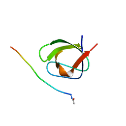
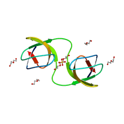
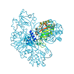
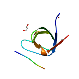
 | | Crystal structure of a chimeric protein mimic of SARS-CoV-2 Spike HR1 in complex with HR2 | | Descriptor: | Spike protein S2', Spike protein S2',Chimeric protein mimic of SARS-CoV-2 Spike HR1 | | Authors: | Camara-Artigas, A, Gavira, J.A, Cano-Munoz, M, Polo-Megias, D, Conejero-Lara, F. | | Deposit date: | 2022-05-03 | | Release date: | 2022-11-09 | | Last modified: | 2024-10-09 | | Method: | X-RAY DIFFRACTION (1.45 Å) | | Cite: | Novel chimeric proteins mimicking SARS-CoV-2 spike epitopes with broad inhibitory activity.
Int.J.Biol.Macromol., 222, 2022
|
|
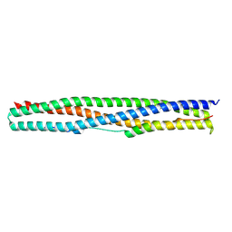
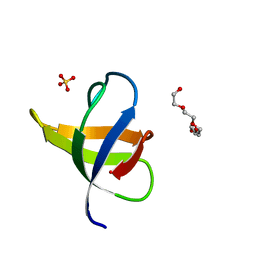
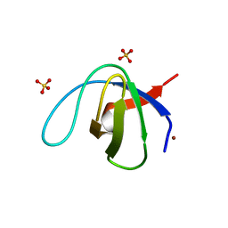
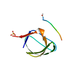
9HS7
 | | Anti-HIV-1 chimeric miniprotein mimicking the N-terminal half of gp41 NHR with an extended region targeting the MPER | | Descriptor: | DI(HYDROXYETHYL)ETHER, Transmembrane protein gp41 | | Authors: | Camara-Artigas, A, Gavira, J.A, Conejero-Lara, F, Polo-Megias, D, Salinas-Garcia, M.C. | | Deposit date: | 2024-12-18 | | Release date: | 2025-04-30 | | Method: | X-RAY DIFFRACTION (1.698 Å) | | Cite: | Potent HIV-1 miniprotein inhibitors targeting highly conserved gp41 epitopes.
Int.J.Biol.Macromol., 310, 2025
|
|
2O88
 | |
7NET
 | |
7NES
 | |
7NER
 | |
4Y92
 | |
4YC1
 | |
4ZNX
 | |
4ZNY
 | |
2HKI
 | |
1Z9K
 | |
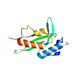
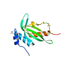
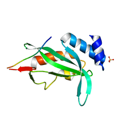
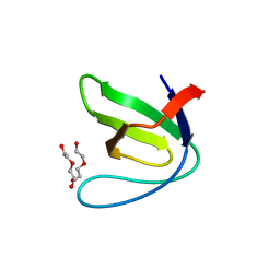
2HDA
 | | Yes SH3 domain | | Descriptor: | Proto-oncogene tyrosine-protein kinase Yes, SULFATE ION | | Authors: | Camara-Artigas, A, Luque, I, Ruiz-Sanz, J, Mateo, P.L, Martin-Garcia, J.M. | | Deposit date: | 2006-06-20 | | Release date: | 2007-04-17 | | Last modified: | 2023-08-30 | | Method: | X-RAY DIFFRACTION (1.9 Å) | | Cite: | Crystallographic structure of the SH3 domain of the human c-Yes tyrosine kinase: Loop flexibility and amyloid aggregation.
Febs Lett., 581, 2007
|
|
4RTX
 | |
4RTU
 | |
4RTZ
 | |
4RTW
 | |
5EC7
 | |
5DK8
 | |
5ECA
 | |
8AH5
 | |


