Loading
PDBj
MenuPDBj@FacebookPDBj@TwitterPDBj@YouTubewwPDB FoundationwwPDB
RCSB PDBPDBeBMRBAdv. SearchSearch help
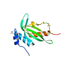
PDB: 186 results

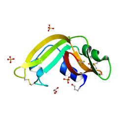
3HG6
DownloadVisualize
BU of 3hg6 by Molmil
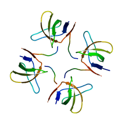
Crystal Structure of the Recombinant Onconase from Rana pipiens
Descriptor: GLYCEROL, Onconase, SULFATE ION
Authors:Camara-Artigas, A, Gavira, J.A, Casares-Atienza, S, Weininger, U, Balbach, J, Garcia-Mira, M.M.
Deposit date:2009-05-13
Release date:2010-05-19
Last modified:2023-11-08
Method:X-RAY DIFFRACTION (1.7 Å)
Cite:Three-state thermal unfolding of onconase.
Biophys.Chem., 159, 2011
3V58
DownloadVisualize
BU of 3v58 by Molmil
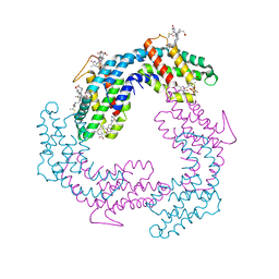
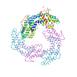
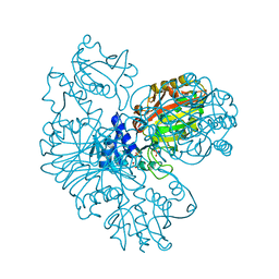
Crystal Structure of the B-phycoerythrin from the red algae Porphyridium Cruentum at pH5
Descriptor: PHYCOERYTHROBILIN, Phycoerythrin alpha subunit, Phycoerythrin beta subunit, ...
Authors:Camara-Artigas, A.
Deposit date:2011-12-16
Release date:2012-10-03
Last modified:2023-09-13
Method:X-RAY DIFFRACTION (1.85 Å)
Cite:pH-dependent structural conformations of B-phycoerythrin from Porphyridium cruentum
Febs J., 279, 2012
6TG7
DownloadVisualize
BU of 6tg7 by Molmil
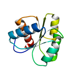
Crystal structure of the CheY in presence of magnesium
Descriptor: Chemotaxis protein CheY, MAGNESIUM ION
Authors:Camara-Artigas, A, Salinas-Garcia, M.C, Alba-Elena, D.
Deposit date:2019-11-15
Release date:2019-12-04
Last modified:2024-01-24
Method:X-RAY DIFFRACTION (1.65 Å)
Cite:Crystal structure of the CheY in presence of magnesium
To be published
2O88
DownloadVisualize
BU of 2o88 by Molmil
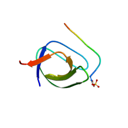
Crystal structure of the N114A mutant of ABL-SH3 domain complexed with a designed high-affinity peptide ligand: implications for SH3-ligand interactions
Descriptor: P41 peptide, Proto-oncogene tyrosine-protein kinase ABL1, SULFATE ION
Authors:Camara-Artigas, A.
Deposit date:2006-12-12
Release date:2007-05-01
Last modified:2023-08-30
Method:X-RAY DIFFRACTION (1.75 Å)
Cite:Crystallization by capillary counter-diffusion and structure determination of the N114A mutant of the SH3 domain of Abl tyrosine kinase complexed with a high-affinity peptide ligand.
Acta Crystallogr.,Sect.D, 63, 2007
4Y92
DownloadVisualize
BU of 4y92 by Molmil
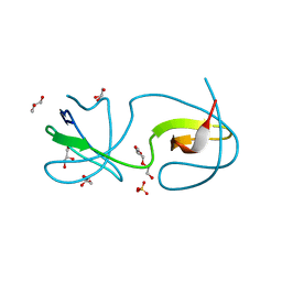
Crystal structure of the intertwined form of the Src tyrosine kinase SH3 domain E97T-Q128R mutant
Descriptor: GLYCEROL, Protein-tyrosine kinase, SULFATE ION, ...
Authors:Camara-Artigas, A, Bacarizo, J.
Deposit date:2015-02-16
Release date:2016-03-02
Last modified:2024-01-10
Method:X-RAY DIFFRACTION (2.1 Å)
Cite:Crystal structure of the intertwined form of the Src tyrosine kinase SH3 domain E97T-Q128R mutant
to be published
4YC1
DownloadVisualize
BU of 4yc1 by Molmil
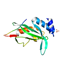
Structure of the human TSG101-UEV domain in the P321 space group
Descriptor: SULFATE ION, Tumor susceptibility gene 101 protein
Authors:Camara-Artigas, A.
Deposit date:2015-02-19
Release date:2016-03-09
Last modified:2024-01-10
Method:X-RAY DIFFRACTION (2 Å)
Cite:Structure of the human TSG101-UEV Domain
to be published
7NER
DownloadVisualize
BU of 7ner by Molmil
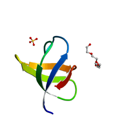
Crystal structure of the v-Src SH3 domain Q128R mutant
Descriptor: SULFATE ION, TETRAETHYLENE GLYCOL, v-Src SH3 domain
Authors:Camara-Artigas, A, Salinas-Garcia, M.C.
Deposit date:2021-02-04
Release date:2021-06-02
Last modified:2024-01-31
Method:X-RAY DIFFRACTION (1.55 Å)
Cite:The impact of oncogenic mutations of the viral Src kinase on the structure and stability of the SH3 domain.
Acta Crystallogr D Struct Biol, 77, 2021
7NET
DownloadVisualize
BU of 7net by Molmil
Crystal structure of the v-Src SH3 domain W95R-I96T mutant
Descriptor: DI(HYDROXYETHYL)ETHER, SULFATE ION, TRIETHYLENE GLYCOL, ...
Authors:Camara-Artigas, A, Salinas-Garcia, M.C.
Deposit date:2021-02-04
Release date:2021-06-02
Last modified:2024-01-31
Method:X-RAY DIFFRACTION (1.5 Å)
Cite:The impact of oncogenic mutations of the viral Src kinase on the structure and stability of the SH3 domain.
Acta Crystallogr D Struct Biol, 77, 2021
7NES
DownloadVisualize
BU of 7nes by Molmil
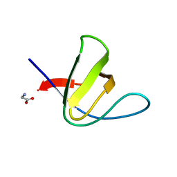
Crystal structure of the v-Src SH3 domain N117D-V124L mutant
Descriptor: GLYCINE, v-Src SH3 domain
Authors:Camara-Artigas, A, Salinas-Garcia, M.C.
Deposit date:2021-02-04
Release date:2021-06-02
Last modified:2024-05-15
Method:X-RAY DIFFRACTION (1.35 Å)
Cite:The impact of oncogenic mutations of the viral Src kinase on the structure and stability of the SH3 domain.
Acta Crystallogr D Struct Biol, 77, 2021
4OMO
DownloadVisualize
BU of 4omo by Molmil
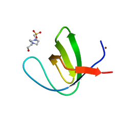
Crystal structure of the c-Src tyrosine kinase SH3 domain mutant Q128E
Descriptor: 4-(2-HYDROXYETHYL)-1-PIPERAZINE ETHANESULFONIC ACID, NICKEL (II) ION, Proto-oncogene tyrosine-protein kinase Src
Authors:Camara-Artigas, A, Bacarizo, J.
Deposit date:2014-01-27
Release date:2014-12-10
Last modified:2023-09-20
Method:X-RAY DIFFRACTION (1.04 Å)
Cite:Electrostatic Effects in the Folding of the SH3 Domain of the c-Src Tyrosine Kinase: pH-Dependence in 3D-Domain Swapping and Amyloid Formation.
Plos One, 9, 2014
4OML
DownloadVisualize
BU of 4oml by Molmil
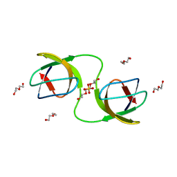
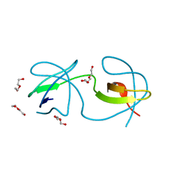
Crystal structure of the intertwined dimer of the c-Src tyrosine kinase SH3 domain mutant Q128R
Descriptor: DI(HYDROXYETHYL)ETHER, Proto-oncogene tyrosine-protein kinase Src, TRIETHYLENE GLYCOL
Authors:Camara-Artigas, A, Bacarizo, J.
Deposit date:2014-01-27
Release date:2014-12-10
Last modified:2023-09-20
Method:X-RAY DIFFRACTION (1.6 Å)
Cite:Electrostatic Effects in the Folding of the SH3 Domain of the c-Src Tyrosine Kinase: pH-Dependence in 3D-Domain Swapping and Amyloid Formation.
Plos One, 9, 2014
4OMN
DownloadVisualize
BU of 4omn by Molmil
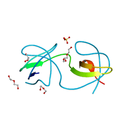
Crystal structure of the intertwined dimer of the c-Src tyrosine kinase SH3 domain mutant Q128E
Descriptor: DI(HYDROXYETHYL)ETHER, GLYCEROL, Proto-oncogene tyrosine-protein kinase Src, ...
Authors:Camara-Artigas, A, Bacarizo, J.
Deposit date:2014-01-27
Release date:2014-12-10
Last modified:2023-09-20
Method:X-RAY DIFFRACTION (1.5 Å)
Cite:Electrostatic Effects in the Folding of the SH3 Domain of the c-Src Tyrosine Kinase: pH-Dependence in 3D-Domain Swapping and Amyloid Formation.
Plos One, 9, 2014
4OMQ
DownloadVisualize
BU of 4omq by Molmil
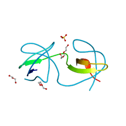
Crystal structure of the intertwined dimer of the c-Src tyrosine kinase SH3 domain mutant S94A
Descriptor: DI(HYDROXYETHYL)ETHER, Proto-oncogene tyrosine-protein kinase Src, SULFATE ION, ...
Authors:Camara-Artigas, A, Bacarizo, J.
Deposit date:2014-01-27
Release date:2015-01-28
Last modified:2023-09-20
Method:X-RAY DIFFRACTION (2 Å)
Cite:The role of Gutamine 128 of the SH3 domain of the c-Src tyrosine kinase in 3D domain swapping and amyloid formation
To be Published
4ZNX
DownloadVisualize
BU of 4znx by Molmil
Crystal structure of the Fyn-SH3 domain in complex with the high affinity peptide APP12
Descriptor: APP12, Tyrosine-protein kinase Fyn
Authors:Camara-Artigas, A.
Deposit date:2015-05-05
Release date:2016-06-29
Last modified:2024-01-10
Method:X-RAY DIFFRACTION (2.1 Å)
Cite:The role of water molecules in the binding of class I and II peptides to the SH3 domain of the Fyn tyrosine kinase.
Acta Crystallogr.,Sect.F, 72, 2016
4ZNY
DownloadVisualize
BU of 4zny by Molmil
Structure of the human TSG101-UEV Domain in complex with the PTAP motif of the p19 gag protein of the Human T-cell Leukemia type I virus
Descriptor: SULFATE ION, T-cell leukemia virus type I, partial gag gene; HTLV1 (human T-lymphotropic virus type I), ...
Authors:Camara-Artigas, A, Bacarizo, J.
Deposit date:2015-05-05
Release date:2016-06-29
Last modified:2024-01-10
Method:X-RAY DIFFRACTION (2.4 Å)
Cite:Structure of the human TSG101-UEV Domain in complex with the PTAP motif of the p19 gag protein of the Human T-cell Leukemia type I virus
to be published
3V57
DownloadVisualize
BU of 3v57 by Molmil
Crystal Structure of the B-phycoerythrin from the red algae Porphyridium Cruentum at pH8
Descriptor: PHYCOERYTHROBILIN, Phycoerythrin alpha subunit, Phycoerythrin beta subunit, ...
Authors:Camara-Artigas, A.
Deposit date:2011-12-16
Release date:2012-10-03
Last modified:2023-09-13
Method:X-RAY DIFFRACTION (1.7 Å)
Cite:pH-dependent structural conformations of B-phycoerythrin from Porphyridium cruentum
Febs J., 279, 2012
1Z9K
DownloadVisualize
BU of 1z9k by Molmil
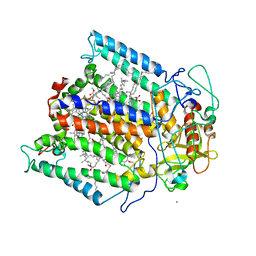
Photosynthetic Reaction Center from Rhodobacter sphaeroides
Descriptor: BACTERIOCHLOROPHYLL A, BACTERIOPHEOPHYTIN A, FE (III) ION, ...
Authors:Camara-Artigas, A, Allen, J.P.
Deposit date:2005-04-02
Release date:2005-06-07
Last modified:2023-08-23
Method:X-RAY DIFFRACTION (4.6 Å)
Cite:Design of a Redox-Linked Active Metal Site: Manganese Bound to Bacterial Reaction Centers at a Site Resembling That of Photosystem II
Biochemistry, 44, 2005
3UA6
DownloadVisualize
BU of 3ua6 by Molmil
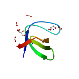
Crystal Structure of the Human Fyn SH3 domain
Descriptor: FORMIC ACID, GLYCEROL, SODIUM ION, ...
Authors:Camara-Artigas, A, Martin-Garcia, J.M.
Deposit date:2011-10-21
Release date:2012-07-25
Last modified:2023-09-13
Method:X-RAY DIFFRACTION (1.85 Å)
Cite:The promiscuous binding of the Fyn SH3 domain to a peptide from the NS5A protein.
Acta Crystallogr.,Sect.D, 68, 2012
2HKI
DownloadVisualize
BU of 2hki by Molmil
Crystal structure of photosynthetic glyceraldehyde-3-phosphate dehydrogenase A4 isoform
Descriptor: Glyceraldehyde-3-phosphate dehydrogenase A, chloroplast, SULFATE ION
Authors:Camara-Artigas, A.
Deposit date:2006-07-04
Release date:2006-11-14
Last modified:2023-08-30
Method:X-RAY DIFFRACTION (3 Å)
Cite:Crystallization and structural analysis of GADPH from Spinacia oleracea in a new form.
Acta Crystallogr.,Sect.F, 62, 2006
6QJJ
DownloadVisualize
BU of 6qjj by Molmil
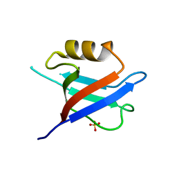
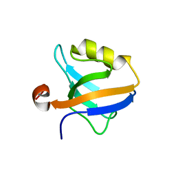
Crystal Structure of the third PDZ domain of PSD-95 protein: space group P3221
Descriptor: Disks large homolog 4, SULFATE ION
Authors:Camara-Artigas, A.
Deposit date:2019-01-24
Release date:2019-04-17
Last modified:2024-01-24
Method:X-RAY DIFFRACTION (1.7 Å)
Cite:Conformational changes in the third PDZ domain of the neuronal postsynaptic density protein 95.
Acta Crystallogr D Struct Biol, 75, 2019
6QJD
DownloadVisualize
BU of 6qjd by Molmil
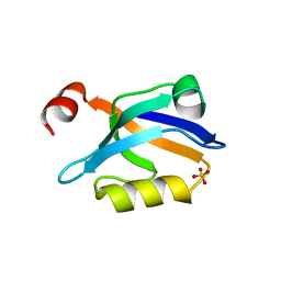
Crystal Structure of the truncated form of the third PDZ domain of PSD-95: residues 302-392
Descriptor: Disks large homolog 4, SULFATE ION
Authors:Camara-Artigas, A.
Deposit date:2019-01-24
Release date:2019-04-17
Last modified:2024-01-24
Method:X-RAY DIFFRACTION (1.551 Å)
Cite:Conformational changes in the third PDZ domain of the neuronal postsynaptic density protein 95.
Acta Crystallogr D Struct Biol, 75, 2019
6QJN
DownloadVisualize
BU of 6qjn by Molmil
Crystal Structure of the third PDZ domain of PSD-95 protein D332G mutant: space group I4122
Descriptor: Disks large homolog 4
Authors:Camara-Artigas, A.
Deposit date:2019-01-24
Release date:2019-04-17
Last modified:2024-01-24
Method:X-RAY DIFFRACTION (1.8 Å)
Cite:Conformational changes in the third PDZ domain of the neuronal postsynaptic density protein 95.
Acta Crystallogr D Struct Biol, 75, 2019
2HDA
DownloadVisualize
BU of 2hda by Molmil
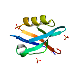
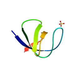
Yes SH3 domain
Descriptor: Proto-oncogene tyrosine-protein kinase Yes, SULFATE ION
Authors:Camara-Artigas, A, Luque, I, Ruiz-Sanz, J, Mateo, P.L, Martin-Garcia, J.M.
Deposit date:2006-06-20
Release date:2007-04-17
Last modified:2023-08-30
Method:X-RAY DIFFRACTION (1.9 Å)
Cite:Crystallographic structure of the SH3 domain of the human c-Yes tyrosine kinase: Loop flexibility and amyloid aggregation.
Febs Lett., 581, 2007
6QJI
DownloadVisualize
BU of 6qji by Molmil
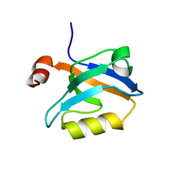
Crystal Structure of the third PDZ domain of PSD-95 protein: space group P3112
Descriptor: Disks large homolog 4, SULFATE ION
Authors:Camara-Artigas, A.
Deposit date:2019-01-24
Release date:2019-04-17
Last modified:2024-01-24
Method:X-RAY DIFFRACTION (1.5 Å)
Cite:Conformational changes in the third PDZ domain of the neuronal postsynaptic density protein 95.
Acta Crystallogr D Struct Biol, 75, 2019
6QJG
DownloadVisualize
BU of 6qjg by Molmil
Crystal Structure of the third PDZ domain of PSD-95 protein D332P mutant: space group C121, structure 2
Descriptor: Disks large homolog 4
Authors:Camara-Artigas, A.
Deposit date:2019-01-24
Release date:2019-04-17
Last modified:2024-01-24
Method:X-RAY DIFFRACTION (2.003 Å)
Cite:Conformational changes in the third PDZ domain of the neuronal postsynaptic density protein 95.
Acta Crystallogr D Struct Biol, 75, 2019

222415

PDB entries from 2024-07-10

PDB statisticsPDBj update infoContact PDBjnumon