Loading
PDBj
MenuPDBj@FacebookPDBj@TwitterPDBj@YouTubewwPDB FoundationwwPDB
RCSB PDBPDBeBMRBAdv. SearchSearch help
PDB: 64 results

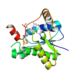
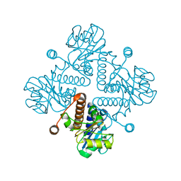
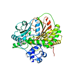
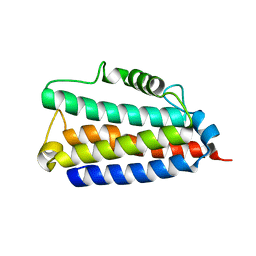
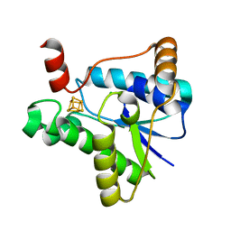
6ANR
DownloadVisualize
BU of 6anr by Molmil
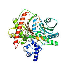
Crystal structure of a self resistance protein ClbS from colibactin biosynthetic gene cluster
Descriptor: Colibactin self-protection protein ClbS
Authors:Tripathi, P, Bruner, S.D.
Deposit date:2017-08-14
Release date:2018-06-27
Last modified:2023-10-04
Method:X-RAY DIFFRACTION (2.098 Å)
Cite:ClbS Is a Cyclopropane Hydrolase That Confers Colibactin Resistance.
J. Am. Chem. Soc., 139, 2017
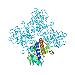
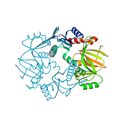
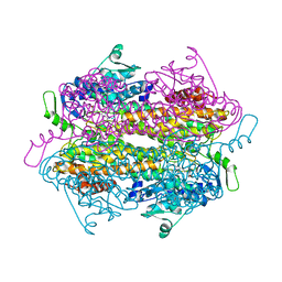
7LC5
DownloadVisualize
BU of 7lc5 by Molmil
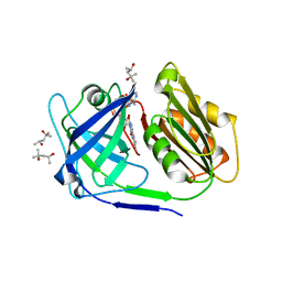
Crystal structure of epoxyqueuosine reductase QueH from Thermotoga maritima
Descriptor: CHLORIDE ION, Epoxyqueuosine reductase QueH, FE (III) ION, ...
Authors:Li, Q, Bruner, S.D.
Deposit date:2021-01-09
Release date:2021-11-03
Last modified:2024-05-22
Method:X-RAY DIFFRACTION (1.5 Å)
Cite:Epoxyqueuosine Reductase QueH in the Biosynthetic Pathway to tRNA Queuosine Is a Unique Metalloenzyme.
Biochemistry, 60, 2021
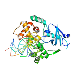
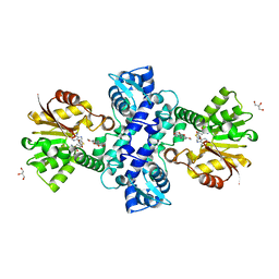
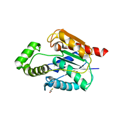
1LWW
DownloadVisualize
BU of 1lww by Molmil
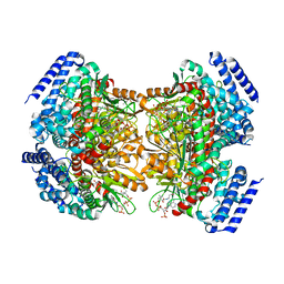
Borohydride-trapped hOgg1 Intermediate Structure Co-Crystallized with 8-bromoguanine
Descriptor: 5'-D(*GP*CP*GP*TP*CP*CP*AP*(PED)P*GP*TP*CP*TP*AP*CP*C)-3', 5'-D(*GP*GP*TP*AP*GP*AP*CP*CP*TP*GP*GP*AP*CP*GP*C)-3', 8-BROMOGUANINE, ...
Authors:Fromme, J.C, Bruner, S.D, Yang, W, Karplus, M, Verdine, G.L.
Deposit date:2002-06-03
Release date:2003-02-25
Last modified:2011-07-13
Method:X-RAY DIFFRACTION (2.1 Å)
Cite:Product-Assisted Catalysis in Base Excision DNA Repair
Nat.Struct.Biol., 10, 2003
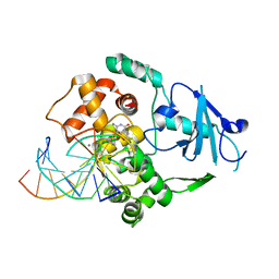
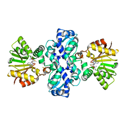
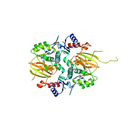
1LWV
DownloadVisualize
BU of 1lwv by Molmil
Borohydride-trapped hOgg1 Intermediate Structure Co-Crystallized with 8-aminoguanine
Descriptor: 5'-D(*GP*CP*GP*TP*CP*CP*AP*(PED)P*GP*TP*CP*TP*AP*CP*C)-3', 5'-D(*GP*GP*TP*AP*GP*AP*CP*CP*TP*GP*GP*AP*CP*GP*C)-3', 8-AMINOGUANINE, ...
Authors:Fromme, J.C, Bruner, S.D, Yang, W, Karplus, M, Verdine, G.L.
Deposit date:2002-06-03
Release date:2003-02-25
Last modified:2011-07-13
Method:X-RAY DIFFRACTION (2.3 Å)
Cite:Product-Assisted Catalysis in Base Excision DNA Repair
Nat.Struct.Biol., 10, 2003
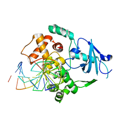
1LWY
DownloadVisualize
BU of 1lwy by Molmil
hOgg1 Borohydride-Trapped Intermediate without 8-oxoguanine
Descriptor: 5'-D(*GP*CP*GP*TP*CP*CP*AP*(PED)P*GP*TP*CP*TP*AP*CP*C)-3', 5'-D(*GP*GP*TP*AP*GP*AP*CP*CP*TP*GP*GP*AP*CP*GP*C)-3', 8-OXOGUANINE DNA GLYCOSYLASE
Authors:Fromme, J.C, Bruner, S.D, Yang, W, Karplus, M, Verdine, G.L.
Deposit date:2002-06-03
Release date:2003-02-25
Last modified:2011-07-13
Method:X-RAY DIFFRACTION (2.01 Å)
Cite:Product-Assisted Catalysis in Base Excision DNA Repair
Nat.Struct.Biol., 10, 2003
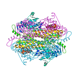
7LC7
DownloadVisualize
BU of 7lc7 by Molmil
Crystal structure of epoxyqueuosine reductase QueH in complex with GMP from Thermotoga maritima
Descriptor: CHLORIDE ION, Epoxyqueuosine reductase QueH, FE (III) ION, ...
Authors:Li, Q, Bruner, S.D.
Deposit date:2021-01-09
Release date:2022-01-12
Last modified:2023-10-18
Method:X-RAY DIFFRACTION (1.58 Å)
Cite:The epoxyqueuosine reductase QueH in the biosynthesis of tRNA queuosine is a unique metalloenzyme
To Be Published
6BTG
DownloadVisualize
BU of 6btg by Molmil
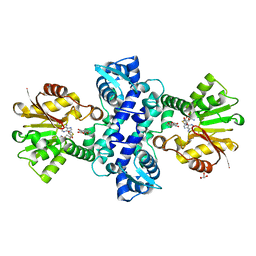
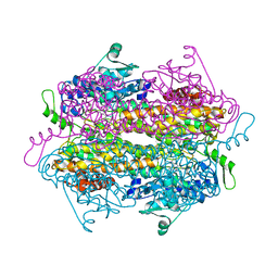
Crystal structure of deoxyribose-phosphate aldolase bound with DHAP from Bacillus Thuringiensis
Descriptor: 1,3-DIHYDROXYACETONEPHOSPHATE, Fuculose phosphate aldolase, MANGANESE (II) ION
Authors:Li, Q, Bruner, S.D.
Deposit date:2017-12-06
Release date:2018-07-04
Last modified:2023-10-04
Method:X-RAY DIFFRACTION (1.698 Å)
Cite:Salvage of the 5-deoxyribose byproduct of radical SAM enzymes.
Nat Commun, 9, 2018
6BTD
DownloadVisualize
BU of 6btd by Molmil
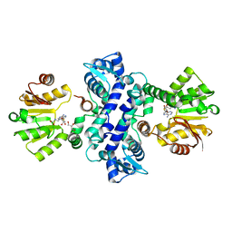
Crystal structure of deoxyribose-phosphate aldolase from Bacillus Thuringiensis involved in dispatching the ubiquitous radical SAM enzyme byproduct 5-deoxyribose
Descriptor: Fuculose phosphate aldolase, MANGANESE (II) ION, SULFATE ION
Authors:Li, Q, Bruner, S.D.
Deposit date:2017-12-06
Release date:2018-07-04
Last modified:2023-10-04
Method:X-RAY DIFFRACTION (1.551 Å)
Cite:Salvage of the 5-deoxyribose byproduct of radical SAM enzymes.
Nat Commun, 9, 2018
3I58
DownloadVisualize
BU of 3i58 by Molmil
Crystal structure of an O-methyltransferase (NcsB1) from neocarzinostatin biosynthesis in complex with S-adenosyl-L-homocysteine (SAH) and 2-hydroxy-7-methoxy-5-methyl naphthoic acid (NA)
Descriptor: 2-hydroxy-7-methoxy-5-methyl naphthoic acid, GLYCEROL, O-methyltransferase, ...
Authors:Cooke, H.A, Bruner, S.D.
Deposit date:2009-07-03
Release date:2009-09-01
Last modified:2023-09-06
Method:X-RAY DIFFRACTION (2.69 Å)
Cite:Molecular basis of substrate promiscuity for the SAM-dependent O-methyltransferase NcsB1, involved in the biosynthesis of the enediyne antitumor antibiotic neocarzinostatin.
Biochemistry, 48, 2009
3I5U
DownloadVisualize
BU of 3i5u by Molmil
Crystal structure of an O-methyltransferase (NcsB1) from neocarzinostatin biosynthesis in complex with S-adenosylmethionine (SAM) and 2-hydroxy-5-methyl naphthoic acid (MNA)
Descriptor: 2-hydroxy-5-methyl naphthoic acid, GLYCEROL, O-methyltransferase, ...
Authors:Cooke, H.A, Bruner, S.D.
Deposit date:2009-07-06
Release date:2009-09-01
Last modified:2023-09-06
Method:X-RAY DIFFRACTION (2.6 Å)
Cite:Molecular basis of substrate promiscuity for the SAM-dependent O-methyltransferase NcsB1, involved in the biosynthesis of the enediyne antitumor antibiotic neocarzinostatin.
Biochemistry, 48, 2009
3I64
DownloadVisualize
BU of 3i64 by Molmil
Crystal structure of an O-methyltransferase (NcsB1) from neocarzinostatin biosynthesis in complex with S-adenosyl-L-homocysteine (SAH) and 1,4-dihydroxy-2-naphthoic acid (DHN)
Descriptor: 1,4-dihydroxy-2-naphthoic acid, GLYCEROL, O-methyltransferase, ...
Authors:Cooke, H.A, Bruner, S.D.
Deposit date:2009-07-06
Release date:2009-09-01
Last modified:2023-09-06
Method:X-RAY DIFFRACTION (3 Å)
Cite:Molecular basis of substrate promiscuity for the SAM-dependent O-methyltransferase NcsB1, involved in the biosynthesis of the enediyne antitumor antibiotic neocarzinostatin.
Biochemistry, 48, 2009
3I53
DownloadVisualize
BU of 3i53 by Molmil
Crystal structure of an O-methyltransferase (NcsB1) from neocarzinostatin biosynthesis in complex with S-adenosyl-L-homocysteine (SAH)
Descriptor: GLYCEROL, O-methyltransferase, S-ADENOSYL-L-HOMOCYSTEINE
Authors:Cooke, H.A, Bruner, S.D.
Deposit date:2009-07-03
Release date:2009-09-01
Last modified:2023-09-06
Method:X-RAY DIFFRACTION (2.08 Å)
Cite:Molecular basis of substrate promiscuity for the SAM-dependent O-methyltransferase NcsB1, involved in the biosynthesis of the enediyne antitumor antibiotic neocarzinostatin.
Biochemistry, 48, 2009
5V6J
DownloadVisualize
BU of 5v6j by Molmil
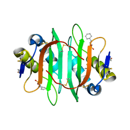
Glycan binding protein Y3 from mushroom Coprinus comatus possesses anti-leukemic activity
Descriptor: 2-[N-CYCLOHEXYLAMINO]ETHANE SULFONIC ACID, TMV resistance protein Y3
Authors:Li, K, Zhang, P, Gang, Y, Xia, C, Polston, J.E, Li, G, Li, S, Lin, Z, Yang, L.-J, Bruner, S.D, Ding, Y.
Deposit date:2017-03-16
Release date:2017-08-16
Last modified:2024-10-23
Method:X-RAY DIFFRACTION (1.18 Å)
Cite:Cytotoxic protein from the mushroom Coprinus comatus possesses a unique mode for glycan binding and specificity.
Proc. Natl. Acad. Sci. U.S.A., 114, 2017
5V6I
DownloadVisualize
BU of 5v6i by Molmil
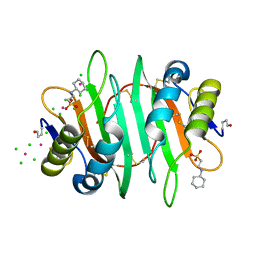
Glycan binding protein Y3 from mushroom Coprinus comatus possesses anti-leukemic activity - Pt derivative
Descriptor: 2-[N-CYCLOHEXYLAMINO]ETHANE SULFONIC ACID, CHLORIDE ION, PLATINUM (II) ION, ...
Authors:Li, K, Zhang, P, Gang, Y, Xia, C, Polston, J.E, Li, G, Li, S, Lin, Z, Yang, L.-J, Bruner, S.D, Ding, Y.
Deposit date:2017-03-16
Release date:2017-08-16
Last modified:2024-10-09
Method:X-RAY DIFFRACTION (1.7 Å)
Cite:Cytotoxic protein from the mushroom Coprinus comatus possesses a unique mode for glycan binding and specificity.
Proc. Natl. Acad. Sci. U.S.A., 114, 2017
5ISX
DownloadVisualize
BU of 5isx by Molmil
Structure of the holo PCP-E didomain of the gramicidin S synthetase A
Descriptor: 4'-PHOSPHOPANTETHEINE, GLYCEROL, Gramicidin S synthase 1
Authors:Chen, W.-H, Li, K, Bruner, S.D.
Deposit date:2016-03-15
Release date:2016-06-29
Last modified:2019-11-27
Method:X-RAY DIFFRACTION (2.335 Å)
Cite:Interdomain and Intermodule Organization in Epimerization Domain Containing Nonribosomal Peptide Synthetases.
Acs Chem.Biol., 11, 2016
5IG8
DownloadVisualize
BU of 5ig8 by Molmil
Crystal structure of macrocyclase MdnB from Microcystis aeruginosa MRC
Descriptor: ATP grasp ligase
Authors:Li, K, Condurso, H.L, Bruner, S.D.
Deposit date:2016-02-27
Release date:2016-09-21
Last modified:2024-10-23
Method:X-RAY DIFFRACTION (2.278 Å)
Cite:Structural basis for precursor protein-directed ribosomal peptide macrocyclization.
Nat.Chem.Biol., 12, 2016
5UGZ
DownloadVisualize
BU of 5ugz by Molmil
Crystal structure of ClbQ from the colibactin NRPS/PKS pathway
Descriptor: BETA-MERCAPTOETHANOL, Putative thioesterase
Authors:Guntaka, N.S, Bruner, S.D.
Deposit date:2017-01-10
Release date:2017-09-20
Last modified:2023-10-04
Method:X-RAY DIFFRACTION (1.983 Å)
Cite:Structure and Functional Analysis of ClbQ, an Unusual Intermediate-Releasing Thioesterase from the Colibactin Biosynthetic Pathway.
ACS Chem. Biol., 12, 2017
5IG9
DownloadVisualize
BU of 5ig9 by Molmil
Crystal structure of macrocyclase MdnC bound with precursor peptide MdnA from Microcystis aeruginosa MRC
Descriptor: ATP grasp ligase, Microviridin
Authors:Li, K, Condurso, H.L, Bruner, S.D.
Deposit date:2016-02-27
Release date:2016-09-21
Last modified:2024-03-06
Method:X-RAY DIFFRACTION (2.665 Å)
Cite:Structural basis for precursor protein-directed ribosomal peptide macrocyclization.
Nat.Chem.Biol., 12, 2016
5ISW
DownloadVisualize
BU of 5isw by Molmil
Structure of the apo PCP-E didomain of the gramicidin S synthetase A
Descriptor: GLYCEROL, Gramicidin S synthase 1
Authors:Chen, W.-H, Li, K, Bruner, S.D.
Deposit date:2016-03-15
Release date:2016-06-29
Last modified:2024-03-06
Method:X-RAY DIFFRACTION (1.75 Å)
Cite:Interdomain and Intermodule Organization in Epimerization Domain Containing Nonribosomal Peptide Synthetases.
Acs Chem.Biol., 11, 2016
4YHB
DownloadVisualize
BU of 4yhb by Molmil
Crystal structure of a siderophore utilization protein from T. fusca
Descriptor: (4S)-2-METHYL-2,4-PENTANEDIOL, FLAVIN-ADENINE DINUCLEOTIDE, Iron-chelator utilization protein, ...
Authors:Li, K, Bruner, S.D.
Deposit date:2015-02-27
Release date:2015-07-15
Last modified:2023-09-27
Method:X-RAY DIFFRACTION (1.8892 Å)
Cite:Structure and Mechanism of the Siderophore-Interacting Protein from the Fuscachelin Gene Cluster of Thermobifida fusca.
Biochemistry, 54, 2015
4YLH
DownloadVisualize
BU of 4ylh by Molmil
Crystal structure of DpgC with bound substrate analog and Xe on oxygen diffusion pathway
Descriptor: DpgC, XENON, [(2R,3S,4R,5R)-5-(6-AMINO-9H-PURIN-9-YL)-4-HYDROXY-3-(PHOSPHONOOXY)TETRAHYDROFURAN-2-YL]METHYL (3R)-4-({3-[(2-{[(3,5-DIHYDROXYPHENYL)ACETYL]AMINO}ETHYL)AMINO]-3-OXOPROPYL}AMINO)-3-HYDROXY-2,2-DIMETHYL-4-OXOBUTYL DIHYDROGEN DIPHOSPHATE
Authors:Li, K, Di Russo, N.V, Condurso, H.L, Roitberg, A.E, Bruner, S.D.
Deposit date:2015-03-05
Release date:2015-08-05
Last modified:2023-09-27
Method:X-RAY DIFFRACTION (2.58 Å)
Cite:Oxygen diffusion pathways in a cofactor-independent dioxygenase.
Chem Sci, 6, 2015
2QVE
DownloadVisualize
BU of 2qve by Molmil
Crystal Structure of SgTAM bound to mechanism based inhibitor
Descriptor: (3R)-3-amino-2,2-difluoro-3-(4-hydroxyphenyl)propanoic acid, Tyrosine Aminomutase
Authors:Christianson, C.V, Montavon, T.J, Bruner, S.D.
Deposit date:2007-08-08
Release date:2008-07-22
Last modified:2023-11-15
Method:X-RAY DIFFRACTION (2 Å)
Cite:Design and characterization of mechanism-based inhibitors for the tyrosine aminomutase SgTAM
Bioorg.Med.Chem.Lett., 18, 2008
2RJS
DownloadVisualize
BU of 2rjs by Molmil
SgTAM bound to substrate mimic
Descriptor: (3R)-3-amino-2,2-difluoro-3-(4-methoxyphenyl)propanoic acid, Tyrosine aminomutase
Authors:Montavon, T.J, Christianson, C.V, Bruner, S.D.
Deposit date:2007-10-15
Release date:2008-01-15
Last modified:2024-10-30
Method:X-RAY DIFFRACTION (2.4 Å)
Cite:Design and characterization of mechanism-based inhibitors for the tyrosine aminomutase SgTAM.
Bioorg.Med.Chem.Lett., 18, 2008
2RJR
DownloadVisualize
BU of 2rjr by Molmil
Substrate mimic bound to SgTAM
Descriptor: (2S,3S)-3-(4-fluorophenyl)-2,3-dihydroxypropanoic acid, Tyrosine aminomutase
Authors:Montavon, T.J, Christianson, C.V, Bruner, S.D.
Deposit date:2007-10-15
Release date:2008-01-15
Last modified:2024-10-16
Method:X-RAY DIFFRACTION (2.1 Å)
Cite:Design and characterization of mechanism-based inhibitors for the tyrosine aminomutase SgTAM.
Bioorg.Med.Chem.Lett., 18, 2008
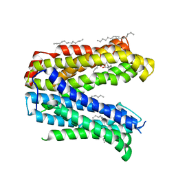
4Z3N
DownloadVisualize
BU of 4z3n by Molmil
Crystal structure of the MATE transporter ClbM
Descriptor: (2R)-2,3-dihydroxypropyl (9Z)-octadec-9-enoate, CACODYLATE ION, Putative drug/sodium antiporter
Authors:Mousa, J.J, Bruner, S.D.
Deposit date:2015-03-31
Release date:2016-01-13
Last modified:2023-09-27
Method:X-RAY DIFFRACTION (2.7 Å)
Cite:MATE transport of the E. coli-derived genotoxin colibactin.
Nat Microbiol, 1, 2016

226707

PDB entries from 2024-10-30

PDB statisticsPDBj update infoContact PDBjnumon