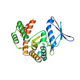
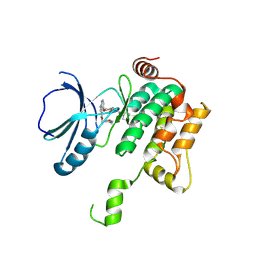
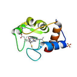
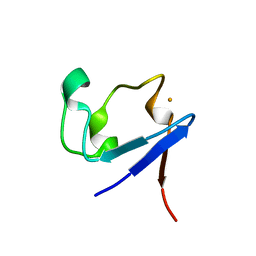
6DI3
 
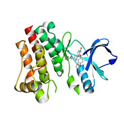 | | CRYSTAL STRUCTURE OF BTK IN COMPLEX WITH FRAGMENT LIGAND | | 分子名称: | 6-[(3S)-3-(acryloylamino)pyrrolidin-1-yl]-2-(4-phenoxyphenoxy)pyridine-3-carboxamide, Tyrosine-protein kinase BTK | | 著者 | GARDBERG, A. | | 登録日 | 2018-05-22 | | 公開日 | 2018-09-05 | | 最終更新日 | 2024-03-13 | | 実験手法 | X-RAY DIFFRACTION (2 Å) | | 主引用文献 | Discovery of potent, highly selective covalent irreversible BTK inhibitors from a fragment hit.
Bioorg. Med. Chem. Lett., 28, 2018
|
|
6V8B
 | |
5VYL
 | |
7PGQ
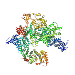 | | GAP-SecPH region of human neurofibromin isoform 2 in closed conformation. | | 分子名称: | (1S)-2-{[(2-AMINOETHOXY)(HYDROXY)PHOSPHORYL]OXY}-1-[(PALMITOYLOXY)METHYL]ETHYL STEARATE, Neurofibromin, ZINC ION | | 著者 | Naschberger, A, Baradaran, R, Carroni, M, Rupp, B. | | 登録日 | 2021-08-15 | | 公開日 | 2022-10-26 | | 最終更新日 | 2024-07-17 | | 実験手法 | ELECTRON MICROSCOPY (3.5 Å) | | 主引用文献 | The structure of neurofibromin isoform 2 reveals different functional states.
Nature, 599, 2021
|
|
5IGU
 | |
5E96
 | | Crystal structure of aminoglycoside 6'-acetyltransferase type Ii | | 分子名称: | 1,2-ETHANEDIOL, Aminoglycoside 6'-acetyltransferase, PHOSPHATE ION | | 著者 | Berghuis, A.M, Burk, D.L, Baettig, O.M, Shi, K. | | 登録日 | 2015-10-14 | | 公開日 | 2016-07-06 | | 最終更新日 | 2023-09-27 | | 実験手法 | X-RAY DIFFRACTION (2.1 Å) | | 主引用文献 | Comprehensive characterization of ligand-induced plasticity changes in a dimeric enzyme.
Febs J., 283, 2016
|
|
6TAM
 | |
8PAW
 | | Crystal structure of MST1 with a MAP4K1 SMOL inhibitor | | 分子名称: | 1-[3,5-bis(fluoranyl)-4-[[3-(1-propan-2-ylpyrazol-3-yl)-1~{H}-pyrrolo[2,3-b]pyridin-4-yl]oxy]phenyl]-3-(2-methoxyethyl)urea, 3-CYCLOHEXYL-1-PROPYLSULFONIC ACID, ASPARTIC ACID, ... | | 著者 | Friberg, A. | | 登録日 | 2023-06-08 | | 公開日 | 2024-06-26 | | 実験手法 | X-RAY DIFFRACTION (2.14 Å) | | 主引用文献 | Identification and optimization of Azaindole based MAP4K1 Inhibitors and the discovery of BAY-405
To Be Published
|
|
8PAS
 | | Crystal structure of MAP4K1 with a SMOL inhibitor | | 分子名称: | 4-[2,6-bis(fluoranyl)-4-(3-morpholin-4-ylpropylcarbamoylamino)phenoxy]-~{N}-[(4-methyl-1,2,5-oxadiazol-3-yl)methyl]-1~{H}-pyrrolo[2,3-b]pyridine-3-carboxamide, Mitogen-activated protein kinase kinase kinase kinase 1 | | 著者 | Friberg, A. | | 登録日 | 2023-06-08 | | 公開日 | 2024-06-26 | | 実験手法 | X-RAY DIFFRACTION (2.7 Å) | | 主引用文献 | Identification and optimization of Azaindole based MAP4K1 Inhibitors and the discovery of BAY-405
To Be Published
|
|
8PAV
 | | Crystal structure of MST1 with a MAP4K1 SMOL inhibitor | | 分子名称: | 1-[3,5-bis(fluoranyl)-4-[[3-(1,3-thiazol-5-yl)-1~{H}-pyrrolo[2,3-b]pyridin-4-yl]oxy]phenyl]-3-(2-methoxyethyl)urea, 3-CYCLOHEXYL-1-PROPYLSULFONIC ACID, GLYCEROL, ... | | 著者 | Friberg, A. | | 登録日 | 2023-06-08 | | 公開日 | 2024-06-26 | | 実験手法 | X-RAY DIFFRACTION (1.9 Å) | | 主引用文献 | Identification and optimization of Azaindole based MAP4K1 Inhibitors and the discovery of BAY-405
To Be Published
|
|
2GBV
 | | C6A/C111A/C57A/C146A holo CuZn Superoxide dismutase | | 分子名称: | COPPER (I) ION, Superoxide dismutase [Cu-Zn], ZINC ION | | 著者 | Hornberg, A, Logan, D.T, Marklund, S.L, Oliveberg, M. | | 登録日 | 2006-03-11 | | 公開日 | 2007-01-02 | | 最終更新日 | 2023-10-25 | | 実験手法 | X-RAY DIFFRACTION (2 Å) | | 主引用文献 | The Coupling between Disulphide Status, Metallation and Dimer Interface Strength in Cu/Zn Superoxide Dismutase
J.Mol.Biol., 365, 2007
|
|
2GBT
 | | C6A/C111A CuZn Superoxide dismutase | | 分子名称: | COPPER (I) ION, Superoxide dismutase [Cu-Zn], ZINC ION | | 著者 | Hornberg, A, Logan, D.T, Marklund, S.L, Oliveberg, M. | | 登録日 | 2006-03-11 | | 公開日 | 2007-01-02 | | 最終更新日 | 2023-10-25 | | 実験手法 | X-RAY DIFFRACTION (1.7 Å) | | 主引用文献 | The Coupling between Disulphide Status, Metallation and Dimer Interface Strength in Cu/Zn Superoxide Dismutase
J.Mol.Biol., 365, 2007
|
|
2GBU
 | | C6A/C111A/C57A/C146A apo CuZn Superoxide dismutase | | 分子名称: | Superoxide dismutase [Cu-Zn] | | 著者 | Hornberg, A, Logan, D.T, Marklund, S.L, Oliveberg, M. | | 登録日 | 2006-03-11 | | 公開日 | 2007-01-02 | | 最終更新日 | 2023-10-25 | | 実験手法 | X-RAY DIFFRACTION (2 Å) | | 主引用文献 | The Coupling between Disulphide Status, Metallation and Dimer Interface Strength in Cu/Zn Superoxide Dismutase
J.Mol.Biol., 365, 2007
|
|
3KYU
 | |
3KYX
 | |
4KCC
 | | Crystal Structure of the NMDA Receptor GluN1 Ligand Binding Domain Apo State | | 分子名称: | Glutamate receptor ionotropic, NMDA 1, PHOSPHATE ION | | 著者 | Berger, A.J, Lau, A.Y, Mayer, M.L. | | 登録日 | 2013-04-24 | | 公開日 | 2013-07-31 | | 最終更新日 | 2023-09-20 | | 実験手法 | X-RAY DIFFRACTION (1.894 Å) | | 主引用文献 | Conformational Analysis of NMDA Receptor GluN1, GluN2, and GluN3 Ligand-Binding Domains Reveals Subtype-Specific Characteristics.
Structure, 21, 2013
|
|
2YCC
 | |
8PAU
 | | Crystal structure of MAP4K1 with a SMOL inhibitor | | 分子名称: | Mitogen-activated protein kinase kinase kinase kinase 1, [(5~{R})-2-[[3,5-bis(fluoranyl)-4-[[3-(trifluoromethyl)-1~{H}-pyrrolo[2,3-b]pyridin-4-yl]oxy]phenyl]amino]-5-fluoranyl-4,6-dihydro-1,3-oxazin-5-yl]methanol | | 著者 | Friberg, A. | | 登録日 | 2023-06-08 | | 公開日 | 2024-06-26 | | 実験手法 | X-RAY DIFFRACTION (2.8 Å) | | 主引用文献 | Identification and optimization of Azaindole based MAP4K1 Inhibitors and the discovery of BAY-405
To Be Published
|
|
3KYV
 | |
3KYW
 | |
3KYY
 | |
2WAC
 | |
3EYS
 | |
2WU3
 | | CRYSTAL STRUCTURE OF MOUSE ACETYLCHOLINESTERASE IN COMPLEX WITH FENAMIPHOS AND HI-6 | | 分子名称: | 2-acetamido-2-deoxy-beta-D-glucopyranose, 4-(AMINOCARBONYL)-1-[({2-[(E)-(HYDROXYIMINO)METHYL]PYRIDINIUM-1-YL}METHOXY)METHYL]PYRIDINIUM, ACETYLCHOLINESTERASE, ... | | 著者 | Hornberg, A, Artursson, E, Warme, R, Pang, Y.-P, Ekstrom, F. | | 登録日 | 2009-09-28 | | 公開日 | 2009-10-20 | | 最終更新日 | 2023-12-20 | | 実験手法 | X-RAY DIFFRACTION (2.7 Å) | | 主引用文献 | Crystal Structures of Oxime-Bound Fenamiphos-Acetylcholinesterases: Reactivation Involving Flipping of the His447 Ring to Form a Reactive Glu334-His447-Oxime Triad.
Biochem.Pharm., 79, 2010
|
|
2WU4
 | | CRYSTAL STRUCTURE OF MOUSE ACETYLCHOLINESTERASE IN COMPLEX WITH FENAMIPHOS AND ORTHO-7 | | 分子名称: | 1,7-HEPTYLENE-BIS-N,N'-SYN-2-PYRIDINIUMALDOXIME, 2-acetamido-2-deoxy-beta-D-glucopyranose, ACETYLCHOLINESTERASE, ... | | 著者 | Hornberg, A, Artursson, E, Warme, R, Pang, Y.-P, Ekstrom, F. | | 登録日 | 2009-09-28 | | 公開日 | 2009-10-20 | | 最終更新日 | 2023-12-20 | | 実験手法 | X-RAY DIFFRACTION (2.4 Å) | | 主引用文献 | Crystal Structures of Oxime-Bound Fenamiphos-Acetylcholinesterases: Reactivation Involving Flipping of the His447 Ring to Form a Reactive Glu334-His447-Oxime Triad.
Biochem.Pharm., 79, 2010
|
|


