4X3F
 
 | |
8AR7
 | |
8AR8
 | |
8QN5
 | | M. tuberculosis salicylate synthase MbtI in complex with methyl-AMT (new crystal form) | | 分子名称: | 3-{[(1Z)-1-carboxyprop-1-en-1-yl]oxy}-2-hydroxybenzoic acid, AMMONIUM ION, CITRATE ANION, ... | | 著者 | Mori, M, Villa, S, Meneghetti, M, Bellinzoni, M. | | 登録日 | 2023-09-25 | | 公開日 | 2023-11-15 | | 最終更新日 | 2023-12-06 | | 実験手法 | X-RAY DIFFRACTION (1.544 Å) | | 主引用文献 | Structural Study of a New MbtI-Inhibitor Complex: Towards an Optimized Model for Structure-Based Drug Discovery.
Pharmaceuticals, 16, 2023
|
|
8QC4
 | | M. tuberculosis salicylate synthase MbtI in complex with 5-(3-carboxyphenyl)furan-2-carboxylic acid | | 分子名称: | 5-(3-carboxyphenyl)furan-2-carboxylic acid, GLYCEROL, SULFATE ION, ... | | 著者 | Mori, M, Villa, S, Meneghetti, F, Bellinzoni, M. | | 登録日 | 2023-08-25 | | 公開日 | 2023-11-15 | | 最終更新日 | 2023-12-06 | | 実験手法 | X-RAY DIFFRACTION (1.578 Å) | | 主引用文献 | Structural Study of a New MbtI-Inhibitor Complex: Towards an Optimized Model for Structure-Based Drug Discovery.
Pharmaceuticals, 16, 2023
|
|
6R2A
 | | Crystal structure of the SucA domain of Mycobacterium smegmatis KGD cocrystallized with succinylphosphonate phosphonoethyl ester (PESP) | | 分子名称: | (4S)-2-METHYL-2,4-PENTANEDIOL, (4~{S})-4-[(2~{R})-3-[(4-azanyl-2-methyl-pyrimidin-5-yl)methyl]-4-methyl-5-[2-[oxidanyl(phosphonooxy)phosphoryl]oxyethyl]-2~{H}-1,3-thiazol-2-yl]-4-[ethoxy(oxidanyl)phosphoryl]-4-oxidanyl-butanoic acid, MAGNESIUM ION, ... | | 著者 | Wagner, T, Alzari, P.M, Bellinzoni, M. | | 登録日 | 2019-03-15 | | 公開日 | 2019-09-11 | | 最終更新日 | 2024-01-24 | | 実験手法 | X-RAY DIFFRACTION (1.7 Å) | | 主引用文献 | Conformational transitions in the active site of mycobacterial 2-oxoglutarate dehydrogenase upon binding phosphonate analogues of 2-oxoglutarate: From a Michaelis-like complex to ThDP adducts.
J.Struct.Biol., 208, 2019
|
|
6R2D
 | | Crystal structure of the SucA domain of Mycobacterium smegmatis KGD after soaking with succinylphosphonate phosphonoethyl ester, followed by temperature increase | | 分子名称: | (4~{S})-4-[(2~{R})-3-[(4-azanyl-2-methyl-pyrimidin-5-yl)methyl]-4-methyl-5-[2-[oxidanyl(phosphonooxy)phosphoryl]oxyethyl]-2~{H}-1,3-thiazol-2-yl]-4-[ethoxy(oxidanyl)phosphoryl]-4-oxidanyl-butanoic acid, CALCIUM ION, MAGNESIUM ION, ... | | 著者 | Wagner, T, Alzari, P.M, Bellinzoni, M. | | 登録日 | 2019-03-15 | | 公開日 | 2019-09-11 | | 最終更新日 | 2024-01-24 | | 実験手法 | X-RAY DIFFRACTION (2.3 Å) | | 主引用文献 | Conformational transitions in the active site of mycobacterial 2-oxoglutarate dehydrogenase upon binding phosphonate analogues of 2-oxoglutarate: From a Michaelis-like complex to ThDP adducts.
J.Struct.Biol., 208, 2019
|
|
6R2C
 | | Crystal structure of the SucA domain of Mycobacterium smegmatis KGD after soaking with succinylphosphonate phosphonoethyl ester (PESP) | | 分子名称: | 4-[ethoxy(oxidanyl)phosphoryl]-4-oxidanylidene-butanoic acid, CALCIUM ION, MAGNESIUM ION, ... | | 著者 | Wagner, T, Alzari, P.M, Bellinzoni, M. | | 登録日 | 2019-03-15 | | 公開日 | 2019-09-11 | | 最終更新日 | 2024-01-24 | | 実験手法 | X-RAY DIFFRACTION (2.09 Å) | | 主引用文献 | Conformational transitions in the active site of mycobacterial 2-oxoglutarate dehydrogenase upon binding phosphonate analogues of 2-oxoglutarate: From a Michaelis-like complex to ThDP adducts.
J.Struct.Biol., 208, 2019
|
|
6R2B
 | | Crystal structure of the SucA domain of Mycobacterium smegmatis KGD after soaking with succinylphosphonate | | 分子名称: | (4~{S})-4-[(2~{R})-3-[(4-azanyl-2-methyl-pyrimidin-5-yl)methyl]-4-methyl-5-[2-[oxidanyl(phosphonooxy)phosphoryl]oxyethyl]-2~{H}-1,3-thiazol-2-yl]-4-oxidanyl-4-phosphono-butanoic acid, CALCIUM ION, MAGNESIUM ION, ... | | 著者 | Wagner, T, Alzari, P.M, Bellinzoni, M. | | 登録日 | 2019-03-15 | | 公開日 | 2019-09-11 | | 最終更新日 | 2024-01-24 | | 実験手法 | X-RAY DIFFRACTION (1.96 Å) | | 主引用文献 | Conformational transitions in the active site of mycobacterial 2-oxoglutarate dehydrogenase upon binding phosphonate analogues of 2-oxoglutarate: From a Michaelis-like complex to ThDP adducts.
J.Struct.Biol., 208, 2019
|
|
6R29
 | | Crystal structure of the SucA domain of Mycobacterium smegmatis KGD cocrystallized with succinylphosphonate | | 分子名称: | (4S)-2-METHYL-2,4-PENTANEDIOL, (4~{S})-4-[(2~{R})-3-[(4-azanyl-2-methyl-pyrimidin-5-yl)methyl]-4-methyl-5-[2-[oxidanyl(phosphonooxy)phosphoryl]oxyethyl]-2~{H}-1,3-thiazol-2-yl]-4-oxidanyl-4-phosphono-butanoic acid, MAGNESIUM ION, ... | | 著者 | Wagner, T, Alzari, P.M, Bellinzoni, M. | | 登録日 | 2019-03-15 | | 公開日 | 2019-09-11 | | 最終更新日 | 2024-01-24 | | 実験手法 | X-RAY DIFFRACTION (1.67 Å) | | 主引用文献 | Conformational transitions in the active site of mycobacterial 2-oxoglutarate dehydrogenase upon binding phosphonate analogues of 2-oxoglutarate: From a Michaelis-like complex to ThDP adducts.
J.Struct.Biol., 208, 2019
|
|
8P5R
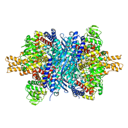
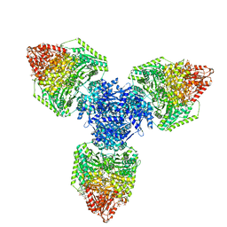
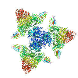
 | | Crystal structure of full-length, homohexameric 2-oxoglutarate dehydrogenase KGD from Mycobacterium smegmatis in complex with GarA | | 分子名称: | CALCIUM ION, Glycogen accumulation regulator GarA, MAGNESIUM ION, ... | | 著者 | Wagner, T, Mechaly, A.M, Alzari, P.M, Bellinzoni, M. | | 登録日 | 2023-05-24 | | 公開日 | 2023-08-16 | | 最終更新日 | 2023-08-23 | | 実験手法 | X-RAY DIFFRACTION (4.562 Å) | | 主引用文献 | High resolution cryo-EM and crystallographic snapshots of the actinobacterial two-in-one 2-oxoglutarate dehydrogenase.
Nat Commun, 14, 2023
|
|
6ZA4
 | | M. tuberculosis salicylate synthase MbtI in complex with 5-(3-cyanophenyl)furan-2-carboxylate | | 分子名称: | 5-(3-cyanophenyl)furan-2-carboxylic acid, CHLORIDE ION, Salicylate synthase | | 著者 | Mori, M, Villa, S, Meneghetti, F, Bellinzoni, M. | | 登録日 | 2020-06-04 | | 公開日 | 2020-07-01 | | 最終更新日 | 2024-01-24 | | 実験手法 | X-RAY DIFFRACTION (2.092 Å) | | 主引用文献 | Shedding X-ray Light on the Role of Magnesium in the Activity ofMycobacterium tuberculosisSalicylate Synthase (MbtI) for Drug Design.
J.Med.Chem., 63, 2020
|
|
6ZA6
 | | M. tuberculosis salicylate synthase MbtI in complex with Ba2+ | | 分子名称: | BARIUM ION, CHLORIDE ION, GLYCEROL, ... | | 著者 | Mori, M, Villa, S, Meneghetti, F, Bellinzoni, M. | | 登録日 | 2020-06-04 | | 公開日 | 2020-07-01 | | 最終更新日 | 2024-01-24 | | 実験手法 | X-RAY DIFFRACTION (1.804 Å) | | 主引用文献 | Shedding X-ray Light on the Role of Magnesium in the Activity ofMycobacterium tuberculosisSalicylate Synthase (MbtI) for Drug Design.
J.Med.Chem., 63, 2020
|
|
6ZA5
 | | M. tuberculosis salicylate synthase MbtI in complex with salicylate and Mg2+ | | 分子名称: | 2-HYDROXYBENZOIC ACID, ACETATE ION, MAGNESIUM ION, ... | | 著者 | Mori, M, Villa, S, Meneghetti, F, Bellinzoni, M. | | 登録日 | 2020-06-04 | | 公開日 | 2020-07-15 | | 最終更新日 | 2024-01-24 | | 実験手法 | X-RAY DIFFRACTION (2.109 Å) | | 主引用文献 | Shedding X-ray Light on the Role of Magnesium in the Activity ofMycobacterium tuberculosisSalicylate Synthase (MbtI) for Drug Design.
J.Med.Chem., 63, 2020
|
|
8P5W
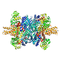
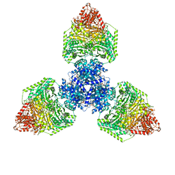
 | | Single particle cryo-EM structure of homohexameric 2-oxoglutarate dehydrogenase OdhA from Corynebacterium glutamicum following reaction with the 2-oxoglutarate analogue succinyl phosphonate | | 分子名称: | (4~{S})-4-[(2~{R})-3-[(4-azanyl-2-methyl-pyrimidin-5-yl)methyl]-4-methyl-5-[2-[oxidanyl(phosphonooxy)phosphoryl]oxyethyl]-2~{H}-1,3-thiazol-2-yl]-4-oxidanyl-4-phosphono-butanoic acid, 2-oxoglutarate dehydrogenase E1/E2 component, ACETYL COENZYME *A, ... | | 著者 | Yang, L, Mechaly, A.M, Bellinzoni, M. | | 登録日 | 2023-05-24 | | 公開日 | 2023-08-16 | | 最終更新日 | 2023-08-23 | | 実験手法 | ELECTRON MICROSCOPY (2.26 Å) | | 主引用文献 | High resolution cryo-EM and crystallographic snapshots of the actinobacterial two-in-one 2-oxoglutarate dehydrogenase.
Nat Commun, 14, 2023
|
|
8P5U
 | | Single particle cryo-EM structure of homohexameric 2-oxoglutarate dehydrogenase OdhA from Corynebacterium glutamicum with Coenzyme A bound to the E2o domain | | 分子名称: | 2-oxoglutarate dehydrogenase E1/E2 component, ACETYL COENZYME *A, COENZYME A, ... | | 著者 | Yang, L, Mechaly, A.M, Bellinzoni, M. | | 登録日 | 2023-05-24 | | 公開日 | 2023-08-16 | | 最終更新日 | 2023-08-23 | | 実験手法 | ELECTRON MICROSCOPY (2.17 Å) | | 主引用文献 | High resolution cryo-EM and crystallographic snapshots of the actinobacterial two-in-one 2-oxoglutarate dehydrogenase.
Nat Commun, 14, 2023
|
|
8P5T
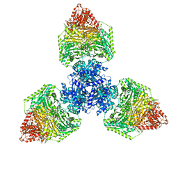 | | Single particle cryo-EM structure of the homohexameric 2-oxoglutarate dehydrogenase OdhA from Corynebacterium glutamicum | | 分子名称: | 2-oxoglutarate dehydrogenase E1/E2 component, ACETYL COENZYME *A, MAGNESIUM ION, ... | | 著者 | Yang, L, Mechaly, A.M, Bellinzoni, M. | | 登録日 | 2023-05-24 | | 公開日 | 2023-08-16 | | 最終更新日 | 2023-08-23 | | 実験手法 | ELECTRON MICROSCOPY (2.17 Å) | | 主引用文献 | High resolution cryo-EM and crystallographic snapshots of the actinobacterial two-in-one 2-oxoglutarate dehydrogenase.
Nat Commun, 14, 2023
|
|
8P5V
 | | Single particle cryo-EM structure of homohexameric 2-oxoglutarate dehydrogenase OdhA from Corynebacterium glutamicum in complex with the product succinyl-CoA | | 分子名称: | 2-oxoglutarate dehydrogenase E1/E2 component, ACETYL COENZYME *A, MAGNESIUM ION, ... | | 著者 | Yang, L, Mechaly, A.M, Bellinzoni, M. | | 登録日 | 2023-05-24 | | 公開日 | 2023-08-16 | | 最終更新日 | 2023-08-23 | | 実験手法 | ELECTRON MICROSCOPY (2.07 Å) | | 主引用文献 | High resolution cryo-EM and crystallographic snapshots of the actinobacterial two-in-one 2-oxoglutarate dehydrogenase.
Nat Commun, 14, 2023
|
|
8P5X
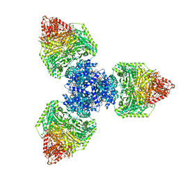
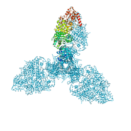
 | | Single particle cryo-EM structure of the complex between Corynebacterium glutamicum homohexameric 2-oxoglutarate dehydrogenase OdhA and the FHA-protein inhibitor OdhI | | 分子名称: | 2-oxoglutarate dehydrogenase E1/E2 component, MAGNESIUM ION, Oxoglutarate dehydrogenase inhibitor, ... | | 著者 | Yang, L, Mechaly, A.M, Bellinzoni, M. | | 登録日 | 2023-05-24 | | 公開日 | 2023-08-16 | | 最終更新日 | 2023-08-23 | | 実験手法 | ELECTRON MICROSCOPY (2.29 Å) | | 主引用文献 | High resolution cryo-EM and crystallographic snapshots of the actinobacterial two-in-one 2-oxoglutarate dehydrogenase.
Nat Commun, 14, 2023
|
|
8P5S
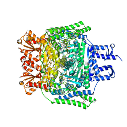
 | | Crystal structure of the homohexameric 2-oxoglutarate dehydrogenase OdhA from Corynebacterium glutamicum | | 分子名称: | 2-oxoglutarate dehydrogenase E1/E2 component, 4-(2-HYDROXYETHYL)-1-PIPERAZINE ETHANESULFONIC ACID, ACETYL COENZYME *A, ... | | 著者 | Yang, L, Boyko, A, Bellinzoni, M. | | 登録日 | 2023-05-24 | | 公開日 | 2023-08-16 | | 最終更新日 | 2023-08-23 | | 実験手法 | X-RAY DIFFRACTION (2.459 Å) | | 主引用文献 | High resolution cryo-EM and crystallographic snapshots of the actinobacterial two-in-one 2-oxoglutarate dehydrogenase.
Nat Commun, 14, 2023
|
|
6Z0F
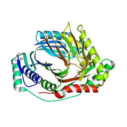
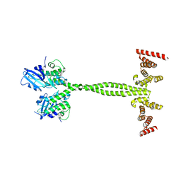 | | Crystal structure of the membrane pseudokinase YukC/EssB from Bacillus subtilis T7SS | | 分子名称: | ESX secretion system protein YukC | | 著者 | Tassinari, M, Bellinzoni, M, Alzari, P.M, Fronzes, R, Gubellini, F. | | 登録日 | 2020-05-08 | | 公開日 | 2021-05-19 | | 最終更新日 | 2024-02-07 | | 実験手法 | X-RAY DIFFRACTION (2.553 Å) | | 主引用文献 | The Antibacterial Type VII Secretion System of Bacillus subtilis: Structure and Interactions of the Pseudokinase YukC/EssB.
Mbio, 13, 2022
|
|
5M07
 | | Crystal structure of Mycobacterium tuberculosis PknI kinase domain, C20A mutant | | 分子名称: | SODIUM ION, Serine/threonine-protein kinase PknI | | 著者 | Lisa, M.N, Wagner, T, Alexandre, M, Barilone, N, Raynal, B, Alzari, P.M, Bellinzoni, M. | | 登録日 | 2016-10-03 | | 公開日 | 2017-01-11 | | 最終更新日 | 2024-01-17 | | 実験手法 | X-RAY DIFFRACTION (2.5 Å) | | 主引用文献 | The crystal structure of PknI from Mycobacterium tuberculosis shows an inactive, pseudokinase-like conformation.
FEBS J., 284, 2017
|
|
5M08
 | | Crystal structure of Mycobacterium tuberculosis PknI kinase domain, C20A_R136A double mutant | | 分子名称: | Serine/threonine-protein kinase PknI | | 著者 | Lisa, M.N, Wagner, T, Alexandre, M, Barilone, N, Raynal, B, Alzari, P.M, Bellinzoni, M. | | 登録日 | 2016-10-03 | | 公開日 | 2017-01-11 | | 最終更新日 | 2024-01-17 | | 実験手法 | X-RAY DIFFRACTION (3.03 Å) | | 主引用文献 | The crystal structure of PknI from Mycobacterium tuberculosis shows an inactive, pseudokinase-like conformation.
FEBS J., 284, 2017
|
|
5M09
 | | Crystal structure of Mycobacterium tuberculosis PknI kinase domain, C20A_R136N double mutant | | 分子名称: | SODIUM ION, Serine/threonine-protein kinase PknI | | 著者 | Lisa, M.N, Wagner, T, Alexandre, M, Barilone, N, Raynal, B, Alzari, P.M, Bellinzoni, M. | | 登録日 | 2016-10-03 | | 公開日 | 2017-01-11 | | 最終更新日 | 2024-01-17 | | 実験手法 | X-RAY DIFFRACTION (2.98 Å) | | 主引用文献 | The crystal structure of PknI from Mycobacterium tuberculosis shows an inactive, pseudokinase-like conformation.
FEBS J., 284, 2017
|
|
5M06
 | | Crystal structure of Mycobacterium tuberculosis PknI kinase domain | | 分子名称: | ADENOSINE-5'-DIPHOSPHATE, CALCIUM ION, Serine/threonine-protein kinase PknI | | 著者 | Wagner, T, Lisa, M.N, Alexandre, M, Barilone, N, Raynal, B, Alzari, P.M, Bellinzoni, M. | | 登録日 | 2016-10-03 | | 公開日 | 2017-01-11 | | 最終更新日 | 2024-05-01 | | 実験手法 | X-RAY DIFFRACTION (2 Å) | | 主引用文献 | The crystal structure of PknI from Mycobacterium tuberculosis shows an inactive, pseudokinase-like conformation.
FEBS J., 284, 2017
|
|


