Loading
PDBj
MenuPDBj@FacebookPDBj@TwitterPDBj@YouTubewwPDB FoundationwwPDB
RCSB PDBPDBeBMRBAdv. SearchSearch help
PDB: 71 results

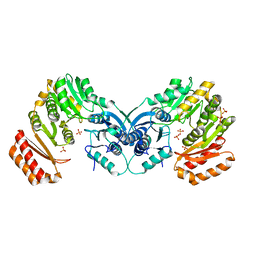
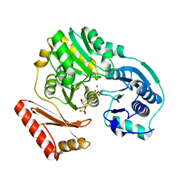
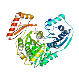
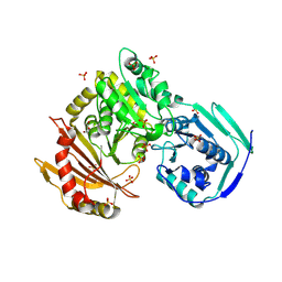
3NA5
DownloadVisualize
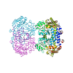
BU of 3na5 by Molmil
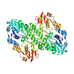
Crystal structure of a bacterial phosphoglucomutase, an enzyme important in the virulence of several human pathogens.
Descriptor: 2-[BIS-(2-HYDROXY-ETHYL)-AMINO]-2-HYDROXYMETHYL-PROPANE-1,3-DIOL, MAGNESIUM ION, Phosphoglucomutase
Authors:Mehra-Chaudhary, R, Beamer, L.J.
Deposit date:2010-06-01
Release date:2011-02-16
Last modified:2023-11-22
Method:X-RAY DIFFRACTION (1.7 Å)
Cite:Crystal structure of a bacterial phosphoglucomutase, an enzyme involved in the virulence of multiple human pathogens.
Proteins, 79, 2011
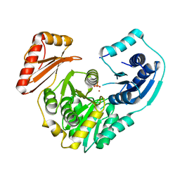
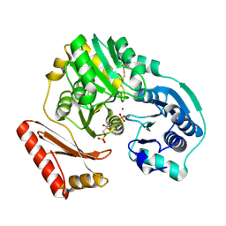
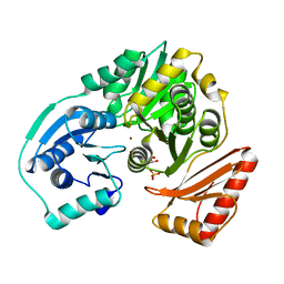
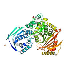
3PDK
DownloadVisualize
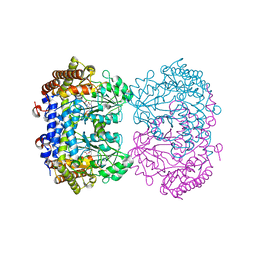
BU of 3pdk by Molmil
crystal structure of phosphoglucosamine mutase from B. anthracis
Descriptor: PHOSPHATE ION, Phosphoglucosamine mutase
Authors:Mehra-Chaudhary, R, Mick, J, Tanner, J.J, Henzl, M, Beamer, L.J.
Deposit date:2010-10-22
Release date:2011-08-24
Last modified:2023-09-06
Method:X-RAY DIFFRACTION (2.7 Å)
Cite:Crystal Structure of Bacillus anthracis Phosphoglucosamine Mutase, an Enzyme in the Peptidoglycan Biosynthetic Pathway.
J.Bacteriol., 193, 2011
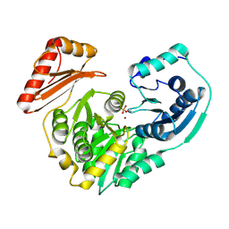
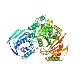
1U6D
DownloadVisualize
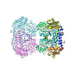
BU of 1u6d by Molmil
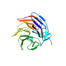
Crystal structure of the Kelch domain of human Keap1
Descriptor: kelch-like ECH-associated protein 1
Authors:Li, X, Zhang, D, Hannink, M, Beamer, L.J.
Deposit date:2004-07-29
Release date:2004-10-12
Last modified:2024-02-14
Method:X-RAY DIFFRACTION (1.85 Å)
Cite:Crystal structure of the kelch domain of human keap1
J.Biol.Chem., 279, 2004
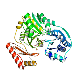
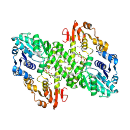
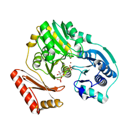
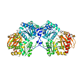
3RSM
DownloadVisualize
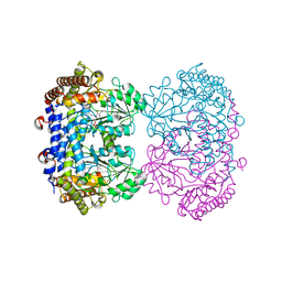
BU of 3rsm by Molmil
Crystal structure of S108C mutant of PMM/PGM
Descriptor: PHOSPHATE ION, Phosphomannomutase/phosphoglucomutase, ZINC ION
Authors:Akella, A, Anbanandam, A, Kelm, A, Wei, Y, Mehra-Chaudhary, R, Beamer, L, Van Doren, S.
Deposit date:2011-05-02
Release date:2012-02-29
Last modified:2023-09-13
Method:X-RAY DIFFRACTION (2.1 Å)
Cite:Solution NMR of a 463-residue phosphohexomutase: domain 4 mobility, substates, and phosphoryl transfer defect.
Biochemistry, 51, 2012
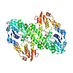
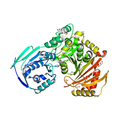
3C04
DownloadVisualize
BU of 3c04 by Molmil
Structure of the P368G mutant of PMM/PGM from P. aeruginosa
Descriptor: Phosphomannomutase/phosphoglucomutase, ZINC ION
Authors:Mehra-Chaudhary, R, Beamer, L.J.
Deposit date:2008-01-18
Release date:2008-09-09
Last modified:2023-08-30
Method:X-RAY DIFFRACTION (2.2 Å)
Cite:Backbone flexibility, conformational change, and catalysis in a phosphohexomutase from Pseudomonas aeruginosa.
Biochemistry, 47, 2008
3BKQ
DownloadVisualize
BU of 3bkq by Molmil
Structure of the P368G mutant of PMM/PGM in complex with its substrate
Descriptor: 1-O-phosphono-alpha-D-glucopyranose, Phosphomannomutase/phosphoglucomutase, ZINC ION
Authors:Mehra-Chaudhary, R, Beamer, L.J.
Deposit date:2007-12-07
Release date:2008-09-09
Last modified:2023-08-30
Method:X-RAY DIFFRACTION (2.05 Å)
Cite:Backbone flexibility, conformational change, and catalysis in a phosphohexomutase from Pseudomonas aeruginosa.
Biochemistry, 47, 2008
1PCJ
DownloadVisualize
BU of 1pcj by Molmil
Enzyme-ligand complex of P. aeruginosa PMM/PGM
Descriptor: 1-O-phosphono-alpha-D-mannopyranose, Phosphomannomutase, ZINC ION
Authors:Regni, C, Tipton, P.A, Beamer, L.J.
Deposit date:2003-05-16
Release date:2004-01-20
Last modified:2023-08-16
Method:X-RAY DIFFRACTION (2 Å)
Cite:Structural basis of diverse substrate recognition by the enzyme PMM/PGM from P. aeruginosa.
Structure, 12, 2004
1PCM
DownloadVisualize
BU of 1pcm by Molmil
Enzyme-ligand complex of P. aeruginosa PMM/PGM
Descriptor: 6-O-phosphono-alpha-D-mannopyranose, Phosphomannomutase, ZINC ION
Authors:Regni, C, Tipton, P.A, Beamer, L.J.
Deposit date:2003-05-16
Release date:2004-01-20
Last modified:2023-08-16
Method:X-RAY DIFFRACTION (1.9 Å)
Cite:Structural basis of diverse substrate recognition by the enzyme PMM/PGM from P. aeruginosa.
Structure, 12, 2004
1MFZ
DownloadVisualize
BU of 1mfz by Molmil
Partially refined 2.8 A Crystal structure of GDP-mannose dehydrogenase from P. aeruginosa
Descriptor: GDP-mannose 6-dehydrogenase, GUANOSINE 5'-(TRIHYDROGEN DIPHOSPHATE), P'-D-MANNOPYRANOSYL ESTER
Authors:Snook, C.F, Tipton, P.A, Beamer, L.J.
Deposit date:2002-08-14
Release date:2003-05-06
Last modified:2014-11-12
Method:X-RAY DIFFRACTION (2.8 Å)
Cite:The crystal structure of GDP-mannose dehydrogenase: A key enzyme in alginate biosynthesis of P. aeruginosa
Biochemistry, 42, 2003
1MV8
DownloadVisualize
BU of 1mv8 by Molmil
1.55 A crystal structure of a ternary complex of GDP-mannose dehydrogenase from Psuedomonas aeruginosa
Descriptor: (4S)-2-METHYL-2,4-PENTANEDIOL, ACETIC ACID, GDP-mannose 6-dehydrogenase, ...
Authors:Snook, C.F, Tipton, P.A, Beamer, L.J.
Deposit date:2002-09-24
Release date:2003-05-06
Last modified:2024-02-14
Method:X-RAY DIFFRACTION (1.55 Å)
Cite:The crystal structure of GDP-mannose dehydrogenase: A key enzyme in alginate biosynthesis of P. aeruginosa
Biochemistry, 42, 2003
1MUU
DownloadVisualize
BU of 1muu by Molmil
2.0 A crystal structure of GDP-mannose dehydrogenase
Descriptor: GDP-mannose 6-dehydrogenase, GUANOSINE 5'-(TRIHYDROGEN DIPHOSPHATE), P'-D-MANNOPYRANOSYL ESTER, ...
Authors:Snook, C.F, Tipton, P.A, Beamer, L.J.
Deposit date:2002-09-24
Release date:2003-05-06
Last modified:2020-07-29
Method:X-RAY DIFFRACTION (2.02 Å)
Cite:Crystal structure of GDP-mannose dehydrogenase: A key enzyme of alginate biosynthesis in P. aeruginosa
Biochemistry, 42, 2003
2FLU
DownloadVisualize
BU of 2flu by Molmil
Crystal Structure of the Kelch-Neh2 Complex
Descriptor: Kelch-like ECH-associated protein 1, Nrf2
Authors:Li, X, Lo, J, Beamer, L, Hannink, M.
Deposit date:2006-01-06
Release date:2006-08-15
Last modified:2023-08-30
Method:X-RAY DIFFRACTION (1.5 Å)
Cite:Structure of the Keap1:Nrf2 interface provides mechanistic insight into Nrf2 signaling.
Embo J., 25, 2006
6UXJ
DownloadVisualize
BU of 6uxj by Molmil
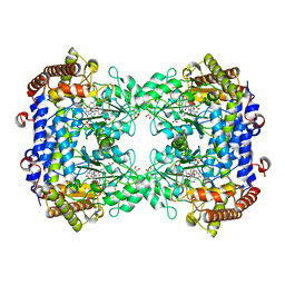
Structure of serine hydroxymethyltransferase 8 from Glycine max cultivar Essex complexed with PLP-glycine and 5-formyltetrahydrofolate
Descriptor: 1,2-ETHANEDIOL, N-GLYCINE-[3-HYDROXY-2-METHYL-5-PHOSPHONOOXYMETHYL-PYRIDIN-4-YL-METHANE], N-[4-({[(6S)-2-amino-5-formyl-4-oxo-3,4,5,6,7,8-hexahydropteridin-6-yl]methyl}amino)benzoyl]-L-glutamic acid, ...
Authors:Korasick, D.A, Tanner, J.J, Beamer, L.J.
Deposit date:2019-11-07
Release date:2020-02-12
Last modified:2023-10-11
Method:X-RAY DIFFRACTION (1.4 Å)
Cite:Impaired folate binding of serine hydroxymethyltransferase 8 from soybean underlies resistance to the soybean cyst nematode.
J.Biol.Chem., 295, 2020
5HSH
DownloadVisualize
BU of 5hsh by Molmil
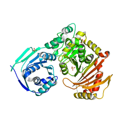
Crystal structure of the G291R mutant of human phosphoglucomutase 1
Descriptor: Phosphoglucomutase-1, SULFATE ION
Authors:Stiers, K.M, Beamer, L.J.
Deposit date:2016-01-25
Release date:2016-04-27
Last modified:2023-09-27
Method:X-RAY DIFFRACTION (2.65 Å)
Cite:Induced Structural Disorder as a Molecular Mechanism for Enzyme Dysfunction in Phosphoglucomutase 1 Deficiency.
J.Mol.Biol., 428, 2016
6UXL
DownloadVisualize
BU of 6uxl by Molmil
Structure of serine hydroxymethyltransferase 8 from Glycine max cultivar Forrest complexed with PLP-Glycine
Descriptor: N-GLYCINE-[3-HYDROXY-2-METHYL-5-PHOSPHONOOXYMETHYL-PYRIDIN-4-YL-METHANE], Serine hydroxymethyltransferase
Authors:Korasick, D.A, Tanner, J.J, Beamer, L.J.
Deposit date:2019-11-07
Release date:2020-02-12
Last modified:2023-10-11
Method:X-RAY DIFFRACTION (2.35 Å)
Cite:Impaired folate binding of serine hydroxymethyltransferase 8 from soybean underlies resistance to the soybean cyst nematode.
J.Biol.Chem., 295, 2020
6UXI
DownloadVisualize
BU of 6uxi by Molmil
Structure of serine hydroxymethyltransferase 8 from Glycine max cultivar Essex complexed with PLP-Glycine
Descriptor: 1,2-ETHANEDIOL, N-GLYCINE-[3-HYDROXY-2-METHYL-5-PHOSPHONOOXYMETHYL-PYRIDIN-4-YL-METHANE], Serine hydroxymethyltransferase
Authors:Korasick, D.A, Tanner, J.J, Beamer, L.J.
Deposit date:2019-11-07
Release date:2020-02-12
Last modified:2023-10-11
Method:X-RAY DIFFRACTION (2.1 Å)
Cite:Impaired folate binding of serine hydroxymethyltransferase 8 from soybean underlies resistance to the soybean cyst nematode.
J.Biol.Chem., 295, 2020
6UXK
DownloadVisualize
BU of 6uxk by Molmil
Structure of serine hydroxymethyltransferase 8 from Glycine max cultivar Forrest complexed with PLP
Descriptor: 1,2-ETHANEDIOL, Serine hydroxymethyltransferase
Authors:Korasick, D.A, Tanner, J.J, Beamer, L.J.
Deposit date:2019-11-07
Release date:2020-02-12
Last modified:2023-11-15
Method:X-RAY DIFFRACTION (2.1 Å)
Cite:Impaired folate binding of serine hydroxymethyltransferase 8 from soybean underlies resistance to the soybean cyst nematode.
J.Biol.Chem., 295, 2020
6UXH
DownloadVisualize
BU of 6uxh by Molmil
Structure of serine hydroxymethyltransferase 8 from Glycine max cultivar Essex complexed with PLP
Descriptor: 1,2-ETHANEDIOL, Serine hydroxymethyltransferase
Authors:Korasick, D.A, Tanner, J.J, Beamer, L.J.
Deposit date:2019-11-07
Release date:2020-02-12
Last modified:2023-11-15
Method:X-RAY DIFFRACTION (1.858 Å)
Cite:Impaired folate binding of serine hydroxymethyltransferase 8 from soybean underlies resistance to the soybean cyst nematode.
J.Biol.Chem., 295, 2020
2FKF
DownloadVisualize
BU of 2fkf by Molmil
Phosphomannomutase/Phosphoglucomutase from Pseudomonas aeruginosa with alpha-D-glucose 1,6-bisphosphate bound
Descriptor: 1,6-di-O-phosphono-alpha-D-glucopyranose, Phosphomannomutase/phosphoglucomutase, ZINC ION
Authors:Regni, C.A, Beamer, L.J.
Deposit date:2006-01-04
Release date:2006-04-04
Last modified:2023-08-30
Method:X-RAY DIFFRACTION (2 Å)
Cite:The reaction of phosphohexomutase from Pseudomonas aeruginosa: structural insights into a simple processive enzyme.
J.Biol.Chem., 281, 2006
2FKM
DownloadVisualize
BU of 2fkm by Molmil
PMM/PGM S108D mutant with alpha-d-glucose 1,6-bisphosphate bound
Descriptor: 1,6-di-O-phosphono-alpha-D-glucopyranose, Phosphomannomutase/phosphoglucomutase, ZINC ION
Authors:Regni, C.A, Beamer, L.J.
Deposit date:2006-01-04
Release date:2006-04-04
Last modified:2023-08-30
Method:X-RAY DIFFRACTION (1.9 Å)
Cite:The reaction of phosphohexomutase from Pseudomonas aeruginosa: structural insights into a simple processive enzyme.
J.Biol.Chem., 281, 2006
2H5A
DownloadVisualize
BU of 2h5a by Molmil
Complex of the enzyme PMM/PGM with xylose 1-phosphate
Descriptor: 1-O-phosphono-alpha-D-xylopyranose, Phosphomannomutase/phosphoglucomutase, ZINC ION
Authors:Regni, C, Shackelford, G.S, Beamer, L.J.
Deposit date:2006-05-25
Release date:2006-08-08
Last modified:2020-07-29
Method:X-RAY DIFFRACTION (1.72 Å)
Cite:Complexes of the enzyme phosphomannomutase/phosphoglucomutase with a slow substrate and an inhibitor.
Acta Crystallogr.,Sect.F, 62, 2006
5VBI
DownloadVisualize
BU of 5vbi by Molmil
Crystal Structure of the R515W missense variant of human PGM1
Descriptor: GLYCEROL, MAGNESIUM ION, Phosphoglucomutase-1, ...
Authors:Stiers, K.M, Beamer, L.J.
Deposit date:2017-03-29
Release date:2018-06-20
Last modified:2023-10-04
Method:X-RAY DIFFRACTION (1.75000083 Å)
Cite:A Hotspot for Disease-Associated Variants of Human PGM1 Is Associated with Impaired Ligand Binding and Loop Dynamics.
Structure, 26, 2018
5VG7
DownloadVisualize
BU of 5vg7 by Molmil
Crystal Structure of the R503Q missense variant of human PGM1
Descriptor: GLYCEROL, MAGNESIUM ION, Phosphoglucomutase-1, ...
Authors:Stiers, K.M, Beamer, L.J.
Deposit date:2017-04-10
Release date:2018-06-20
Last modified:2023-10-04
Method:X-RAY DIFFRACTION (1.95000327 Å)
Cite:A Hotspot for Disease-Associated Variants of Human PGM1 Is Associated with Impaired Ligand Binding and Loop Dynamics.
Structure, 26, 2018
5VIN
DownloadVisualize
BU of 5vin by Molmil
Crystal Structure of the R515Q missense variant of human PGM1
Descriptor: COBALT (II) ION, GLYCEROL, Phosphoglucomutase-1, ...
Authors:Stiers, K.M, Beamer, L.J.
Deposit date:2017-04-17
Release date:2018-06-27
Last modified:2023-10-04
Method:X-RAY DIFFRACTION (2.60004282 Å)
Cite:A Hotspot for Disease-Associated Variants of Human PGM1 Is Associated with Impaired Ligand Binding and Loop Dynamics.
Structure, 26, 2018
5VEC
DownloadVisualize
BU of 5vec by Molmil
Crystal Structure of the R515L missense variant of human PGM1
Descriptor: GLYCEROL, MAGNESIUM ION, Phosphoglucomutase-1, ...
Authors:Stiers, K.M, Beamer, L.J.
Deposit date:2017-04-04
Release date:2018-06-27
Last modified:2023-10-04
Method:X-RAY DIFFRACTION (2.20001626 Å)
Cite:A Hotspot for Disease-Associated Variants of Human PGM1 Is Associated with Impaired Ligand Binding and Loop Dynamics.
Structure, 26, 2018

219869

PDB entries from 2024-05-15

PDB statisticsPDBj update infoContact PDBjnumon