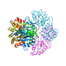
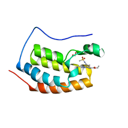
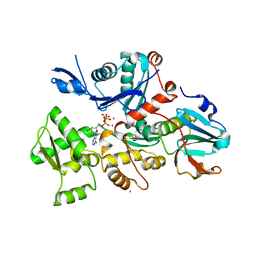
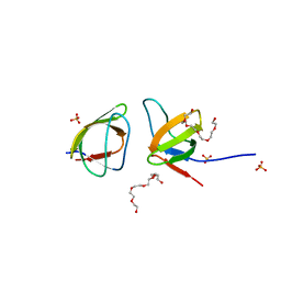
6V1O
 
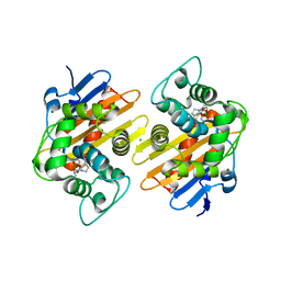 | | Structure of OXA-48 bound to QPX7728 at 1.80 A | | 分子名称: | (1aR,7bS)-5-fluoro-2-hydroxy-1,1a,2,7b-tetrahydrocyclopropa[c][1,2]benzoxaborinine-4-carboxylic acid, CHLORIDE ION, MAGNESIUM ION, ... | | 著者 | Pemberton, O.A, Chen, Y. | | 登録日 | 2019-11-20 | | 公開日 | 2020-03-25 | | 最終更新日 | 2023-11-15 | | 実験手法 | X-RAY DIFFRACTION (1.8 Å) | | 主引用文献 | Discovery of Cyclic Boronic Acid QPX7728, an Ultrabroad-Spectrum Inhibitor of Serine and Metallo-beta-lactamases.
J.Med.Chem., 63, 2020
|
|
5MOO
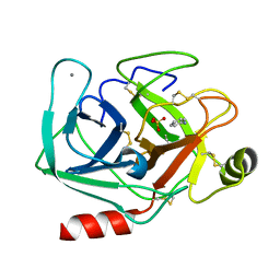 | | Joint X-ray/neutron structure of cationic trypsin in complex with aniline | | 分子名称: | CALCIUM ION, Cationic trypsin, SULFATE ION, ... | | 著者 | Schiebel, J, Schrader, T.E, Ostermann, A, Heine, A, Klebe, G. | | 登録日 | 2016-12-14 | | 公開日 | 2017-05-24 | | 最終更新日 | 2024-05-01 | | 実験手法 | NEUTRON DIFFRACTION (1.441 Å), X-RAY DIFFRACTION | | 主引用文献 | Charges Shift Protonation: Neutron Diffraction Reveals that Aniline and 2-Aminopyridine Become Protonated Upon Binding to Trypsin.
Angew. Chem. Int. Ed. Engl., 56, 2017
|
|
6SD8
 | | Bd2924 apo-form | | 分子名称: | FLAVIN-ADENINE DINUCLEOTIDE, Probable acyl-CoA dehydrogenase | | 著者 | Lovering, A.L, Harding, C.J. | | 登録日 | 2019-07-26 | | 公開日 | 2019-09-11 | | 最終更新日 | 2024-05-15 | | 実験手法 | X-RAY DIFFRACTION (1.51 Å) | | 主引用文献 | Target highlights in CASP13: Experimental target structures through the eyes of their authors.
Proteins, 87, 2019
|
|
7TBP
 | | X-ray structure of the HIV-1 myristoylated matrix protein | | 分子名称: | CITRIC ACID, Capsid protein p24, MYRISTIC ACID, ... | | 著者 | Green, T.J, Saad, J.S, Samal, A.B. | | 登録日 | 2021-12-22 | | 公開日 | 2022-06-22 | | 最終更新日 | 2023-10-18 | | 実験手法 | X-RAY DIFFRACTION (2.151 Å) | | 主引用文献 | Atomic view of the HIV-1 matrix lattice; implications on virus assembly and envelope incorporation.
Proc.Natl.Acad.Sci.USA, 119, 2022
|
|
6V28
 | |
6XEO
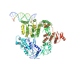 | | Structure of Mfd bound to dsDNA | | 分子名称: | DNA (5'-D(P*AP*GP*GP*AP*TP*AP*CP*TP*TP*AP*CP*AP*GP*CP*CP*AP*TP*C)-3'), DNA (5'-D(P*GP*AP*TP*GP*GP*CP*TP*GP*TP*AP*AP*GP*TP*AP*TP*CP*CP*T)-3'), Transcription-repair-coupling factor | | 著者 | Brugger, C, Deaconescu, A. | | 登録日 | 2020-06-12 | | 公開日 | 2020-08-19 | | 最終更新日 | 2024-03-06 | | 実験手法 | ELECTRON MICROSCOPY (5.5 Å) | | 主引用文献 | Molecular determinants for dsDNA translocation by the transcription-repair coupling and evolvability factor Mfd.
Nat Commun, 11, 2020
|
|
7QYI
 | |
7SQ1
 | | BG505.MD39TS Env trimer in complex with Fab from antibody C05 | | 分子名称: | 2-acetamido-2-deoxy-beta-D-glucopyranose, 2-acetamido-2-deoxy-beta-D-glucopyranose-(1-4)-2-acetamido-2-deoxy-beta-D-glucopyranose, C05 Fab Light chain, ... | | 著者 | Moore, A, Du, J, Xu, Z, Walker, S, Kulp, D.W, Pallesen, J. | | 登録日 | 2021-11-04 | | 公開日 | 2022-06-22 | | 最終更新日 | 2022-12-07 | | 実験手法 | ELECTRON MICROSCOPY (3.8 Å) | | 主引用文献 | Induction of tier-2 neutralizing antibodies in mice with a DNA-encoded HIV envelope native like trimer.
Nat Commun, 13, 2022
|
|
6V0U
 | | Crystal structure of the first bromodomain (BD1) of human BRD4 bound to bromosporine | | 分子名称: | 1,2-ETHANEDIOL, Bromodomain-containing protein 4, Bromosporine | | 著者 | Chan, A, Karim, M.R, Schonbrunn, E. | | 登録日 | 2019-11-19 | | 公開日 | 2020-03-11 | | 最終更新日 | 2023-10-11 | | 実験手法 | X-RAY DIFFRACTION (1.4 Å) | | 主引用文献 | Structural Basis of Inhibitor Selectivity in the BRD7/9 Subfamily of Bromodomains.
J.Med.Chem., 63, 2020
|
|
6V0Z
 | | Structure of ALDH7A1 mutant R441C complexed with NAD | | 分子名称: | 1,2-ETHANEDIOL, Alpha-aminoadipic semialdehyde dehydrogenase, NICOTINAMIDE-ADENINE-DINUCLEOTIDE | | 著者 | Korasick, D.A, Tanner, J.J. | | 登録日 | 2019-11-19 | | 公開日 | 2020-11-25 | | 最終更新日 | 2024-04-24 | | 実験手法 | X-RAY DIFFRACTION (2.02 Å) | | 主引用文献 | Biochemical, structural, and computational analyses of two new clinically identified missense mutations of ALDH7A1.
Chem.Biol.Interact., 2024
|
|
6V1V
 | | VIP3B (VIP3B_2160) adapted for crystallization | | 分子名称: | Vegetative insecticidal protein | | 著者 | Evdokimov, A.G, Zheng, M, Moshiri, F, Haas, J, Lowder, C. | | 登録日 | 2019-11-21 | | 公開日 | 2019-12-18 | | 最終更新日 | 2024-04-03 | | 実験手法 | X-RAY DIFFRACTION (3.189 Å) | | 主引用文献 | Crystal structure of a Vip3B family insecticidal protein reveals a new fold and a unique tetrameric assembly.
Protein Sci., 29, 2020
|
|
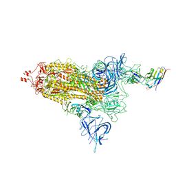
7OB9
 | | Cryo-EM structure of human RNA Polymerase I in elongation state | | 分子名称: | DNA non-template strand, DNA template strand, DNA-directed RNA polymerase I subunit RPA1, ... | | 著者 | Misiaszek, A.D, Girbig, M, Mueller, C.W. | | 登録日 | 2021-04-21 | | 公開日 | 2021-12-08 | | 最終更新日 | 2024-07-10 | | 実験手法 | ELECTRON MICROSCOPY (2.7 Å) | | 主引用文献 | Cryo-EM structures of human RNA polymerase I.
Nat.Struct.Mol.Biol., 28, 2021
|
|
5MVV
 | | Crystal structure of Plasmodium falciparum actin I- gelsolin segment 1 -CdATP complex | | 分子名称: | ADENOSINE-5'-TRIPHOSPHATE, Actin-1, CADMIUM ION, ... | | 著者 | Panneerselvam, S, Kumpula, E.-P, Kursula, I, Burkhardt, A, Meents, A. | | 登録日 | 2017-01-17 | | 公開日 | 2017-07-12 | | 最終更新日 | 2024-05-08 | | 実験手法 | X-RAY DIFFRACTION (1.4 Å) | | 主引用文献 | Rapid cadmium SAD phasing at the standard wavelength (1 angstrom ).
Acta Crystallogr D Struct Biol, 73, 2017
|
|
8TTO
 | |
5MV8
 | | Structure of human Myosin 7b C-terminal MyTH4-FERM domain in complex with harmonin-a PDZ3 domain | | 分子名称: | GLYCEROL, MAGNESIUM ION, Unconventional myosin-VIIb, ... | | 著者 | Yu, I, Sourigues, Y, Titus, M.A, Houdusse, A. | | 登録日 | 2017-01-16 | | 公開日 | 2017-07-05 | | 最終更新日 | 2024-05-08 | | 実験手法 | X-RAY DIFFRACTION (1.88 Å) | | 主引用文献 | Myosin 7 and its adaptors link cadherins to actin.
Nat Commun, 8, 2017
|
|
7NYK
 | |
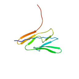
7NYM
 | | Mutant V517A - SH3 domain of JNK-interacting Protein 1 (JIP1) | | 分子名称: | HEXAETHYLENE GLYCOL, PHOSPHATE ION, SH3 domain of JNK-interacting Protein 1 (JIP1), ... | | 著者 | Perez, L.M, Ielasi, F.S, Palencia, A, Jensen, M.R. | | 登録日 | 2021-03-23 | | 公開日 | 2021-12-22 | | 最終更新日 | 2024-01-31 | | 実験手法 | X-RAY DIFFRACTION (1.614 Å) | | 主引用文献 | Visualizing protein breathing motions associated with aromatic ring flipping.
Nature, 602, 2022
|
|
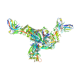
8U7I
 | | Structure of the phage immune evasion protein Gad1 bound to the Gabija GajAB complex | | 分子名称: | Endonuclease GajA, Gabija Anti-Defense 1, Gabija protein GajB | | 著者 | Antine, S.P, Johnson, A.G, Mooney, S.E, Mayer, M.L, Kranzusch, P.J. | | 登録日 | 2023-09-15 | | 公開日 | 2023-11-22 | | 最終更新日 | 2024-06-05 | | 実験手法 | ELECTRON MICROSCOPY (2.57 Å) | | 主引用文献 | Structural basis of Gabija anti-phage defence and viral immune evasion.
Nature, 625, 2024
|
|
5JR8
 | | Disposal of Iron by a Mutant form of Siderocalin NGAL | | 分子名称: | GLYCEROL, Neutrophil gelatinase-associated lipocalin, PHOSPHATE ION | | 著者 | Rupert, P.B, Strong, R.K, Barasch, J, Hollman, M, Deng, R, Hod, E.A, Abergel, R, Allred, B, Xu, K, Darrah, S, Tekabe, Y, Perlstein, A, Bruck, E, Stauber, J, Corbin, K, Buchen, C, Slavkovich, V, Graziano, J, Spitalnik, S, Qiu, A. | | 登録日 | 2016-05-05 | | 公開日 | 2016-09-28 | | 最終更新日 | 2023-09-27 | | 実験手法 | X-RAY DIFFRACTION (2.65 Å) | | 主引用文献 | Disposal of iron by a mutant form of lipocalin 2.
Nat Commun, 7, 2016
|
|
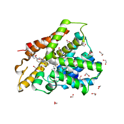
5MVO
 | | FoxE P43212 crystal structure of Rhodopseudomonas ferrooxidans SW2 putative iron oxidase | | 分子名称: | COPPER (II) ION, FoxE, HEME C, ... | | 著者 | Pereira, L, Saraiva, I.H, Oliveira, A.S, Soares, C, Gomes, R.O, Frazao, C. | | 登録日 | 2017-01-17 | | 公開日 | 2018-02-28 | | 最終更新日 | 2024-01-17 | | 実験手法 | X-RAY DIFFRACTION (2.668 Å) | | 主引用文献 | Crystallization and preliminary crystallographic studies of FoxE from Rhodobacter ferrooxidans SW2, an Fe(II) oxidoreductase involved in photoferrotrophy.
Acta Crystallogr. Sect. F Struct. Biol. Cryst. Commun., 68, 2012
|
|
7BNN
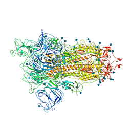 | | Open conformation of D614G SARS-CoV-2 spike with 1 Erect RBD | | 分子名称: | 2-acetamido-2-deoxy-beta-D-glucopyranose, 2-acetamido-2-deoxy-beta-D-glucopyranose-(1-4)-2-acetamido-2-deoxy-beta-D-glucopyranose, 2-acetamido-2-deoxy-beta-D-glucopyranose-(1-4)-[alpha-L-fucopyranose-(1-6)]2-acetamido-2-deoxy-beta-D-glucopyranose, ... | | 著者 | Benton, D.J, Wrobel, A.G, Rosenthal, P.B, Gamblin, S.J. | | 登録日 | 2021-01-22 | | 公開日 | 2021-02-03 | | 最終更新日 | 2021-03-10 | | 実験手法 | ELECTRON MICROSCOPY (3.5 Å) | | 主引用文献 | The effect of the D614G substitution on the structure of the spike glycoprotein of SARS-CoV-2.
Proc.Natl.Acad.Sci.USA, 118, 2021
|
|
6MWV
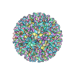 | | CryoEM structure of Chimeric Eastern Equine Encephalitis Virus with Fab of EEEV-58 Antibody | | 分子名称: | E1, E2, EEEV-58 antibody heavy chain, ... | | 著者 | Hasan, S.S, Sun, C, Kim, A.S, Watanabe, Y, Chen, C.L, Klose, T, Buda, G, Crispin, M, Diamond, M.S, Klimstra, W.B, Rossmann, M.G. | | 登録日 | 2018-10-30 | | 公開日 | 2018-12-19 | | 最終更新日 | 2019-12-18 | | 実験手法 | ELECTRON MICROSCOPY (7.3 Å) | | 主引用文献 | Cryo-EM Structures of Eastern Equine Encephalitis Virus Reveal Mechanisms of Virus Disassembly and Antibody Neutralization.
Cell Rep, 25, 2018
|
|
8EYG
 | |
6FET
 | | Crystal structure of human phosphodiesterase 4D2 catalytic domain with inhibitor NPD-1439 | | 分子名称: | 1,2-ETHANEDIOL, 1-(2-{4-[(4aS,8aR)-4-[3,4-bis(difluoromethoxy)phenyl]-1-oxo-1,2,4a,5,8,8a-hexahydrophthalazin-2-yl]piperidin-1-yl}-2-oxoethyl)-4,4-dimethylpiperidine-2,6-dione, 4-(2-HYDROXYETHYL)-1-PIPERAZINE ETHANESULFONIC ACID, ... | | 著者 | Singh, A.K, Brown, D.G. | | 登録日 | 2018-01-03 | | 公開日 | 2019-04-24 | | 最終更新日 | 2024-05-08 | | 実験手法 | X-RAY DIFFRACTION (1.88 Å) | | 主引用文献 | hPDE4D2 structure with inhibitor NPD-1439
To be published
|
|
7BTE
 | | Lifeact-F-actin complex | | 分子名称: | ADENOSINE-5'-DIPHOSPHATE, Actin, alpha skeletal muscle, ... | | 著者 | Kumari, A, Ragunath, V.K, Sirajuddin, M. | | 登録日 | 2020-04-01 | | 公開日 | 2020-05-20 | | 最終更新日 | 2024-03-27 | | 実験手法 | ELECTRON MICROSCOPY (4.2 Å) | | 主引用文献 | Structural insights into actin filament recognition by commonly used cellular actin markers.
Embo J., 39, 2020
|
|


