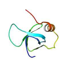
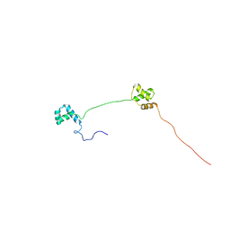
2K16
 
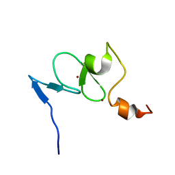 | | Solution structure of the free TAF3 PHD domain | | 分子名称: | Transcription initiation factor TFIID subunit 3, ZINC ION | | 著者 | van Ingen, H, van Schaik, F.M.A, Wienk, H, Timmers, M, Boelens, R. | | 登録日 | 2008-02-22 | | 公開日 | 2008-07-15 | | 最終更新日 | 2023-06-14 | | 実験手法 | SOLUTION NMR | | 主引用文献 | Recognition of the H3K4me3 mark by the TAF3-PHD finger
To be Published
|
|
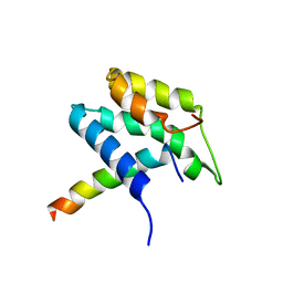
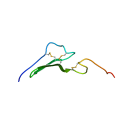
2K17
 | | Solution structure of the TAF3 PHD domain in complex with a H3K4me3 peptide | | 分子名称: | H3K4me3 peptide, Transcription initiation factor TFIID subunit 3, ZINC ION | | 著者 | van Ingen, H, van Schaik, F.M.A, Wienk, H, Timmers, M, Boelens, R. | | 登録日 | 2008-02-22 | | 公開日 | 2008-07-15 | | 最終更新日 | 2023-06-14 | | 実験手法 | SOLUTION NMR | | 主引用文献 | Recognition of the H3K4me3 mark by the TAF3-PHD finger
To be Published
|
|
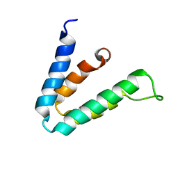
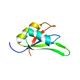
1PD7
 | | Extended SID of Mad1 bound to the PAH2 domain of mSin3B | | 分子名称: | Mad1, Sin3b protein | | 著者 | Van Ingen, H, Lasonder, E, Jansen, J.F, Kaan, A.M, Spronk, C.A, Stunnenberg, H.G, Vuister, G.W. | | 登録日 | 2003-05-19 | | 公開日 | 2004-01-20 | | 最終更新日 | 2022-02-23 | | 実験手法 | SOLUTION NMR | | 主引用文献 | Extension of the binding motif of the sin3 interacting domain of the mad family proteins(,).
Biochemistry, 43, 2004
|
|
2F05
 | |
7PJ1
 | |
3ZEH
 | |
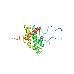
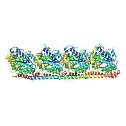
6YN1
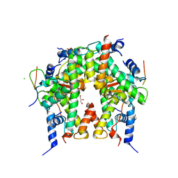 | | Crystal structure of histone chaperone APLF acidic domain bound to the histone H2A-H2B-H3-H4 octamer | | 分子名称: | Aprataxin and PNK-like factor, CHLORIDE ION, GLYCEROL, ... | | 著者 | Corbeski, I, Guo, X, Van Ingen, H, Sixma, T.K. | | 登録日 | 2020-04-10 | | 公開日 | 2021-11-17 | | 最終更新日 | 2024-01-24 | | 実験手法 | X-RAY DIFFRACTION (2.35 Å) | | 主引用文献 | Chaperoning of the histone octamer by the acidic domain of DNA repair factor APLF.
Sci Adv, 8, 2022
|
|
1P9J
 | | Solution structure and dynamics of the EGF/TGF-alpha chimera T1E | | 分子名称: | chimera of Epidermal growth factor(EGF) and Transforming growth factor alpha (TGF-alpha) | | 著者 | Wingens, M, Walma, T, Van Ingen, H, Stortelers, C, Van Leeuwen, J.E, Van Zoelen, E.J, Vuister, G.W. | | 登録日 | 2003-05-12 | | 公開日 | 2003-10-07 | | 最終更新日 | 2023-06-14 | | 実験手法 | SOLUTION NMR | | 主引用文献 | Structural Analysis of an Epidermal Growth Factor/Transforming Growth Factor-alpha Chimera with Unique ErbB Binding Specificity.
J.Biol.Chem., 278, 2003
|
|
4C7Q
 | |
2K27
 | | Solution structure of Human Pax8 Paired Box Domain | | 分子名称: | Paired box protein Pax-8 | | 著者 | Codutti, L, Esposito, G, Corazza, A, Fogolari, F, Tell, G, Vascotto, C, van Ingen, H, Boelens, R, Viglino, P, Quadrifoglio, F. | | 登録日 | 2008-03-26 | | 公開日 | 2008-09-30 | | 最終更新日 | 2023-06-14 | | 実験手法 | SOLUTION NMR | | 主引用文献 | The Solution Structure of DNA-free Pax-8 Paired Box Domain Accounts for Redox Regulation of Transcriptional Activity in the Pax Protein Family.
J.Biol.Chem., 283, 2008
|
|
8RC1
 | |
5U3A
 | |
5VA9
 | |
3KGR
 | |


