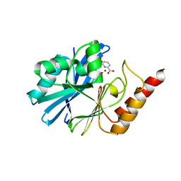
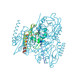
4E7N
 
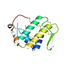
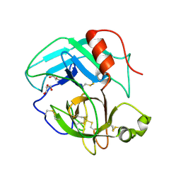 | | Crystal Structure of AhV_TL-I, a Glycosylated Snake-venom Thrombin-like Enzyme from Agkistrodon halys | | 分子名称: | 2-acetamido-2-deoxy-beta-D-glucopyranose-(1-4)-2-acetamido-2-deoxy-beta-D-glucopyranose, GLYCEROL, Snake-venom Thrombin-like Enzyme | | 著者 | Zeng, F, Li, X, Teng, M, Niu, L. | | 登録日 | 2012-03-18 | | 公開日 | 2012-04-04 | | 最終更新日 | 2023-11-08 | | 実験手法 | X-RAY DIFFRACTION (1.75 Å) | | 主引用文献 | Crystal Structure of AhV_TL-I, a Glycosylated Snake-venom Thrombin-like Enzyme from Agkistrodon halys
to be published
|
|
4RFP
 | |
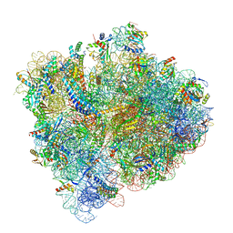
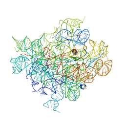
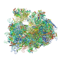
7RR5
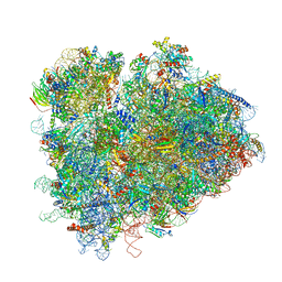 | | Structure of ribosomal complex bound with Rbg1/Tma46 | | 分子名称: | 18S rRNA, 25S rRNA, 40S ribosomal protein S0, ... | | 著者 | Zeng, F, Li, X, Pires-Alves, M, Chen, X, Hawk, C.W, Jin, H. | | 登録日 | 2021-08-09 | | 公開日 | 2021-11-10 | | 実験手法 | ELECTRON MICROSCOPY (3.23 Å) | | 主引用文献 | Conserved heterodimeric GTPase Rbg1/Tma46 promotes efficient translation in eukaryotic cells.
Cell Rep, 37, 2021
|
|
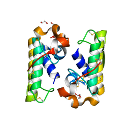
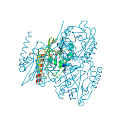
4HG9
 | | Crystal structure of AhV_bPA, a basic PLA2 from Agkistrodon halys pallas venom | | 分子名称: | Basic phospholipase A2 B, CALCIUM ION, CITRIC ACID, ... | | 著者 | Zeng, F, Niu, L, Li, X, Teng, M. | | 登録日 | 2012-10-07 | | 公開日 | 2012-10-17 | | 最終更新日 | 2023-09-20 | | 実験手法 | X-RAY DIFFRACTION (1.6 Å) | | 主引用文献 | Crystal structure of AhV_bPA, a basic PLA2 from Agkistrodon halys pallas venom
TO BE PUBLISHED
|
|
4H0S
 | |
5U4I
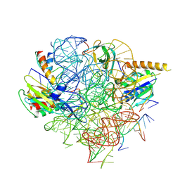
 | | Structural Basis of Co-translational Quality Control by ArfA and RF2 Bound to Ribosome | | 分子名称: | 16S rRNA, 23S rRNA, 30S ribosomal protein S10, ... | | 著者 | Zeng, F, Chen, Y, Remis, J, Shekhar, M, Phillips, J.C, Tajkhorshid, E, Jin, H. | | 登録日 | 2016-12-04 | | 公開日 | 2017-01-11 | | 最終更新日 | 2019-12-18 | | 実験手法 | ELECTRON MICROSCOPY (3.5 Å) | | 主引用文献 | Structural basis of co-translational quality control by ArfA and RF2 bound to ribosome.
Nature, 541, 2017
|
|
5U4J
 | | Structural Basis of Co-translational Quality Control by ArfA and RF2 Bound to Ribosome | | 分子名称: | 16S rRNA, 23S rRNA, 30S ribosomal protein S12, ... | | 著者 | Zeng, F, Chen, Y, Remis, J, Shekhar, M, Phillips, J.C, Tajkhorshid, E, Jin, H. | | 登録日 | 2016-12-04 | | 公開日 | 2017-01-11 | | 最終更新日 | 2024-03-13 | | 実験手法 | ELECTRON MICROSCOPY (3.7 Å) | | 主引用文献 | Structural basis of co-translational quality control by ArfA and RF2 bound to ribosome.
Nature, 541, 2017
|
|
6C4I
 | |
6C4H
 | |
6C5L
 | |
8J1Z
 | |
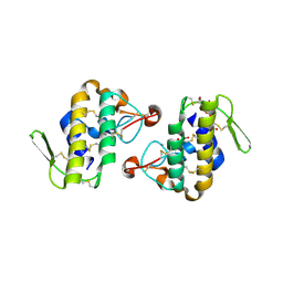
4ED5
 | | Crystal structure of the two N-terminal RRM domains of HuR complexed with RNA | | 分子名称: | 1,2-ETHANEDIOL, 1-METHOXY-2-(2-METHOXYETHOXY)ETHANE, 5'-R(*A*UP*UP*UP*UP*UP*AP*UP*UP*UP*U)-3', ... | | 著者 | Wang, H, Zeng, F, Liu, Q, Niu, L, Teng, M, Li, X. | | 登録日 | 2012-03-27 | | 公開日 | 2012-05-23 | | 最終更新日 | 2024-03-20 | | 実験手法 | X-RAY DIFFRACTION (2 Å) | | 主引用文献 | The structure of the ARE-binding domains of Hu antigen R (HuR) undergoes conformational changes during RNA binding.
Acta Crystallogr.,Sect.D, 69, 2013
|
|
8Y0W
 | |
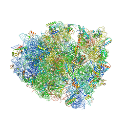
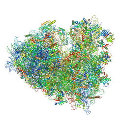
8Y0U
 | | dormant ribosome with STM1 | | 分子名称: | 18S rRNA, 25S rRNA, 40S ribosomal protein S1-A, ... | | 著者 | Du, M, Zeng, F. | | 登録日 | 2024-01-23 | | 公開日 | 2024-02-07 | | 実験手法 | ELECTRON MICROSCOPY (3.59 Å) | | 主引用文献 | dormant ribosome with STM1
To Be Published
|
|
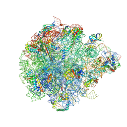
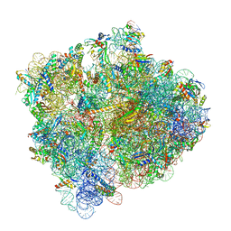
8Y0X
 | | Dormant ribosome with SERBP1 | | 分子名称: | 18S rRNA, 28S rRNA, 40S ribosomal protein S10, ... | | 著者 | Du, M, Zeng, F. | | 登録日 | 2024-01-23 | | 公開日 | 2024-02-07 | | 実験手法 | ELECTRON MICROSCOPY (3.3 Å) | | 主引用文献 | The global structure of dormant ribosome
To Be Published
|
|
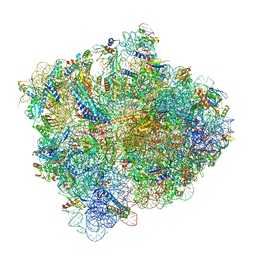
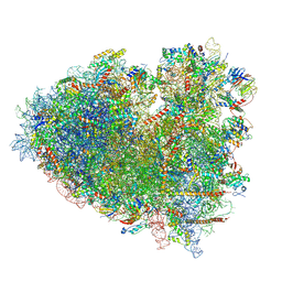
7SA4
 | | Damaged 70S ribosome with PrfH bound | | 分子名称: | 16S ribosomal RNA, 23S ribosomal RNA, 30S ribosomal protein S10, ... | | 著者 | Tian, Y, Zeng, F, Raybarman, A, Carruthers, A, Li, Q, Fatma, S, Huang, R.H. | | 登録日 | 2021-09-22 | | 公開日 | 2022-08-03 | | 実験手法 | ELECTRON MICROSCOPY (2.55 Å) | | 主引用文献 | Sequential rescue and repair of stalled and damaged ribosome by bacterial PrfH and RtcB.
Proc.Natl.Acad.Sci.USA, 119, 2022
|
|
4EGL
 | | Crystal structure of two tandem RNA recognition motifs of Human antigen R | | 分子名称: | ELAV-like protein 1, GLYCEROL, SULFATE ION | | 著者 | Wang, H, Zeng, F, Liu, H, Teng, M, Li, X. | | 登録日 | 2012-03-31 | | 公開日 | 2012-05-30 | | 最終更新日 | 2023-11-08 | | 実験手法 | X-RAY DIFFRACTION (2.9 Å) | | 主引用文献 | Crystal structure of two tandem RNA recognition motifs of Human antigen R
To be Published
|
|
3R27
 | | Crystal structure of the first RRM domain of heterogeneous nuclear ribonucleoprotein L (HnRNP L) | | 分子名称: | GLYCEROL, Heterogeneous nuclear ribonucleoprotein L | | 著者 | Zhang, W, Liu, Y, Zeng, F, Niu, L, Teng, M, Li, X. | | 登録日 | 2011-03-14 | | 公開日 | 2011-09-14 | | 最終更新日 | 2023-09-13 | | 実験手法 | X-RAY DIFFRACTION (2.04 Å) | | 主引用文献 | Crystal structure of the first RRM domain of heterogeneous nuclear ribonucleoprotein L (HnRNP L)
To be Published
|
|
5HH4
 | |
5HH6
 | |
5HH5
 | |
1F2S
 | | CRYSTAL STRUCTURE OF THE COMPLEX FORMED BETWEEN BOVINE BETA-TRYPSIN AND MCTI-A, A TRYPSIN INHIBITOR OF SQUASH FAMILY AT 1.8 A RESOLUTION | | 分子名称: | CALCIUM ION, TRYPSIN, TRYPSIN INHIBITOR A | | 著者 | Zhu, Y, Huang, Q, Qian, M, Jia, Y, Tang, Y. | | 登録日 | 2000-05-29 | | 公開日 | 2000-06-05 | | 最終更新日 | 2023-08-09 | | 実験手法 | X-RAY DIFFRACTION (1.79 Å) | | 主引用文献 | Crystal structure of the complex formed between bovine beta-trypsin and MCTI-A, a trypsin inhibitor of squash family, at 1.8-A resolution.
J.Protein Chem., 18, 1999
|
|
1MCT
 | | THE REFINED 1.6 ANGSTROMS RESOLUTION CRYSTAL STRUCTURE OF THE COMPLEX FORMED BETWEEN PORCINE BETA-TRYPSIN AND MCTI-A, A TRYPSIN INHIBITOR OF SQUASH FAMILY | | 分子名称: | BETA-TRYPSIN, CALCIUM ION, TRYPSIN INHIBITOR A | | 著者 | Huang, Q, Liu, S, Tang, Y. | | 登録日 | 1992-10-24 | | 公開日 | 1994-01-31 | | 最終更新日 | 2011-07-13 | | 実験手法 | X-RAY DIFFRACTION (1.6 Å) | | 主引用文献 | Refined 1.6 A resolution crystal structure of the complex formed between porcine beta-trypsin and MCTI-A, a trypsin inhibitor of the squash family. Detailed comparison with bovine beta-trypsin and its complex.
J.Mol.Biol., 229, 1993
|
|
6KZI
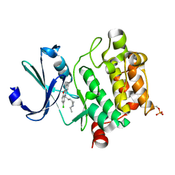 | | Crystal structure of Ser/Thr kinase Pim1 in complex with thioridazine derivatives | | 分子名称: | 4-(2-chloro-10H-phenoxazin-10-yl)-N,N-diethylbutan-1-amine, Serine/threonine-protein kinase pim-1 | | 著者 | Zhang, W, Wan, X, Li, W, Xie, Y, Huang, N. | | 登録日 | 2019-09-24 | | 公開日 | 2020-03-04 | | 最終更新日 | 2021-03-17 | | 実験手法 | X-RAY DIFFRACTION (2.8 Å) | | 主引用文献 | Structure-Based Optimization of 10-DEBC Derivatives as Potent and Selective Pim-1 Kinase Inhibitors.
J.Chem.Inf.Model., 60, 2020
|
|


