Loading
PDBj
MenuPDBj@FacebookPDBj@TwitterPDBj@YouTubewwPDB FoundationwwPDB
RCSB PDBPDBeBMRBAdv. SearchSearch help
Search by PDB author
5I2U
DownloadVisualize
BU of 5i2u by Molmil
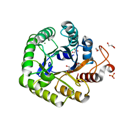
Crystal structure of a novel Halo-Tolerant Cellulase from Soil Metagenome
Descriptor: Cellulase, GLYCEROL, MAGNESIUM ION, ...
Authors:Garg, R, Brahma, V, Srivastava, R, Verma, L, Karthikeyan, S, Sahni, G.
Deposit date:2016-02-09
Release date:2017-01-18
Last modified:2023-11-08
Method:X-RAY DIFFRACTION (2.2 Å)
Cite:Biochemical and structural characterization of a novel halotolerant cellulase from soil metagenome
Sci Rep, 6, 2016
2D82
DownloadVisualize
BU of 2d82 by Molmil
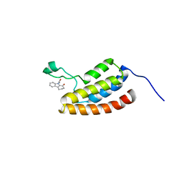
Target Structure-Based Discovery of Small Molecules that Block Human p53 and CREB Binding Protein (CBP) Association
Descriptor: 9-ACETYL-2,3,4,9-TETRAHYDRO-1H-CARBAZOL-1-ONE, CREB-binding protein
Authors:Sachchidanand, Resnick-Silverman, L, Yan, S, Mujtaba, S, Liu, W.J, Zeng, L, Manfredi, J.J, Zhou, M.M.
Deposit date:2005-12-01
Release date:2006-04-04
Last modified:2024-05-29
Method:SOLUTION NMR
Cite:Target structure-based discovery of small molecules that block human p53 and CREB binding protein association
Chem.Biol., 13, 2006
5I95
DownloadVisualize
BU of 5i95 by Molmil
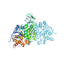
Crystal Structure of Human Mitochondrial Isocitrate Dehydrogenase R140Q Mutant Homodimer bound to NADPH and alpha-Ketoglutaric acid
Descriptor: 2-OXOGLUTARIC ACID, ACETATE ION, CALCIUM ION, ...
Authors:Zhang, B, Jin, L, Wu, W, Jiang, F, DeLaBarre, B, Travins, J.A, Padyana, A.K.
Deposit date:2016-02-19
Release date:2017-03-01
Last modified:2023-09-27
Method:X-RAY DIFFRACTION (1.54 Å)
Cite:AG-221, a First-in-Class Therapy Targeting Acute Myeloid Leukemia Harboring Oncogenic IDH2 Mutations.
Cancer Discov, 7, 2017
5I96
DownloadVisualize
BU of 5i96 by Molmil
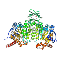
Crystal Structure of Human Mitochondrial Isocitrate Dehydrogenase (IDH2) R140Q Mutant Homodimer in Complex with AG-221 (Enasidenib) Inhibitor.
Descriptor: 2-methyl-1-[(4-[6-(trifluoromethyl)pyridin-2-yl]-6-{[2-(trifluoromethyl)pyridin-4-yl]amino}-1,3,5-triazin-2-yl)amino]propan-2-ol, ACETATE ION, CALCIUM ION, ...
Authors:Wei, W, Zhang, B, Jin, L, Jiang, F, DeLaBarre, B, Travins, J.A, Padyana, A.K.
Deposit date:2016-02-19
Release date:2017-03-08
Last modified:2023-09-27
Method:X-RAY DIFFRACTION (1.55 Å)
Cite:AG-221, a First-in-Class Therapy Targeting Acute Myeloid Leukemia Harboring Oncogenic IDH2 Mutations.
Cancer Discov, 7, 2017
1SHB
DownloadVisualize
BU of 1shb by Molmil
CRYSTAL STRUCTURE OF THE PHOSPHOTYROSINE RECOGNITION DOMAIN SH2 OF V-SRC COMPLEXED WITH TYROSINE-PHOSPHORYLATED PEPTIDES
Descriptor: PHOSPHOPEPTIDE B, V-SRC SH2 DOMAIN
Authors:Waksman, G, Kuriyan, J.
Deposit date:1992-08-18
Release date:1993-10-31
Last modified:2024-06-05
Method:X-RAY DIFFRACTION (2 Å)
Cite:Crystal structure of the phosphotyrosine recognition domain SH2 of v-src complexed with tyrosine-phosphorylated peptides.
Nature, 358, 1992
1SHA
DownloadVisualize
BU of 1sha by Molmil
CRYSTAL STRUCTURE OF THE PHOSPHOTYROSINE RECOGNITION DOMAIN SH2 OF V-SRC COMPLEXED WITH TYROSINE-PHOSPHORYLATED PEPTIDES
Descriptor: PHOSPHOPEPTIDE A, V-SRC SH2 DOMAIN
Authors:Waksman, G, Kuriyan, J.
Deposit date:1992-08-18
Release date:1993-10-31
Last modified:2024-06-05
Method:X-RAY DIFFRACTION (1.5 Å)
Cite:Crystal structure of the phosphotyrosine recognition domain SH2 of v-src complexed with tyrosine-phosphorylated peptides.
Nature, 358, 1992
1AYA
DownloadVisualize
BU of 1aya by Molmil
CRYSTAL STRUCTURES OF PEPTIDE COMPLEXES OF THE AMINO-TERMINAL SH2 DOMAIN OF THE SYP TYROSINE PHOSPHATASE
Descriptor: PEPTIDE PDGFR-1009, PROTEIN-TYROSINE PHOSPHATASE SYP (N-TERMINAL SH2 DOMAIN)
Authors:Lee, C.-H, Kuriyan, J.
Deposit date:1994-05-15
Release date:1994-08-31
Last modified:2024-06-05
Method:X-RAY DIFFRACTION (2.05 Å)
Cite:Crystal structures of peptide complexes of the amino-terminal SH2 domain of the Syp tyrosine phosphatase.
Structure, 2, 1994
1AYC
DownloadVisualize
BU of 1ayc by Molmil
CRYSTAL STRUCTURES OF PEPTIDE COMPLEXES OF THE AMINO-TERMINAL SH2 DOMAIN OF THE SYP TYROSINE PHOSPHATASE
Descriptor: PEPTIDE PDGFR-740, PROTEIN-TYROSINE PHOSPHATASE SYP (N-TERMINAL SH2 DOMAIN)
Authors:Lee, C.-H, Kuriyan, J.
Deposit date:1994-05-15
Release date:1994-08-31
Last modified:2024-06-05
Method:X-RAY DIFFRACTION (2.3 Å)
Cite:Crystal structures of peptide complexes of the amino-terminal SH2 domain of the Syp tyrosine phosphatase.
Structure, 2, 1994
1AYD
DownloadVisualize
BU of 1ayd by Molmil
CRYSTAL STRUCTURES OF PEPTIDE COMPLEXES OF THE AMINO-TERMINAL SH2 DOMAIN OF THE SYP TYROSINE PHOSPHATASE
Descriptor: PROTEIN-TYROSINE PHOSPHATASE SYP (N-TERMINAL SH2 DOMAIN)
Authors:Lee, C.-H, Kuriyan, J.
Deposit date:1994-05-15
Release date:1994-08-31
Last modified:2024-02-07
Method:X-RAY DIFFRACTION (2.2 Å)
Cite:Crystal structures of peptide complexes of the amino-terminal SH2 domain of the Syp tyrosine phosphatase.
Structure, 2, 1994
1AYB
DownloadVisualize
BU of 1ayb by Molmil
CRYSTAL STRUCTURES OF PEPTIDE COMPLEXES OF THE AMINO-TERMINAL SH2 DOMAIN OF THE SYP TYROSINE PHOSPHATASE
Descriptor: PEPTIDE IRS-1-895, PROTEIN-TYROSINE PHOSPHATASE SYP (N-TERMINAL SH2 DOMAIN)
Authors:Lee, C.-H, Kuriyan, J.
Deposit date:1994-05-15
Release date:1994-08-31
Last modified:2024-06-05
Method:X-RAY DIFFRACTION (3 Å)
Cite:Crystal structures of peptide complexes of the amino-terminal SH2 domain of the Syp tyrosine phosphatase.
Structure, 2, 1994

222624

PDB entries from 2024-07-17

PDB statisticsPDBj update infoContact PDBjnumon