1ZRO
 
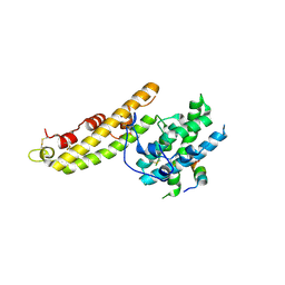
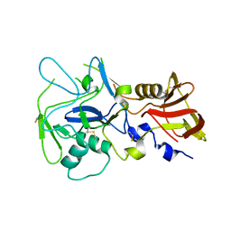
 | | Crystal structure of EBA-175 Region II (RII) crystallized in the presence of (alpha)2,3-sialyllactose | | Descriptor: | CHLORIDE ION, SULFATE ION, erythrocyte binding antigen region II | | Authors: | Tolia, N.H, Enemark, E.J, Sim, B.K, Joshua-Tor, L. | | Deposit date: | 2005-05-19 | | Release date: | 2005-08-09 | | Last modified: | 2023-08-23 | | Method: | X-RAY DIFFRACTION (2.25 Å) | | Cite: | Structural Basis for the EBA-175 Erythrocyte Invasion Pathway of the Malaria Parasite Plasmodium falciparum.
Cell(Cambridge,Mass.), 122, 2005
|
|
1ZRL
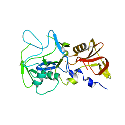
 | | Crystal structure of EBA-175 Region II (RII) | | Descriptor: | CHLORIDE ION, SULFATE ION, erythrocyte binding antigen region II | | Authors: | Tolia, N.H, Enemark, E.J, Sim, B.K, Joshua-Tor, L. | | Deposit date: | 2005-05-19 | | Release date: | 2005-08-09 | | Last modified: | 2021-10-20 | | Method: | X-RAY DIFFRACTION (2.3 Å) | | Cite: | Structural Basis for the EBA-175 Erythrocyte Invasion Pathway of the Malaria Parasite Plasmodium falciparum.
Cell(Cambridge,Mass.), 122, 2005
|
|
4QJB
 | |
3RRC
 | |
4NUU
 | |
4NUV
 | |
4JNO
 | |
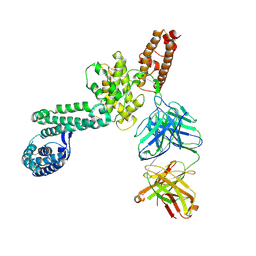
4K2U
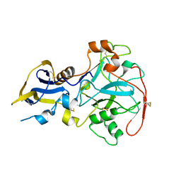
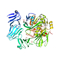
 | | Crystal structure of PfEBA-175 F1 in complex with R218 antibody Fab fragment | | Descriptor: | Antibody Heavy Chain, Antibody Light Chain, Erythrocyte binding antigen 175, ... | | Authors: | Tolia, N.H. | | Deposit date: | 2013-04-09 | | Release date: | 2013-06-12 | | Last modified: | 2017-11-15 | | Method: | X-RAY DIFFRACTION (2.45 Å) | | Cite: | Structural and Functional Basis for Inhibition of Erythrocyte Invasion by Antibodies that Target Plasmodium falciparum EBA-175.
Plos Pathog., 9, 2013
|
|
7K3Z
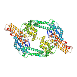
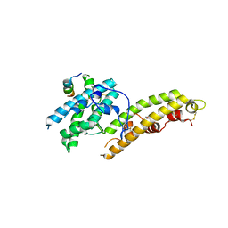
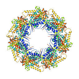 | | P. falciparum Cpn60 D474A mutant bound to ATP | | Descriptor: | 60 kDa chaperonin, ADENOSINE-5'-TRIPHOSPHATE, MAGNESIUM ION | | Authors: | Tolia, N.H, Shi, D, Nguyen, B. | | Deposit date: | 2020-09-14 | | Release date: | 2021-04-21 | | Last modified: | 2023-10-18 | | Method: | X-RAY DIFFRACTION (3.69 Å) | | Cite: | Crystal structure of P. falciparum Cpn60 bound to ATP reveals an open dynamic conformation before substrate binding.
Sci Rep, 11, 2021
|
|
5TSZ
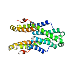 | | Crystal structure of Plasmodium vivax CelTOS | | Descriptor: | Pv cell-traversal protein, SODIUM ION | | Authors: | Tolia, N.H, Jimah, J.R. | | Deposit date: | 2016-10-31 | | Release date: | 2016-12-28 | | Last modified: | 2024-03-06 | | Method: | X-RAY DIFFRACTION (3.002 Å) | | Cite: | Malaria parasite CelTOS targets the inner leaflet of cell membranes for pore-dependent disruption.
Elife, 5, 2016
|
|
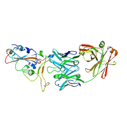
4GF2
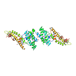
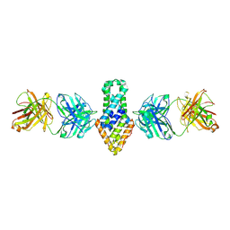
 | | Crystal structure of Plasmodium falciparum Erythrocyte Binding Antigen 140 (PfEBA-140/BAEBL) | | Descriptor: | Erythrocyte binding antigen 140, GLYCEROL | | Authors: | Lin, D.H, Malpede, B.M, Batchelor, J.D, Tolia, N.H. | | Deposit date: | 2012-08-02 | | Release date: | 2012-09-26 | | Last modified: | 2023-09-13 | | Method: | X-RAY DIFFRACTION (2.4 Å) | | Cite: | Crystal and Solution Structures of Plasmodium falciparum Erythrocyte-binding Antigen 140 Reveal Determinants of Receptor Specificity during Erythrocyte Invasion.
J.Biol.Chem., 287, 2012
|
|
6B2N
 | |
8GIF
 | |
8GIE
 | |
8GID
 | |
6WG9
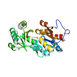
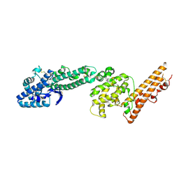
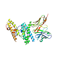
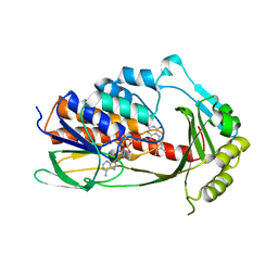 | | Crystal structure of tetracycline destructase Tet(X7) | | Descriptor: | FLAVIN-ADENINE DINUCLEOTIDE, Tetracycline destructase Tet(X7) | | Authors: | Kumar, H, Tolia, N.H. | | Deposit date: | 2020-04-05 | | Release date: | 2021-02-10 | | Last modified: | 2023-10-18 | | Method: | X-RAY DIFFRACTION (2.55 Å) | | Cite: | Tetracycline-inactivating enzymes from environmental, human commensal, and pathogenic bacteria cause broad-spectrum tetracycline resistance.
Commun Biol, 3, 2020
|
|
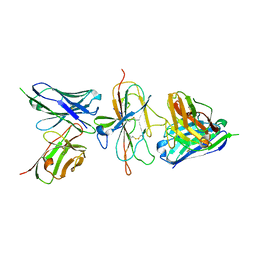
4QEX
 | | Crystal structure of PfEBA-175 RII in complex with a Fab fragment from inhibitory antibody R217 | | Descriptor: | Antibody Heavy Chain, Antibody Light Chain, Erythrocyte-binding antigen-175 | | Authors: | Chen, E, Paing, M.M, Salinas, N, Sim, B.K, Tolia, N.H. | | Deposit date: | 2014-05-19 | | Release date: | 2014-06-04 | | Method: | X-RAY DIFFRACTION (4.5 Å) | | Cite: | Structural and Functional Basis for Inhibition of Erythrocyte Invasion by Antibodies that Target Plasmodium falciparum EBA-175.
Plos Pathog., 9, 2013
|
|
8DCC
 | |
7U9E
 | | Pfs230 D1 domain in complex with 230AL-26 | | Descriptor: | 230AL-26, Gametocyte surface protein P230, LMIV230-01 | | Authors: | Tang, W.K, Tolia, N.H. | | Deposit date: | 2022-03-10 | | Release date: | 2023-02-15 | | Last modified: | 2023-10-25 | | Method: | X-RAY DIFFRACTION (2.39 Å) | | Cite: | A human antibody epitope map of Pfs230D1 derived from analysis of individuals vaccinated with a malaria transmission-blocking vaccine.
Immunity, 56, 2023
|
|
5F3J
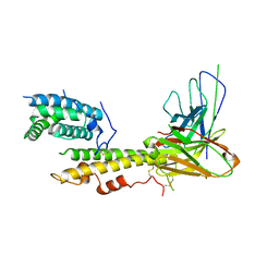 | |
6O38
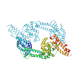
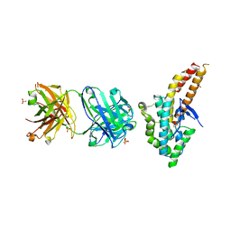
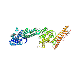
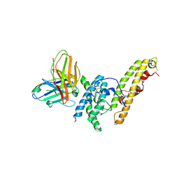
 | | Structure of a chaperone-substrate complex | | Descriptor: | Acinetobacter secreted protease CpaA, SULFATE ION, Type II secretion chaperone CpaB, ... | | Authors: | Urusova, D.V, Tolia, N.H. | | Deposit date: | 2019-02-26 | | Release date: | 2019-07-24 | | Last modified: | 2019-09-25 | | Method: | X-RAY DIFFRACTION (2.595 Å) | | Cite: | The structure ofAcinetobacter-secreted protease CpaA complexed with its chaperone CpaB reveals a novel mode of a T2SS chaperone-substrate interaction.
J.Biol.Chem., 294, 2019
|
|
6OAN
 | | Structure of DBP in complex with human neutralizing antibody 053054 | | Descriptor: | Antibody 053054 single chain variable fragment, Duffy binding surface protein region II, SULFATE ION | | Authors: | Urusova, D, Tolia, N.H. | | Deposit date: | 2019-03-18 | | Release date: | 2019-06-12 | | Last modified: | 2023-10-11 | | Method: | X-RAY DIFFRACTION (2.9 Å) | | Cite: | Structural basis for neutralization of Plasmodium vivax by naturally acquired human antibodies that target DBP.
Nat Microbiol, 4, 2019
|
|
6OAO
 | | Structure of DBP in complex with human neutralizing antibody 092096 | | Descriptor: | Antibody 092096 single chain variable fragment, Duffy binding surface protein region II, SULFATE ION | | Authors: | Urusova, D, Tolia, N.H. | | Deposit date: | 2019-03-18 | | Release date: | 2019-06-12 | | Last modified: | 2023-10-11 | | Method: | X-RAY DIFFRACTION (3.497 Å) | | Cite: | Structural basis for neutralization of Plasmodium vivax by naturally acquired human antibodies that target DBP.
Nat Microbiol, 4, 2019
|
|
1R6Z
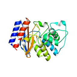
 | | The Crystal Structure of the Argonaute2 PAZ domain (as a MBP fusion) | | Descriptor: | Chimera of Maltose-binding periplasmic protein and Argonaute 2, NICKEL (II) ION, alpha-D-glucopyranose-(1-4)-alpha-D-glucopyranose | | Authors: | Song, J.J, Liu, J, Tolia, N.H, Schneiderman, J, Smith, S.K, Martienssen, R.A, Hannon, G.J, Joshua-Tor, L. | | Deposit date: | 2003-10-17 | | Release date: | 2004-01-13 | | Last modified: | 2023-08-23 | | Method: | X-RAY DIFFRACTION (2.8 Å) | | Cite: | The crystal structure of the Argonaute2 PAZ domain reveals an RNA binding motif in RNAi effector complexes.
Nat.Struct.Biol., 10, 2003
|
|
8ULF
 | |


