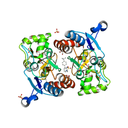
3AA2
 
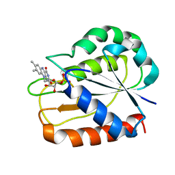
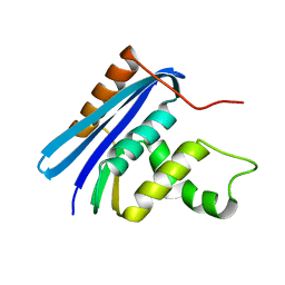 | | A52I E. coli RNase HI | | 分子名称: | Ribonuclease HI | | 著者 | Takano, K. | | 登録日 | 2009-11-11 | | 公開日 | 2010-10-06 | | 最終更新日 | 2024-03-13 | | 実験手法 | X-RAY DIFFRACTION (1.9 Å) | | 主引用文献 | Protein core adaptability: crystal structures of the cavity-filling variants of Escherichia coli RNase HI
Protein Pept.Lett., 17, 2010
|
|
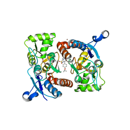
3AA5
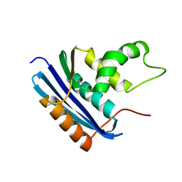 | | A52F E.coli RNase HI | | 分子名称: | Ribonuclease HI | | 著者 | Takano, K. | | 登録日 | 2009-11-11 | | 公開日 | 2010-10-06 | | 最終更新日 | 2024-03-13 | | 実験手法 | X-RAY DIFFRACTION (2.1 Å) | | 主引用文献 | Protein core adaptability: crystal structures of the cavity-filling variants of Escherichia coli RNase HI
Protein Pept.Lett., 17, 2010
|
|
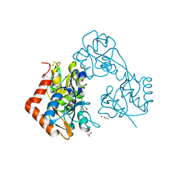
7YUZ
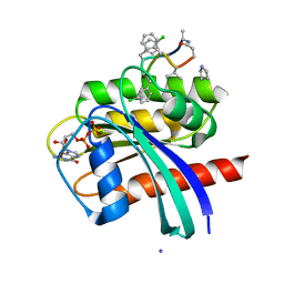 | | Human K-Ras G12D (GDP-bound) in complex with cyclic peptide inhibitor AP8784 | | 分子名称: | AP8784, GUANOSINE-5'-DIPHOSPHATE, IODIDE ION, ... | | 著者 | Irie, M, Fukami, T.A, Tanada, M, Ohta, A, Torizawa, T. | | 登録日 | 2022-08-18 | | 公開日 | 2023-07-26 | | 最終更新日 | 2023-11-22 | | 実験手法 | X-RAY DIFFRACTION (1.878 Å) | | 主引用文献 | Validation of a New Methodology to Create Oral Drugs beyond the Rule of 5 for Intracellular Tough Targets.
J.Am.Chem.Soc., 145, 2023
|
|
7YV1
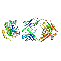 | | Human K-Ras G12D (GDP-bound) in complex with cyclic peptide inhibitor LUNA18 and KA30L Fab | | 分子名称: | GUANOSINE-5'-DIPHOSPHATE, Isoform 2B of GTPase KRas, KA30L Fab H-chain, ... | | 著者 | Irie, M, Fukami, T.A, Matsuo, A, Saka, K, Nishimura, M, Saito, H, Torizawa, T, Tanada, M, Ohta, A. | | 登録日 | 2022-08-18 | | 公開日 | 2023-07-26 | | 最終更新日 | 2023-11-22 | | 実験手法 | X-RAY DIFFRACTION (1.454 Å) | | 主引用文献 | Validation of a New Methodology to Create Oral Drugs beyond the Rule of 5 for Intracellular Tough Targets.
J.Am.Chem.Soc., 145, 2023
|
|
6E7D
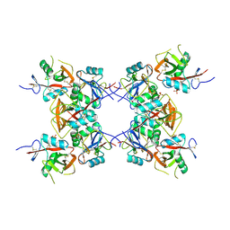 | | Structure of the inhibitory NKR-P1B receptor bound to the host-encoded ligand, Clr-b | | 分子名称: | 2-acetamido-2-deoxy-beta-D-glucopyranose, C-type lectin domain family 2 member D, Killer cell lectin-like receptor subfamily B member 1B allele B, ... | | 著者 | Balaji, G.R, Rossjohn, J, Berry, R. | | 登録日 | 2018-07-26 | | 公開日 | 2018-10-24 | | 最終更新日 | 2023-10-11 | | 実験手法 | X-RAY DIFFRACTION (2.9 Å) | | 主引用文献 | Recognition of host Clr-b by the inhibitory NKR-P1B receptor provides a basis for missing-self recognition.
Nat Commun, 9, 2018
|
|
3W9Z
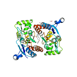
 | | Crystal structure of DusC | | 分子名称: | FLAVIN MONONUCLEOTIDE, tRNA-dihydrouridine synthase C | | 著者 | Chen, M, Yu, J, Tanaka, Y, Tanaka, I, Yao, M. | | 登録日 | 2013-04-19 | | 公開日 | 2013-07-31 | | 最終更新日 | 2024-03-20 | | 実験手法 | X-RAY DIFFRACTION (2.1 Å) | | 主引用文献 | Structure of dihydrouridine synthase C (DusC) from Escherichia coli
Acta Crystallogr.,Sect.F, 69, 2013
|
|
6KS2
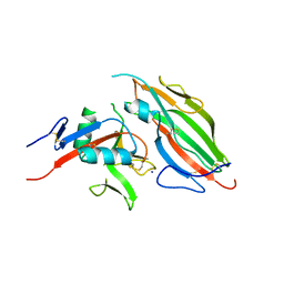
 | | Structure of anti-Ghrelin receptor antibody | | 分子名称: | Fab7881 Heavy Chain (FabH), Fab7881 Light Chain (FabL) | | 著者 | Shiimura, Y, Horita, S, Asada, H, Hirata, K, Iwata, S, Kojima, M. | | 登録日 | 2019-08-23 | | 公開日 | 2020-08-12 | | 最終更新日 | 2023-11-22 | | 実験手法 | X-RAY DIFFRACTION (1.753 Å) | | 主引用文献 | Structure of an antagonist-bound ghrelin receptor reveals possible ghrelin recognition mode.
Nat Commun, 11, 2020
|
|
6KO5
 | | Complex structure of Ghrelin receptor with Fab | | 分子名称: | 6-(4-bromanyl-2-fluoranyl-phenoxy)-2-methyl-3-[[(3~{S})-1-propan-2-ylpiperidin-3-yl]methyl]pyrido[3,2-d]pyrimidin-4-one, Chimera of Soluble cytochrome b562 and Growth hormone secretagogue receptor type 1, Fab7881 Heavy Chain, ... | | 著者 | Shiimura, Y, Horita, S, Asada, H, Hirata, K, Iwata, S, Kojima, M. | | 登録日 | 2019-08-08 | | 公開日 | 2020-08-12 | | 最終更新日 | 2023-11-22 | | 実験手法 | X-RAY DIFFRACTION (3.3 Å) | | 主引用文献 | Structure of an antagonist-bound ghrelin receptor reveals possible ghrelin recognition mode.
Nat Commun, 11, 2020
|
|
5TZN
 | |
1AHL
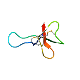 | | ANTHOPLEURIN-A,NMR, 20 STRUCTURES | | 分子名称: | ANTHOPLEURIN-A | | 著者 | Pallaghy, P.K, Scanlon, M.J, Monks, S.A, Norton, R.S. | | 登録日 | 1994-10-28 | | 公開日 | 1995-11-14 | | 最終更新日 | 2017-11-29 | | 実験手法 | SOLUTION NMR | | 主引用文献 | Three-dimensional structure in solution of the polypeptide cardiac stimulant anthopleurin-A.
Biochemistry, 34, 1995
|
|
6AI6
 | | Crystal structure of SpCas9-NG | | 分子名称: | 1,2-ETHANEDIOL, CRISPR-associated endonuclease Cas9/Csn1, DNA (28-MER), ... | | 著者 | Nishimasu, H, Hirano, S, Ishitani, R, Nureki, O. | | 登録日 | 2018-08-21 | | 公開日 | 2018-10-31 | | 最終更新日 | 2023-11-22 | | 実験手法 | X-RAY DIFFRACTION (2.7 Å) | | 主引用文献 | Engineered CRISPR-Cas9 nuclease with expanded targeting space
Science, 361, 2018
|
|
3CPP
 | |
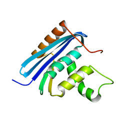
1VEC
 | | Crystal structure of the N-terminal domain of rck/p54, a human DEAD-box protein | | 分子名称: | ATP-dependent RNA helicase p54, L(+)-TARTARIC ACID, ZINC ION | | 著者 | Hogetsu, K, Matsui, T, Yukihiro, Y, Tanaka, M, Sato, T, Kumasaka, T, Tanaka, N. | | 登録日 | 2004-03-29 | | 公開日 | 2004-04-13 | | 最終更新日 | 2023-10-25 | | 実験手法 | X-RAY DIFFRACTION (2.01 Å) | | 主引用文献 | Structural insight of human DEAD-box protein rck/p54 into its substrate recognition with conformational changes
Genes Cells, 11, 2006
|
|
5ZG3
 | | Crystal structure of the GluA2o LBD in complex with glutamate and TAK-137 | | 分子名称: | 9-(4-phenoxyphenyl)-3,4-dihydro-2H-2lambda~6~-pyrido[2,1-c][1,2,4]thiadiazine-2,2-dione, ACETATE ION, GLUTAMIC ACID, ... | | 著者 | Sogabe, S, Igaki, S, Hirokawa, A, Zama, Y, Lane, W, Snell, G. | | 登録日 | 2018-03-07 | | 公開日 | 2019-01-16 | | 最終更新日 | 2023-11-22 | | 実験手法 | X-RAY DIFFRACTION (1.65 Å) | | 主引用文献 | TAK-137, an AMPA-R potentiator with little agonistic effect, has a wide therapeutic window.
Neuropsychopharmacology, 44, 2019
|
|
5ZG1
 | | Crystal structure of the GluA2o LBD in complex with glutamate and Compound-2 | | 分子名称: | 9-(4-~{tert}-butylphenyl)-3,4-dihydropyrido[2,1-c][1,2,4]thiadiazine 2,2-dioxide, GLUTAMIC ACID, GLYCEROL, ... | | 著者 | Sogabe, S, Igaki, S, Hirokawa, A, Zama, Y, Lane, W, Snell, G. | | 登録日 | 2018-03-07 | | 公開日 | 2019-01-16 | | 最終更新日 | 2023-11-22 | | 実験手法 | X-RAY DIFFRACTION (1.32 Å) | | 主引用文献 | TAK-137, an AMPA-R potentiator with little agonistic effect, has a wide therapeutic window.
Neuropsychopharmacology, 44, 2019
|
|
5ZG2
 | | Crystal structure of the GluA2o LBD in complex with ZK200775 and Compound-2 | | 分子名称: | 9-(4-~{tert}-butylphenyl)-3,4-dihydropyrido[2,1-c][1,2,4]thiadiazine 2,2-dioxide, ACETATE ION, GLYCEROL, ... | | 著者 | Sogabe, S, Igaki, S, Hirokawa, A, Zama, Y, Lane, W, Snell, G. | | 登録日 | 2018-03-07 | | 公開日 | 2019-01-16 | | 最終更新日 | 2023-11-22 | | 実験手法 | X-RAY DIFFRACTION (1.25 Å) | | 主引用文献 | TAK-137, an AMPA-R potentiator with little agonistic effect, has a wide therapeutic window.
Neuropsychopharmacology, 44, 2019
|
|
5ZG0
 | | Crystal structure of the GluA2o LBD in complex with glutamate and Compound-1 | | 分子名称: | 9-{4-[(propan-2-yl)oxy]phenyl}-3,4-dihydro-2H-2lambda~6~-pyrido[2,1-c][1,2,4]thiadiazine-2,2-dione, ACETATE ION, GLUTAMIC ACID, ... | | 著者 | Sogabe, S, Igaki, S, Hirokawa, A, Zama, Y, Lane, W, Snell, G. | | 登録日 | 2018-03-07 | | 公開日 | 2019-01-16 | | 最終更新日 | 2023-11-22 | | 実験手法 | X-RAY DIFFRACTION (1.58 Å) | | 主引用文献 | TAK-137, an AMPA-R potentiator with little agonistic effect, has a wide therapeutic window.
Neuropsychopharmacology, 44, 2019
|
|
2YV0
 | | Structural and Thermodynamic Analyses of E. coli ribonuclease HI Variant with Quintuple Thermostabilizing Mutations | | 分子名称: | Ribonuclease HI | | 著者 | Haruki, M, Motegi, T, Tadokoro, T, Koga, Y, Takano, K, Kanaya, S. | | 登録日 | 2007-04-06 | | 公開日 | 2008-03-18 | | 最終更新日 | 2023-10-25 | | 実験手法 | X-RAY DIFFRACTION (1.4 Å) | | 主引用文献 | Structural and thermodynamic analyses of Escherichia coli RNase HI variant with quintuple thermostabilizing mutations.
Febs J., 274, 2007
|
|
1CZN
 | | REFINED STRUCTURES OF OXIDIZED FLAVODOXIN FROM ANACYSTIS NIDULANS | | 分子名称: | FLAVIN MONONUCLEOTIDE, FLAVODOXIN | | 著者 | Smith, W.W, Pattridge, K.A, Luschinsky, C.L, Ludwig, M.L. | | 登録日 | 1999-09-03 | | 公開日 | 1999-12-29 | | 最終更新日 | 2024-02-07 | | 実験手法 | X-RAY DIFFRACTION (1.7 Å) | | 主引用文献 | Refined structures of oxidized flavodoxin from Anacystis nidulans.
J.Mol.Biol., 294, 1999
|
|
5ZFU
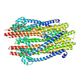 | | Structure of the ExbB/ExbD hexameric complex (ExbB6ExbD3TM) | | 分子名称: | 22-mer peptide from Biopolymer transport protein ExbD, Biopolymer transport protein ExbB | | 著者 | Yonekura, K, Yamashita, Y, Matsuoka, R, Maki-Yonekura, S. | | 登録日 | 2018-03-07 | | 公開日 | 2018-05-09 | | 最終更新日 | 2024-03-27 | | 実験手法 | ELECTRON MICROSCOPY (6.7 Å) | | 主引用文献 | Hexameric and pentameric complexes of the ExbBD energizer in the Ton system.
Elife, 7, 2018
|
|
5ZFP
 | |
5ZFV
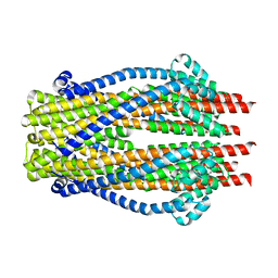
 | | Structure of the ExbB/ExbD pentameric complex (ExbB5ExbD1TM) | | 分子名称: | 22-mer peptide from Biopolymer transport protein ExbD, Biopolymer transport protein ExbB | | 著者 | Yonekura, K, Yamashita, Y, Matsuoka, R, Maki-Yonekura, S. | | 登録日 | 2018-03-07 | | 公開日 | 2018-05-09 | | 実験手法 | ELECTRON MICROSCOPY (7.1 Å) | | 主引用文献 | Hexameric and pentameric complexes of the ExbBD energizer in the Ton system.
Elife, 7, 2018
|
|
7YL5
 | | Cell surface protein YwfG protein complexed with mannose | | 分子名称: | CALCIUM ION, GRAM_POS_ANCHORING domain-containing protein, SULFATE ION, ... | | 著者 | Tsuchiya, W, Fujimoto, Z, Suzuki, C. | | 登録日 | 2022-07-25 | | 公開日 | 2023-06-07 | | 最終更新日 | 2023-11-29 | | 実験手法 | X-RAY DIFFRACTION (3 Å) | | 主引用文献 | Cell-surface protein YwfG of Lactococcus lactis binds to alpha-1,2-linked mannose.
Plos One, 18, 2023
|
|
7YL4
 | | Cell surface protein YwfG protein (apo form) | | 分子名称: | CALCIUM ION, GRAM_POS_ANCHORING domain-containing protein, SULFATE ION | | 著者 | Tsuchiya, W, Fujimoto, Z, Suzuki, C. | | 登録日 | 2022-07-25 | | 公開日 | 2023-06-07 | | 最終更新日 | 2023-11-29 | | 実験手法 | X-RAY DIFFRACTION (2.5 Å) | | 主引用文献 | Cell-surface protein YwfG of Lactococcus lactis binds to alpha-1,2-linked mannose.
Plos One, 18, 2023
|
|
7YL6
 | | Cell surface protein YwfG protein complexed with alpha-1,2-mannobiose | | 分子名称: | CALCIUM ION, GRAM_POS_ANCHORING domain-containing protein, SULFATE ION, ... | | 著者 | Tsuchiya, W, Fujimoto, Z, Suzuki, C. | | 登録日 | 2022-07-25 | | 公開日 | 2023-06-07 | | 最終更新日 | 2023-11-29 | | 実験手法 | X-RAY DIFFRACTION (2.95 Å) | | 主引用文献 | Cell-surface protein YwfG of Lactococcus lactis binds to alpha-1,2-linked mannose.
Plos One, 18, 2023
|
|


