3QVN
 
 | |
3LSU
 | |
4F6E
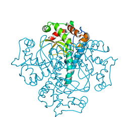
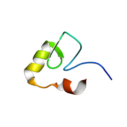
 | | Crystal Structure of the K182R, A183P mutant manganese superoxide dismutase from Sacchromyces cerevisiae | | 分子名称: | 2-AMINO-2-HYDROXYMETHYL-PROPANE-1,3-DIOL, GLYCEROL, MANGANESE (II) ION, ... | | 著者 | Sheng, Y, Cascio, D, Valentine, J.S. | | 登録日 | 2012-05-14 | | 公開日 | 2013-06-12 | | 最終更新日 | 2023-09-20 | | 実験手法 | X-RAY DIFFRACTION (1.6 Å) | | 主引用文献 | Crystal Structure of the K182R, A183P mutant manganese superoxide dismutase from Sacchromyces cerevisiae
to be published
|
|
4GUN
 | |
4E4E
 | |
2JRJ
 | | Solution structure of the human Pirh2 RING-H2 domain. Northeast Structural Genomics Consortium Target HT2B | | 分子名称: | Ring finger and CHY zinc finger domain containing protein 1, ZINC ION | | 著者 | Sheng, Y, Lemak, A, Laister, R.C, Wu, B, Arrowsmith, C.H, Northeast Structural Genomics Consortium (NESG) | | 登録日 | 2007-06-27 | | 公開日 | 2007-07-10 | | 最終更新日 | 2023-12-20 | | 実験手法 | SOLUTION NMR | | 主引用文献 | Molecular basis of Pirh2-mediated p53 ubiquitylation.
Nat.Struct.Mol.Biol., 15, 2008
|
|
2FOO
 | | The Crystal Structure of the N-terminal domain of HAUSP/USP7 complexed with p53 peptide 359-362 | | 分子名称: | Ubiquitin carboxyl-terminal hydrolase 7, p53 peptide | | 著者 | Saridakis, V, Sheng, Y, Sarkari, F, Duan, S, Wu, T, Arrowsmith, C.H, Frappier, L. | | 登録日 | 2006-01-13 | | 公開日 | 2006-02-14 | | 最終更新日 | 2023-08-30 | | 実験手法 | X-RAY DIFFRACTION (2.2 Å) | | 主引用文献 | Molecular recognition of p53 and MDM2 by USP7/HAUSP
Nat.Struct.Mol.Biol., 13, 2006
|
|
2FOP
 | | The Crystal Structure of the N-terminal domain of HAUSP/USP7 complexed with mdm2 peptide 147-150 | | 分子名称: | Ubiquitin carboxyl-terminal hydrolase 7, mdm2 peptide | | 著者 | Saridakis, V, Sheng, Y, Sarkari, F, Duan, S, Wu, T, Arrowsmith, C.H, Frappier, L. | | 登録日 | 2006-01-13 | | 公開日 | 2006-02-14 | | 最終更新日 | 2023-08-30 | | 実験手法 | X-RAY DIFFRACTION (2.1 Å) | | 主引用文献 | Molecular recognition of p53 and MDM2 by USP7/HAUSP
Nat.Struct.Mol.Biol., 13, 2006
|
|
2FOJ
 | | The Crystal Structure of the N-terminal domain of HAUSP/USP7 complexed with p53 peptide 364-367 | | 分子名称: | Ubiquitin carboxyl-terminal hydrolase 7, p53 peptide | | 著者 | Saridakis, V, Sheng, Y, Sarkari, F, Duan, S, Wu, T, Arrowsmith, C.H, Frappier, L. | | 登録日 | 2006-01-13 | | 公開日 | 2006-02-14 | | 最終更新日 | 2023-08-30 | | 実験手法 | X-RAY DIFFRACTION (1.6 Å) | | 主引用文献 | Molecular recognition of p53 and MDM2 by USP7/HAUSP
Nat.Struct.Mol.Biol., 13, 2006
|
|
2K2D
 | | Solution NMR structure of C-terminal domain of human pirh2. Northeast Structural Genomics Consortium (NESG) target HT2C | | 分子名称: | RING finger and CHY zinc finger domain-containing protein 1, ZINC ION | | 著者 | Lemak, A, Sheng, Y, Karra, M, Srisailam, S, Laister, R.C, Duan, S, Arrowsmith, C.H, Northeast Structural Genomics Consortium (NESG) | | 登録日 | 2008-03-31 | | 公開日 | 2008-04-15 | | 最終更新日 | 2024-05-01 | | 実験手法 | SOLUTION NMR | | 主引用文献 | Molecular basis of Pirh2-mediated p53 ubiquitylation.
Nat.Struct.Mol.Biol., 15, 2008
|
|
2K2C
 | | Solution NMR structure of N-terminal domain of human pirh2. Northeast Structural Genomics Consortium (NESG) target HT2A | | 分子名称: | RING finger and CHY zinc finger domain-containing protein 1, ZINC ION | | 著者 | Wu, B, Lemak, A, Sheng, Y, Karra, M, Srisailam, S, Sunnerhagen, M, Arrowsmith, C.H, Northeast Structural Genomics Consortium (NESG) | | 登録日 | 2008-03-31 | | 公開日 | 2008-04-15 | | 最終更新日 | 2023-06-14 | | 実験手法 | SOLUTION NMR | | 主引用文献 | Molecular basis of Pirh2-mediated p53 ubiquitylation.
Nat.Struct.Mol.Biol., 15, 2008
|
|
2MUL
 | | Solution Structure of the UBM1 domain of human HUWE1/ARF-BP1 | | 分子名称: | E3 ubiquitin-protein ligase HUWE1 | | 著者 | Farhadi, S, Khatun, R, Lemak, A, Kaustov, L, Ramabadran, R, Hunter, H, Sheng, Y. | | 登録日 | 2014-09-12 | | 公開日 | 2015-09-16 | | 最終更新日 | 2024-05-01 | | 実験手法 | SOLUTION NMR | | 主引用文献 | Solution structure of Ubiquitin Binding Motif of human Arf-bp1
To be Published
|
|
2MUM
 | | Solution structure of the PHD domain of Yeast YNG2 | | 分子名称: | Chromatin modification-related protein YNG2, ZINC ION | | 著者 | Taeb, S, Kaustov, L, Lemak, A, Farhadi, S, Sheng, Y. | | 登録日 | 2014-09-12 | | 公開日 | 2014-12-24 | | 最終更新日 | 2023-06-14 | | 実験手法 | SOLUTION NMR | | 主引用文献 | Solution structure of the PHD domain of Yeast YNG2
To be Published
|
|
8X7T
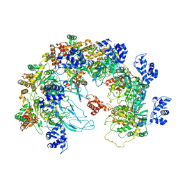 | | MCM in the Apo state. | | 分子名称: | mini-chromosome maintenance complex 3 | | 著者 | Ma, J, Yi, G, Ye, M, MacGregor-Chatwin, C, Sheng, Y, Lu, Y, Li, M, Gilbert, R.J.C, Zhang, P. | | 登録日 | 2023-11-25 | | 公開日 | 2024-01-17 | | 実験手法 | ELECTRON MICROSCOPY (3.26 Å) | | 主引用文献 | MCM in the Apo state
To Be Published
|
|
8X7U
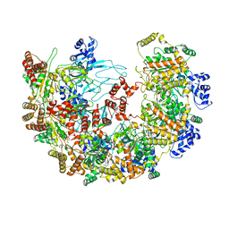 | | MCM in complex with dsDNA in presence of ATP. | | 分子名称: | ADENOSINE-5'-TRIPHOSPHATE, MAGNESIUM ION, mini-chromosome maintenance complex 3 | | 著者 | Ma, J, Yi, G, Ye, M, MacGregor-Chatwin, C, Sheng, Y, Lu, Y, Li, M, Gilbert, R.J.C, Zhang, P. | | 登録日 | 2023-11-25 | | 公開日 | 2024-01-17 | | 実験手法 | ELECTRON MICROSCOPY (3.57 Å) | | 主引用文献 | MCM in complex with dsDNA in presence of ATP
To Be Published
|
|
4Y21
 | | Crystal Structure of Munc13-1 MUN domain | | 分子名称: | Protein unc-13 homolog A | | 著者 | Yang, X.Y, Wang, S, Sheng, Y, Zhang, M, Zou, W.J, Wu, L.J, Kang, L.J, Rizo, J, Zhang, R.G, Xu, T, Ma, C. | | 登録日 | 2015-02-09 | | 公開日 | 2015-06-10 | | 最終更新日 | 2023-11-08 | | 実験手法 | X-RAY DIFFRACTION (2.9 Å) | | 主引用文献 | Syntaxin opening by the MUN domain underlies the function of Munc13 in synaptic-vesicle priming.
Nat.Struct.Mol.Biol., 22, 2015
|
|
1YY6
 | | The Crystal Structure of the N-terminal domain of HAUSP/USP7 complexed with an EBNA1 peptide | | 分子名称: | Epstein-Barr nuclear antigen-1, SODIUM ION, Ubiquitin carboxyl-terminal hydrolase 7 | | 著者 | Saridakis, V, Sheng, Y, Sarkari, F, Holowaty, M, Shire, K, Nguyen, T, Zhang, R, Liao, J, Lee, W, Edwards, A.M, Arrowsmith, C.H, Frappier, L. | | 登録日 | 2005-02-23 | | 公開日 | 2005-04-05 | | 最終更新日 | 2024-02-14 | | 実験手法 | X-RAY DIFFRACTION (1.7 Å) | | 主引用文献 | Structure of the p53 binding domain of HAUSP/USP7 bound to Epstein-Barr nuclear antigen 1 implications for EBV-mediated immortalization.
Mol.Cell, 18, 2005
|
|
1YZE
 | | Crystal structure of the N-terminal domain of USP7/HAUSP. | | 分子名称: | Ubiquitin carboxyl-terminal hydrolase 7 | | 著者 | Saridakis, V, Sheng, Y, Sarkari, F, Holowaty, M.N, Shire, K, Nguyen, T, Zhang, R.G, Liao, J, Lee, W, Edwards, A.M, Arrowsmith, C.H, Frappier, L. | | 登録日 | 2005-02-28 | | 公開日 | 2005-04-05 | | 最終更新日 | 2024-02-14 | | 実験手法 | X-RAY DIFFRACTION (2 Å) | | 主引用文献 | Structure of the p53 binding domain of HAUSP/USP7 bound to Epstein-Barr nuclear antigen 1 implications for EBV-mediated immortalization.
Mol.Cell, 18, 2005
|
|
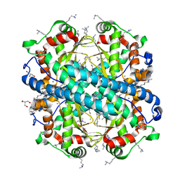
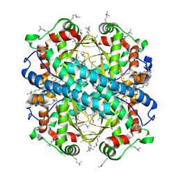
4MCM
 | | Human SOD1 C57S Mutant, As-isolated | | 分子名称: | SULFATE ION, Superoxide dismutase [Cu-Zn], ZINC ION | | 著者 | Sea, K, Sohn, S.H, Durazo, A, Sheng, Y, Shaw, B, Cao, X, Taylor, A.B, Whitson, L.J, Holloway, S.P, Hart, P.J, Cabelli, D.E, Gralla, E.B, Valentine, J.S. | | 登録日 | 2013-08-21 | | 公開日 | 2014-08-27 | | 最終更新日 | 2023-09-20 | | 実験手法 | X-RAY DIFFRACTION (2.2 Å) | | 主引用文献 | Insights into the role of the unusual disulfide bond in copper-zinc superoxide dismutase.
J.Biol.Chem., 290, 2015
|
|
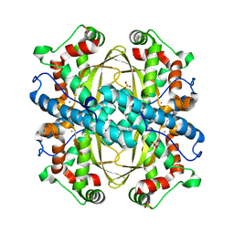
4MCN
 | | Human SOD1 C57S Mutant, Metal-free | | 分子名称: | SULFATE ION, Superoxide dismutase [Cu-Zn] | | 著者 | Sea, K, Sohn, S.H, Durazo, A, Sheng, Y, Shaw, B, Cao, X, Taylor, A.B, Whitson, L.J, Holloway, S.P, Hart, P.J, Cabelli, D.E, Gralla, E.B, Valentine, J.S. | | 登録日 | 2013-08-21 | | 公開日 | 2014-08-27 | | 最終更新日 | 2023-09-20 | | 実験手法 | X-RAY DIFFRACTION (2.6 Å) | | 主引用文献 | Insights into the role of the unusual disulfide bond in copper-zinc superoxide dismutase.
J.Biol.Chem., 290, 2015
|
|
2F4W
 | | Human ubiquitin-conjugating enzyme E2 J2 | | 分子名称: | ubiquitin-conjugating enzyme E2, J2 | | 著者 | Walker, J.R, Avvakumov, G.V, Xue, S, Finerty Jr, P.J, Newman, E.M, Mackenzie, F, Weigelt, J, Sundstrom, M, Arrowsmith, C, Edwards, A, Bochkarev, A, Dhe-Paganon, S, Structural Genomics Consortium (SGC) | | 登録日 | 2005-11-24 | | 公開日 | 2005-12-27 | | 最終更新日 | 2023-08-23 | | 実験手法 | X-RAY DIFFRACTION (2 Å) | | 主引用文献 | A human ubiquitin conjugating enzyme (E2)-HECT E3 ligase structure-function screen.
Mol Cell Proteomics, 11, 2012
|
|
2QGX
 | | Ubiquitin-conjugating enzyme E2Q | | 分子名称: | Ubiquitin-conjugating enzyme E2 Q1 | | 著者 | Neculai, D, Avvakumov, G.V, Xue, S, Walker, J.R, Mackenzie, F, Weigelt, J, Sundstrom, M, Arrowsmith, C.H, Edwards, A.M, Bochkarev, A, Sicheri, F, Dhe-Paganon, S, Structural Genomics Consortium (SGC) | | 登録日 | 2007-06-29 | | 公開日 | 2008-03-18 | | 最終更新日 | 2023-08-30 | | 実験手法 | X-RAY DIFFRACTION (2.56 Å) | | 主引用文献 | A human ubiquitin conjugating enzyme (E2)-HECT E3 ligase structure-function screen.
Mol Cell Proteomics, 11, 2012
|
|
7R5O
 | |
2OB4
 | | Human Ubiquitin-Conjugating Enzyme CDC34 | | 分子名称: | Ubiquitin-conjugating enzyme E2-32 kDa complementing | | 著者 | Neculai, D, Avvakumov, G.V, Xue, S, Walker, J.R, Mackenzie, F, Weigelt, J, Sundstrom, M, Arrowsmith, C.H, Edwards, A.M, Bochkarev, A, Sicheri, F, Dhe-Paganon, S, Structural Genomics Consortium (SGC) | | 登録日 | 2006-12-18 | | 公開日 | 2006-12-26 | | 最終更新日 | 2023-12-27 | | 実験手法 | X-RAY DIFFRACTION (2.4 Å) | | 主引用文献 | A human ubiquitin conjugating enzyme (E2)-HECT E3 ligase structure-function screen.
Mol Cell Proteomics, 11, 2012
|
|
3BZH
 | | Crystal structure of human ubiquitin-conjugating enzyme E2 E1 | | 分子名称: | GLYCEROL, Ubiquitin-conjugating enzyme E2 E1 | | 著者 | Walker, J.R, Avvakumov, G.V, Xue, S, Li, Y, Weigelt, J, Arrowsmith, C.H, Edwards, A.M, Bochkarev, A, Dhe-Paganon, S, Structural Genomics Consortium (SGC) | | 登録日 | 2008-01-18 | | 公開日 | 2008-02-26 | | 最終更新日 | 2023-08-30 | | 実験手法 | X-RAY DIFFRACTION (1.6 Å) | | 主引用文献 | A human ubiquitin conjugating enzyme (E2)-HECT E3 ligase structure-function screen.
Mol Cell Proteomics, 11, 2012
|
|


