6WKO
 
 | |
3VCO
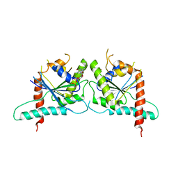
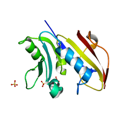 | | Schistosoma mansoni Dihydrofolate reductase | | Descriptor: | Dihydrofolate reductase, SULFATE ION | | Authors: | Serrao, V.H.B, Romanello, L, Cassago, A, DeMarco, R, Pereira, H.M. | | Deposit date: | 2012-01-04 | | Release date: | 2013-03-06 | | Last modified: | 2023-09-13 | | Method: | X-RAY DIFFRACTION (1.946 Å) | | Cite: | Structure and kinetics assays of recombinant Schistosoma mansoni dihydrofolate reductase.
Acta Trop., 170, 2017
|
|
9BH5
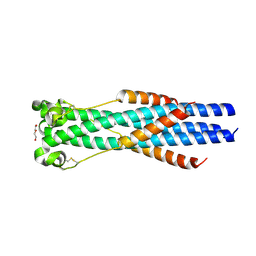
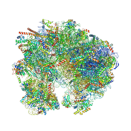 | | High-resolution C. elegans 80S ribosome structure - class 1 | | Descriptor: | 18S rRNA, 28S rRNA, 4-{(2R)-2-[(1S,3S,5S)-3,5-dimethyl-2-oxocyclohexyl]-2-hydroxyethyl}piperidine-2,6-dione, ... | | Authors: | Sehgal, E, Serrao, V.H.B, Arribere, J. | | Deposit date: | 2024-04-19 | | Release date: | 2024-09-04 | | Last modified: | 2024-11-13 | | Method: | ELECTRON MICROSCOPY (2.63 Å) | | Cite: | High-resolution reconstruction of a C. elegans ribosome sheds light on evolutionary dynamics and tissue specificity.
Rna, 30, 2024
|
|
9CAI
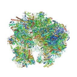 | | High-resolution C. elegans 80S ribosome structure - class 1 | | Descriptor: | 18S rRNA, 28S rRNA, 4-{(2R)-2-[(1S,3S,5S)-3,5-dimethyl-2-oxocyclohexyl]-2-hydroxyethyl}piperidine-2,6-dione, ... | | Authors: | Sehgal, E, Serrao, V.H.B, Arribere, J. | | Deposit date: | 2024-06-17 | | Release date: | 2024-09-11 | | Last modified: | 2024-10-30 | | Method: | ELECTRON MICROSCOPY (2.59 Å) | | Cite: | High-resolution reconstruction of a C. elegans ribosome sheds light on evolutionary dynamics and tissue specificity.
Rna, 30, 2024
|
|
9B7G
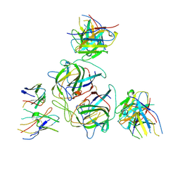
 | | Cryo-EM structure of antibody TJ5-13 bound to H3 COBRA NG2 hemagglutinin | | Descriptor: | 2-acetamido-2-deoxy-beta-D-glucopyranose, 2-acetamido-2-deoxy-beta-D-glucopyranose-(1-4)-2-acetamido-2-deoxy-beta-D-glucopyranose, 2-acetamido-2-deoxy-beta-D-glucopyranose-(1-4)-[alpha-L-fucopyranose-(1-6)]2-acetamido-2-deoxy-beta-D-glucopyranose, ... | | Authors: | Dzimianski, J.V, Cruz, J.M, Serrao, V.H.B, DuBois, R.M. | | Deposit date: | 2024-03-27 | | Release date: | 2025-05-07 | | Method: | ELECTRON MICROSCOPY (2.61 Å) | | Cite: | Assessing the structural boundaries of broadly reactive antibody interactions with diverse H3 influenza hemagglutinin proteins
To Be Published
|
|
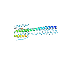
3TW4
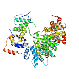
 | | Crystal Structure of Human Septin 7 GTPase Domain | | Descriptor: | GUANOSINE-5'-DIPHOSPHATE, Septin-7 | | Authors: | Serrao, V.H.B, Alessandro, F, Pereira, H.M, Thiemann, O.T, Garratt, R.C. | | Deposit date: | 2011-09-21 | | Release date: | 2011-11-23 | | Last modified: | 2023-09-13 | | Method: | X-RAY DIFFRACTION (3.35 Å) | | Cite: | Promiscuous interactions of human septins: The GTP binding domain of SEPT7 forms filaments within the crystal.
Febs Lett., 585, 2011
|
|
7S94
 | |
9CBN
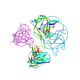
 | | HAstV1 spike in complex with neutralizing Fabs 3H4 and 3B4 | | Descriptor: | 2-acetamido-2-deoxy-beta-D-glucopyranose, HAstV1 neutralizing Fab 3B4 heavy chain, HAstV1 neutralizing Fab 3B4 kappa chain, ... | | Authors: | Lanning, S, DuBois, R.M, Balasco Serrao, V.H. | | Deposit date: | 2024-06-19 | | Release date: | 2024-12-25 | | Last modified: | 2025-03-05 | | Method: | ELECTRON MICROSCOPY (3.33 Å) | | Cite: | Discovery of three novel neutralizing antibody epitopes on the human astrovirus capsid spike and mechanistic insights into virus neutralization.
J.Virol., 99, 2025
|
|
9CB3
 | | E2F1-Cyclin F Interface | | Descriptor: | Cyclin-F, E2F1 peptide, S-phase kinase-associated protein 1 | | Authors: | Ngoi, P, Serrao, V.H, Rubin, S.M. | | Deposit date: | 2024-06-18 | | Release date: | 2025-02-12 | | Last modified: | 2025-07-09 | | Method: | ELECTRON MICROSCOPY (3.47 Å) | | Cite: | Structural mechanism for the recognition of E2F1 by the ubiquitin ligase adaptor Cyclin F.
Proc.Natl.Acad.Sci.USA, 122, 2025
|
|
5CXQ
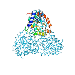 | | Crystal Structure of Isoform 2 of Purine Nucleoside Phosphorylase from Schistosoma mansoni in APO form | | Descriptor: | Purine nucleoside phosphorylase | | Authors: | Torini, J.R, Romanello, L, Bird, L, Owens, R, Brandao-Neto, J, Pereira, H.M. | | Deposit date: | 2015-07-29 | | Release date: | 2016-08-03 | | Last modified: | 2023-09-27 | | Method: | X-RAY DIFFRACTION (1.57 Å) | | Cite: | The molecular structure of Schistosoma mansoni PNP isoform 2 provides insights into the nucleoside selectivity of PNPs.
PLoS ONE, 13, 2018
|
|
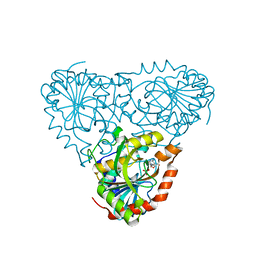
5CXS
 | | Crystal Structure of Isoform 2 of Purine Nucleoside Phosphorylase complexed with MES | | Descriptor: | 2-(N-MORPHOLINO)-ETHANESULFONIC ACID, Purine nucleoside phosphorylase | | Authors: | Torini, J.R, Romanello, L, Bird, L, Owens, R, Brandao-Neto, J, Pereira, H.M. | | Deposit date: | 2015-07-29 | | Release date: | 2016-08-03 | | Last modified: | 2023-09-27 | | Method: | X-RAY DIFFRACTION (1.75 Å) | | Cite: | The molecular structure of Schistosoma mansoni PNP isoform 2 provides insights into the nucleoside selectivity of PNPs.
PLoS ONE, 13, 2018
|
|
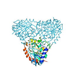
5TBT
 | | Crystal Structure of Isoform 2 of Purine Nucleoside Phosphorylase complexed with Cytidine | | Descriptor: | 4-AMINO-1-BETA-D-RIBOFURANOSYL-2(1H)-PYRIMIDINONE, Purine nucleoside phosphorylase, SULFATE ION | | Authors: | Faheem, M, Torini, J.R, Romanello, L, Brandao-Neto, J, Pereira, H.M. | | Deposit date: | 2016-09-13 | | Release date: | 2017-10-11 | | Last modified: | 2023-10-04 | | Method: | X-RAY DIFFRACTION (2.101 Å) | | Cite: | The molecular structure of Schistosoma mansoni PNP isoform 2 provides insights into the nucleoside selectivity of PNPs.
PLoS ONE, 13, 2018
|
|
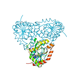
5TBS
 | | Crystal Structure of Isoform 2 of Purine Nucleoside Phosphorylase complexed with adenine | | Descriptor: | ADENINE, DIMETHYL SULFOXIDE, Purine nucleoside phosphorylase | | Authors: | Faheem, M, Torini, J.R, Romanello, L, Brandao-Neto, J, Pereira, H.M. | | Deposit date: | 2016-09-13 | | Release date: | 2017-10-11 | | Last modified: | 2023-10-04 | | Method: | X-RAY DIFFRACTION (1.9 Å) | | Cite: | The molecular structure of Schistosoma mansoni PNP isoform 2 provides insights into the nucleoside selectivity of PNPs.
PLoS ONE, 13, 2018
|
|
5TBU
 | | Crystal Structure of Isoform 2 of Purine Nucleoside Phosphorylase complexed with Hypoxanthine | | Descriptor: | DIMETHYL SULFOXIDE, HYPOXANTHINE, Purine nucleoside phosphorylase | | Authors: | Faheem, M, Torini, J.R, Romanello, L, Brandao-Neto, J, Pereira, H.M. | | Deposit date: | 2016-09-13 | | Release date: | 2017-10-11 | | Last modified: | 2023-10-04 | | Method: | X-RAY DIFFRACTION (2.1 Å) | | Cite: | The molecular structure of Schistosoma mansoni PNP isoform 2 provides insights into the nucleoside selectivity of PNPs.
PLoS ONE, 13, 2018
|
|
5IPF
 | | Crystal structure of Hypoxanthine-guanine phosphoribosyltransferase from Schistosoma mansoni in complex with IMP | | Descriptor: | Hypoxanthine-guanine phosphoribosyltransferase (HGPRT), INOSINIC ACID | | Authors: | Romanello, L, Torini, J.R.S, Bird, L.E, Nettleship, J.E, Owens, R.J, DeMarco, R, Pereira, H.M, Brandao-Neto, J. | | Deposit date: | 2016-03-09 | | Release date: | 2017-03-15 | | Last modified: | 2023-09-27 | | Method: | X-RAY DIFFRACTION (2.8 Å) | | Cite: | In vitro and in vivo characterization of the multiple isoforms of Schistosoma mansoni hypoxanthine-guanine phosphoribosyltransferases.
Mol. Biochem. Parasitol., 229, 2019
|
|
5KO5
 | | Crystal Structure of Isoform 2 of Purine Nucleoside Phosphorylase from Schistosoma mansoni in complex with cytosine | | Descriptor: | 1,2-ETHANEDIOL, 6-AMINOPYRIMIDIN-2(1H)-ONE, Purine nucleoside phosphorylase | | Authors: | Torini, J.R, Romanello, L, Bird, L, Owens, R, Brandao-Neto, J, Pereira, H.M. | | Deposit date: | 2016-06-29 | | Release date: | 2017-08-09 | | Last modified: | 2023-09-27 | | Method: | X-RAY DIFFRACTION (1.36 Å) | | Cite: | The molecular structure of Schistosoma mansoni PNP isoform 2 provides insights into the nucleoside selectivity of PNPs.
PLoS ONE, 13, 2018
|
|
5KO6
 | | Crystal Structure of Isoform 2 of Purine Nucleoside Phosphorylase from Schistosoma mansoni in complex with cytosine and ribose-1-phosphate | | Descriptor: | 1-O-phosphono-alpha-D-ribofuranose, 6-AMINOPYRIMIDIN-2(1H)-ONE, Purine nucleoside phosphorylase | | Authors: | Torini, J.R, Romanello, L, Bird, L, Owens, R, Brandao-Neto, J, Pereira, H.M. | | Deposit date: | 2016-06-29 | | Release date: | 2017-08-09 | | Last modified: | 2023-09-27 | | Method: | X-RAY DIFFRACTION (1.42 Å) | | Cite: | The molecular structure of Schistosoma mansoni PNP isoform 2 provides insights into the nucleoside selectivity of PNPs.
PLoS ONE, 13, 2018
|
|
9CN2
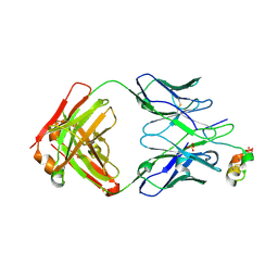
 | | Human astrovirus 2 spike bound to neutralizing scFv 4B6 | | Descriptor: | Neutralizing scFv 4B6, spike protein | | Authors: | Lanning, S, DuBois, R.M. | | Deposit date: | 2024-07-15 | | Release date: | 2024-12-25 | | Last modified: | 2025-07-09 | | Method: | X-RAY DIFFRACTION (2.67 Å) | | Cite: | Discovery of three novel neutralizing antibody epitopes on the human astrovirus capsid spike and mechanistic insights into virus neutralization.
J.Virol., 99, 2025
|
|
9CQA
 | |
9CQB
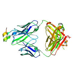 | | Antibody 1G8 bound to the central conserved domain of RSV G | | Descriptor: | Fab 1G8 Heavy Chain, Fab 1G8 Light Chain, Mature secreted glycoprotein G | | Authors: | Juarez, M.J, DuBois, M.J. | | Deposit date: | 2024-07-19 | | Release date: | 2025-02-19 | | Last modified: | 2025-04-02 | | Method: | X-RAY DIFFRACTION (2.5 Å) | | Cite: | Structures of respiratory syncytial virus G bound to broadly reactive antibodies provide insights into vaccine design.
Sci Rep, 15, 2025
|
|
9CQD
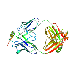 | | Antibody 2B11 bound to the central conserved domain of RSV G | | Descriptor: | Fab 2B11 Heavy Chain, Fab 2B11 Light Chain, Mature secreted glycoprotein G | | Authors: | Juarez, M.G, DuBois, R.M. | | Deposit date: | 2024-07-19 | | Release date: | 2025-02-19 | | Last modified: | 2025-04-02 | | Method: | X-RAY DIFFRACTION (3.1 Å) | | Cite: | Structures of respiratory syncytial virus G bound to broadly reactive antibodies provide insights into vaccine design.
Sci Rep, 15, 2025
|
|


