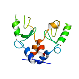
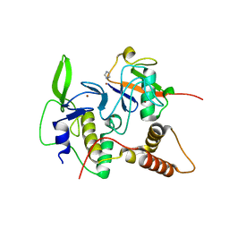
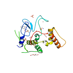
1AM4
 
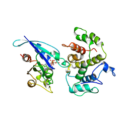 | | COMPLEX BETWEEN CDC42HS.GMPPNP AND P50 RHOGAP (H. SAPIENS) | | 分子名称: | CDC42HS, MAGNESIUM ION, P50-RHOGAP, ... | | 著者 | Rittinger, K, Walker, P, Gamblin, S.J, Smerdon, S.J. | | 登録日 | 1997-06-22 | | 公開日 | 1998-07-15 | | 最終更新日 | 2023-08-02 | | 実験手法 | X-RAY DIFFRACTION (2.7 Å) | | 主引用文献 | Crystal structure of a small G protein in complex with the GTPase-activating protein rhoGAP.
Nature, 388, 1997
|
|
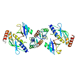
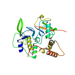
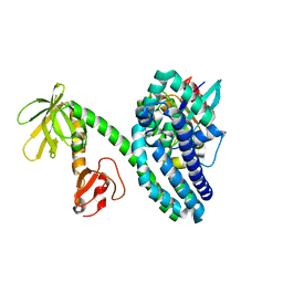
1TX4
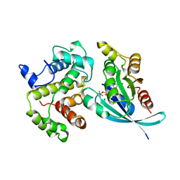 | | RHO/RHOGAP/GDP(DOT)ALF4 COMPLEX | | 分子名称: | GUANOSINE-5'-DIPHOSPHATE, MAGNESIUM ION, P50-RHOGAP, ... | | 著者 | Rittinger, K, Walker, P.A, Smerdon, S.J, Gamblin, S.J. | | 登録日 | 1997-07-29 | | 公開日 | 1998-09-16 | | 最終更新日 | 2023-08-09 | | 実験手法 | X-RAY DIFFRACTION (1.65 Å) | | 主引用文献 | Structure at 1.65 A of RhoA and its GTPase-activating protein in complex with a transition-state analogue.
Nature, 389, 1997
|
|
1QJB
 | | 14-3-3 ZETA/PHOSPHOPEPTIDE COMPLEX (MODE 1) | | 分子名称: | 14-3-3 PROTEIN ZETA/DELTA, PHOSPHOPEPTIDE | | 著者 | Rittinger, K, Budman, J, Xu, J, Volinia, S, Cantley, L.C, Smerdon, S.J, Gamblin, S.J, Yaffe, M.B. | | 登録日 | 1999-06-23 | | 公開日 | 1999-09-15 | | 最終更新日 | 2018-02-28 | | 実験手法 | X-RAY DIFFRACTION (2 Å) | | 主引用文献 | Structural Analysis of 14-3-3 Phosphopeptide Complexes Identifies a Dual Role for the Nuclear Export Signal of 14-3-3 in Ligand Binding
Mol.Cell, 4, 1999
|
|
1QJA
 | | 14-3-3 ZETA/PHOSPHOPEPTIDE COMPLEX (MODE 2) | | 分子名称: | 14-3-3 PROTEIN ZETA, PHOSPHOPEPTIDE | | 著者 | Rittinger, K, Budman, J, Xu, J, Volinia, S, Cantley, L.C, Smerdon, S.J, Gamblin, S.J, Yaffe, M.B. | | 登録日 | 1999-06-23 | | 公開日 | 1999-09-15 | | 最終更新日 | 2018-02-28 | | 実験手法 | X-RAY DIFFRACTION (2 Å) | | 主引用文献 | Structural Analysis of 14-3-3 Phosphopeptide Complexes Identifies a Dual Role for the Nuclear Export Signal of 14-3-3 in Ligand Binding
Mol.Cell, 4, 1999
|
|
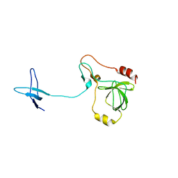
5FEY
 | | TRIM32 RING | | 分子名称: | E3 ubiquitin-protein ligase TRIM32, ZINC ION | | 著者 | Rittinger, K, Esposito, D, Koliopoulos, M.G. | | 登録日 | 2015-12-17 | | 公開日 | 2016-05-18 | | 最終更新日 | 2017-09-13 | | 実験手法 | X-RAY DIFFRACTION (2.23 Å) | | 主引用文献 | Functional role of TRIM E3 ligase oligomerization and regulation of catalytic activity.
Embo J., 35, 2016
|
|
5FER
 | | Complex of TRIM25 RING with UbcH5-Ub | | 分子名称: | E3 ubiquitin/ISG15 ligase TRIM25, Ubiquitin-40S ribosomal protein S27a, Ubiquitin-conjugating enzyme E2 D1, ... | | 著者 | Rittinger, K, Koliopoulos, M.G, Esposito, D. | | 登録日 | 2015-12-17 | | 公開日 | 2016-05-18 | | 最終更新日 | 2019-02-20 | | 実験手法 | X-RAY DIFFRACTION (2.34 Å) | | 主引用文献 | Functional role of TRIM E3 ligase oligomerization and regulation of catalytic activity.
Embo J., 35, 2016
|
|
7ZJ3
 | | Structure of TRIM2 RING domain in complex with UBE2D1~Ub conjugate | | 分子名称: | Polyubiquitin-C, Tripartite motif-containing protein 2, Ubiquitin-conjugating enzyme E2 D1, ... | | 著者 | Esposito, D, Garza-Garcia, A, Dudley-Fraser, J, Rittinger, K. | | 登録日 | 2022-04-08 | | 公開日 | 2022-11-30 | | 最終更新日 | 2024-01-31 | | 実験手法 | X-RAY DIFFRACTION (2.53 Å) | | 主引用文献 | Divergent self-association properties of paralogous proteins TRIM2 and TRIM3 regulate their E3 ligase activity.
Nat Commun, 13, 2022
|
|
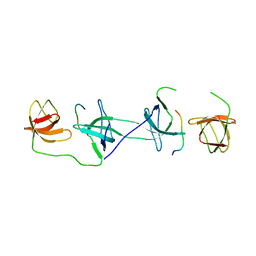
1E96
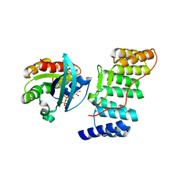 | | Structure of the Rac/p67phox complex | | 分子名称: | GUANOSINE-5'-TRIPHOSPHATE, MAGNESIUM ION, NEUTROPHIL CYTOSOL FACTOR 2 (NCF-2) TPR DOMAIN, ... | | 著者 | Lapouge, K, Smith, S.J.M, Walker, P.A, Gamblin, S.J, Smerdon, S.J, Rittinger, K. | | 登録日 | 2000-10-10 | | 公開日 | 2000-11-17 | | 最終更新日 | 2023-12-13 | | 実験手法 | X-RAY DIFFRACTION (2.4 Å) | | 主引用文献 | Structure of the TPR domain of p67phox in complex with Rac.GTP.
Mol.Cell, 6, 2000
|
|
8BVL
 | |
4EMO
 | | Crystal structure of the PH domain of SHARPIN | | 分子名称: | Sharpin | | 著者 | Stieglitz, B, Haire, L.F, Dikic, I, Rittinger, K. | | 登録日 | 2012-04-12 | | 公開日 | 2012-05-02 | | 最終更新日 | 2012-07-25 | | 実験手法 | X-RAY DIFFRACTION (2 Å) | | 主引用文献 | Structural Analysis of SHARPIN, a Subunit of a Large Multi-protein E3 Ubiquitin Ligase, Reveals a Novel Dimerization Function for the Pleckstrin Homology Superfold.
J.Biol.Chem., 287, 2012
|
|
5NT1
 | |
5NT2
 | |
6I9H
 | |
1OW3
 | | Crystal Structure of RhoA.GDP.MgF3-in Complex with RhoGAP | | 分子名称: | GUANOSINE-5'-DIPHOSPHATE, MAGNESIUM ION, Rho-GTPase-activating protein 1, ... | | 著者 | Graham, D.L, Lowe, P.N, Grime, G.W, Marsh, M, Rittinger, K, Smerdon, S.J, Gamblin, S.J, Eccleston, J.F. | | 登録日 | 2003-03-28 | | 公開日 | 2003-05-06 | | 最終更新日 | 2023-08-16 | | 実験手法 | X-RAY DIFFRACTION (1.8 Å) | | 主引用文献 | MgF(3)(-) as a Transition State Analog of Phosphoryl Transfer
Chem.Biol., 9, 2002
|
|
1NG2
 | | Structure of autoinhibited p47phox | | 分子名称: | Neutrophil cytosolic factor 1 | | 著者 | Groemping, Y, Lapouge, K, Smerdon, S.J, Rittinger, K. | | 登録日 | 2002-12-16 | | 公開日 | 2003-05-20 | | 最終更新日 | 2022-12-21 | | 実験手法 | X-RAY DIFFRACTION (1.7 Å) | | 主引用文献 | Molecular basis of phosphorylation-induced activation of the NADPH oxidase
Cell(Cambridge,Mass.), 113, 2003
|
|
1OV3
 | | Structure of the p22phox-p47phox complex | | 分子名称: | Flavocytochrome b558 alpha polypeptide, Neutrophil cytosol factor 1 | | 著者 | Groemping, Y, Lapouge, K, Smerdon, S.J, Rittinger, K. | | 登録日 | 2003-03-25 | | 公開日 | 2003-05-20 | | 最終更新日 | 2023-09-20 | | 実験手法 | X-RAY DIFFRACTION (1.8 Å) | | 主引用文献 | Molecular basis of phosphorylation-induced activation of the NADPH oxidase
Cell(Cambridge,Mass.), 113, 2003
|
|
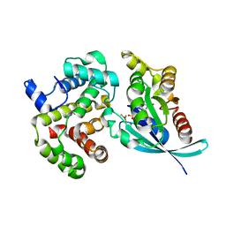
4LJO
 | | Structure of an active ligase (HOIP)/ubiquitin transfer complex | | 分子名称: | E3 ubiquitin-protein ligase RNF31, IMIDAZOLE, Polyubiquitin-C, ... | | 著者 | Rana, R.R, Stieglitz, B, Koliopoulos, M.G, Morris-Davies, A.C, Christodoulou, E, Howell, S, Brown, N.R, Rittinger, K. | | 登録日 | 2013-07-05 | | 公開日 | 2013-10-16 | | 最終更新日 | 2023-09-20 | | 実験手法 | X-RAY DIFFRACTION (1.564 Å) | | 主引用文献 | Structural basis for ligase-specific conjugation of linear ubiquitin chains by HOIP.
Nature, 503, 2013
|
|
4LJP
 | | Structure of an active ligase (HOIP-H889A)/ubiquitin transfer complex | | 分子名称: | E3 ubiquitin-protein ligase RNF31, Polyubiquitin-C, ZINC ION | | 著者 | Rana, R.R, Stieglitz, B, Koliopoulos, M.G, Morris-Davies, A.C, Christodoulou, E, Howell, S, Brown, N.R, Rittinger, K. | | 登録日 | 2013-07-05 | | 公開日 | 2013-10-16 | | 最終更新日 | 2023-09-20 | | 実験手法 | X-RAY DIFFRACTION (2.15 Å) | | 主引用文献 | Structural basis for ligase-specific conjugation of linear ubiquitin chains by HOIP.
Nature, 503, 2013
|
|
4LJQ
 | | Crystal structure of the catalytic core of E3 ligase HOIP | | 分子名称: | E3 ubiquitin-protein ligase RNF31, ZINC ION | | 著者 | Stieglitz, B, Rana, R.R, Koliopoulos, M.G, Morris-Davies, A.C, Christodoulou, E, Howell, S, Brown, N.R, Rittinger, K. | | 登録日 | 2013-07-05 | | 公開日 | 2013-10-16 | | 最終更新日 | 2013-12-18 | | 実験手法 | X-RAY DIFFRACTION (2.45 Å) | | 主引用文献 | Structural basis for ligase-specific conjugation of linear ubiquitin chains by HOIP.
Nature, 503, 2013
|
|
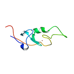
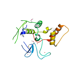
2AK5
 | | beta PIX-SH3 complexed with a Cbl-b peptide | | 分子名称: | 8-residue peptide from a signal transduction protein CBL-B, Rho guanine nucleotide exchange factor 7 | | 著者 | Jozic, D, Cardenes, N, Deribe, Y.L, Moncalian, G, Hoeller, D, Groemping, Y, Dikic, I, Rittinger, K, Bravo, J. | | 登録日 | 2005-08-03 | | 公開日 | 2005-10-11 | | 最終更新日 | 2023-08-23 | | 実験手法 | X-RAY DIFFRACTION (1.85 Å) | | 主引用文献 | Cbl promotes clustering of endocytic adaptor proteins.
Nat.Struct.Mol.Biol., 12, 2005
|
|
6GZY
 | | HOIP-fragment5 complex | | 分子名称: | 1,2-ETHANEDIOL, E3 ubiquitin-protein ligase RNF31, SODIUM ION, ... | | 著者 | Johansson, H, Tsai, Y.C.I, Fantom, K, Chung, C.W, Martino, L, House, D, Rittinger, K. | | 登録日 | 2018-07-05 | | 公開日 | 2019-01-30 | | 最終更新日 | 2024-01-17 | | 実験手法 | X-RAY DIFFRACTION (2.15 Å) | | 主引用文献 | Fragment-Based Covalent Ligand Screening Enables Rapid Discovery of Inhibitors for the RBR E3 Ubiquitin Ligase HOIP.
J. Am. Chem. Soc., 141, 2019
|
|
2VRW
 | |
2W6A
 | |
2W6B
 | |
6EYR
 | |


