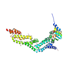
4WHC
 
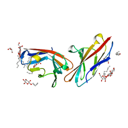
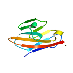 | | Human CEACAM6 N-domain | | 分子名称: | Carcinoembryonic antigen-related cell adhesion molecule 6, ZINC ION | | 著者 | Prive, G.G, Kirouac, K.N. | | 登録日 | 2014-09-22 | | 公開日 | 2015-10-07 | | 最終更新日 | 2023-12-27 | | 実験手法 | X-RAY DIFFRACTION (2.705 Å) | | 主引用文献 | Human CEACAM6 N-domain
To Be Published
|
|
3DNB
 | |
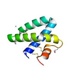
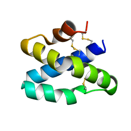
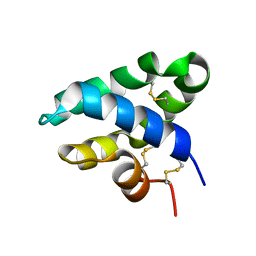
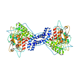
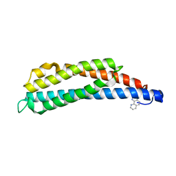
1BYZ
 | | DESIGNED PEPTIDE ALPHA-1, P1 FORM | | 分子名称: | (4R)-2-METHYLPENTANE-2,4-DIOL, (4S)-2-METHYL-2,4-PENTANEDIOL, CHLORIDE ION, ... | | 著者 | Prive, G.G, Anderson, D.H, Wesson, L, Cascio, D, Eisenberg, D. | | 登録日 | 1998-10-20 | | 公開日 | 1998-10-28 | | 最終更新日 | 2024-04-03 | | 実験手法 | X-RAY DIFFRACTION (0.9 Å) | | 主引用文献 | Packed protein bilayers in the 0.90 A resolution structure of a designed alpha helical bundle.
Protein Sci., 8, 1999
|
|
5DNB
 | |
2DOB
 | |
4EOZ
 | |
2GTG
 | |
3BIM
 | |
3BQP
 | |
3BQQ
 | |
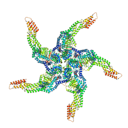
8U7Z
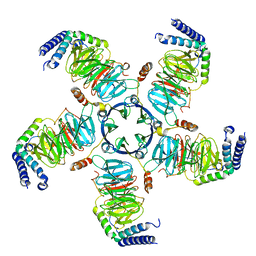 | | KCTD5/Cullin3/Gbeta1gamma2 Complex: Local Refinment of KCTD5(CTD)/Gbeta1gamma2 | | 分子名称: | BTB/POZ domain-containing protein KCTD5, Guanine nucleotide-binding protein G(I)/G(S)/G(O) subunit gamma-2, Guanine nucleotide-binding protein G(I)/G(S)/G(T) subunit beta-1 | | 著者 | Kuntz, D.A, Nguyen, D.M, Narayanan, N, Prive, G.G. | | 登録日 | 2023-09-15 | | 公開日 | 2023-10-11 | | 最終更新日 | 2024-04-24 | | 実験手法 | ELECTRON MICROSCOPY (2.97 Å) | | 主引用文献 | Structure and dynamics of a pentameric KCTD5/Cullin3/G beta gamma E3 ubiquitin ligase complex
Proc.Natl.Acad.Sci.USA, 2024
|
|
8U80
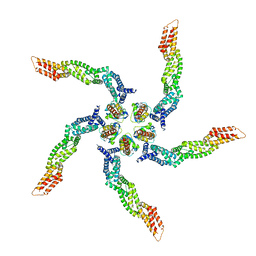 | |
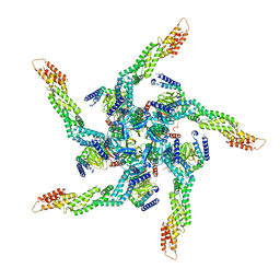
8U83
 | | KCTD5/Cullin3/Gbeta1gamma2 Complex: State C From Composite RELION Multi-body Refinement Map | | 分子名称: | BTB/POZ domain-containing protein KCTD5, Cullin-3, Guanine nucleotide-binding protein G(I)/G(S)/G(O) subunit gamma-2, ... | | 著者 | Kuntz, D.A, Nguyen, D.M, Narayanan, N, Prive, G.G. | | 登録日 | 2023-09-15 | | 公開日 | 2023-10-11 | | 最終更新日 | 2024-04-24 | | 実験手法 | ELECTRON MICROSCOPY (3.975 Å) | | 主引用文献 | Structure and dynamics of a pentameric KCTD5/Cullin3/G beta gamma E3 ubiquitin ligase complex
Proc.Natl.Acad.Sci.USA, 2024
|
|
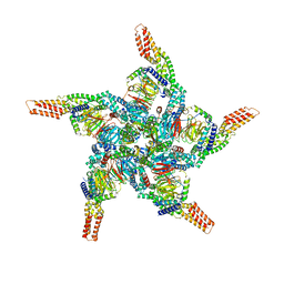
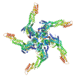
8U84
 | | KCTD5/Cullin3/Gbeta1gamma2 Complex: State D From Composite RELION Multi-body Refinement Map | | 分子名称: | BTB/POZ domain-containing protein KCTD5, Cullin-3, Guanine nucleotide-binding protein G(I)/G(S)/G(O) subunit gamma-2, ... | | 著者 | Kuntz, D.A, Nguyen, D.M, Narayanan, N, Prive, G.G. | | 登録日 | 2023-09-15 | | 公開日 | 2023-10-11 | | 最終更新日 | 2024-04-24 | | 実験手法 | ELECTRON MICROSCOPY (3.88 Å) | | 主引用文献 | Structure and dynamics of a pentameric KCTD5/Cullin3/G beta gamma E3 ubiquitin ligase complex
Proc.Natl.Acad.Sci.USA, 2024
|
|
8U82
 | | KCTD5/Cullin3/Gbeta1gamma2 Complex: State B From Composite RELION Multi-body Refinement Map | | 分子名称: | BTB/POZ domain-containing protein KCTD5, Cullin-3, Guanine nucleotide-binding protein G(I)/G(S)/G(O) subunit gamma-2, ... | | 著者 | Kuntz, D.A, Nguyen, D.M, Narayanan, N, Prive, G.G. | | 登録日 | 2023-09-15 | | 公開日 | 2023-10-11 | | 最終更新日 | 2024-04-24 | | 実験手法 | ELECTRON MICROSCOPY (3.84 Å) | | 主引用文献 | Structure and dynamics of a pentameric KCTD5/Cullin3/G beta gamma E3 ubiquitin ligase complex
Proc.Natl.Acad.Sci.USA, 2024
|
|
8U81
 | | KCTD5/Cullin3/Gbeta1gamma2 Complex: State A From Composite RELION Multi-body Refinement Map | | 分子名称: | BTB/POZ domain-containing protein KCTD5, Cullin-3, Guanine nucleotide-binding protein G(I)/G(S)/G(O) subunit gamma-2, ... | | 著者 | Kuntz, D.A, Nguyen, D.M, Narayanan, N, Prive, G.G. | | 登録日 | 2023-09-15 | | 公開日 | 2023-10-11 | | 最終更新日 | 2024-04-24 | | 実験手法 | ELECTRON MICROSCOPY (3.82 Å) | | 主引用文献 | Structure and dynamics of a pentameric KCTD5/Cullin3/G beta gamma E3 ubiquitin ligase complex
Proc.Natl.Acad.Sci.USA, 2024
|
|
3GP6
 | | Crystal structure of PagP in SDS/MPD | | 分子名称: | (4R)-2-METHYLPENTANE-2,4-DIOL, (4S)-2-METHYL-2,4-PENTANEDIOL, DODECYL SULFATE, ... | | 著者 | Cuesta-Seijo, J.A, Prive, G.G. | | 登録日 | 2009-03-20 | | 公開日 | 2010-06-23 | | 最終更新日 | 2023-09-06 | | 実験手法 | X-RAY DIFFRACTION (1.4 Å) | | 主引用文献 | PagP crystallized from SDS/cosolvent reveals the route for phospholipid access to the hydrocarbon ruler.
Structure, 18, 2010
|
|
5JG8
 | | Crystal structure of human acid sphingomyelinase | | 分子名称: | 2-acetamido-2-deoxy-beta-D-glucopyranose, 2-acetamido-2-deoxy-beta-D-glucopyranose-(1-3)-2-acetamido-2-deoxy-beta-D-glucopyranose, 2-acetamido-2-deoxy-beta-D-glucopyranose-(1-4)-2-acetamido-2-deoxy-beta-D-glucopyranose, ... | | 著者 | Xiong, Z.J, Prive, G.G. | | 登録日 | 2016-04-19 | | 公開日 | 2016-07-06 | | 最終更新日 | 2023-09-27 | | 実験手法 | X-RAY DIFFRACTION (2.8 Å) | | 主引用文献 | Structure of Human Acid Sphingomyelinase Reveals the Role of the Saposin Domain in Activating Substrate Hydrolysis.
J.Mol.Biol., 428, 2016
|
|
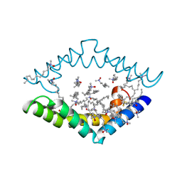
2LIG
 | | THREE-DIMENSIONAL STRUCTURES OF THE LIGAND-BINDING DOMAIN OF THE BACTERIAL ASPARTATE RECEPTOR WITH AND WITHOUT A LIGAND | | 分子名称: | 1,10-PHENANTHROLINE, ASPARTATE RECEPTOR, ASPARTIC ACID, ... | | 著者 | Kim, S.-H, Yeh, J.I, Prive, G.G, Milburn, M, Scott, W, Koshland Junior, D.E. | | 登録日 | 1995-04-18 | | 公開日 | 1995-09-15 | | 最終更新日 | 2017-11-29 | | 実験手法 | X-RAY DIFFRACTION (2 Å) | | 主引用文献 | Three-dimensional structures of the ligand-binding domain of the bacterial aspartate receptor with and without a ligand.
Science, 254, 1991
|
|
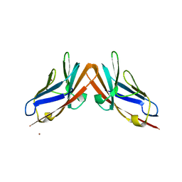
4WHD
 | | Human CEACAM1 N-domain homodimer | | 分子名称: | Carcinoembryonic antigen-related cell adhesion molecule 1, GLYCEROL, octyl beta-D-glucopyranoside | | 著者 | Kirouac, K.N, Prive, G.G. | | 登録日 | 2014-09-22 | | 公開日 | 2015-10-07 | | 最終更新日 | 2023-12-27 | | 実験手法 | X-RAY DIFFRACTION (2.5 Å) | | 主引用文献 | Human CEACAM1 N-domain homodimer
To Be Published
|
|
4WTZ
 | |
4DDJ
 | |
1LIH
 | | THREE-DIMENSIONAL STRUCTURES OF THE LIGAND-BINDING DOMAIN OF THE BACTERIAL ASPARTATE RECEPTOR WITH AND WITHOUT A LIGAND | | 分子名称: | 1,10-PHENANTHROLINE, ASPARTATE RECEPTOR | | 著者 | Kim, S.-H, Scott, W, Yeh, J.I, Prive, G.G, Milburn, M. | | 登録日 | 1995-04-18 | | 公開日 | 1995-09-15 | | 最終更新日 | 2024-02-14 | | 実験手法 | X-RAY DIFFRACTION (2.2 Å) | | 主引用文献 | Three-dimensional structures of the ligand-binding domain of the bacterial aspartate receptor with and without a ligand.
Science, 254, 1991
|
|
4HXI
 | |
7T0U
 | | Crystal structure of the BCL6 BTB domain in complex with OICR-12387 | | 分子名称: | 3-chloro-5-{7-[2-({5-chloro-2-[(2S)-2-methyl-4-(oxetan-3-yl)piperazin-1-yl]pyridin-4-yl}amino)-2-oxoethyl]-3-methyl-4-oxo-2-(trifluoromethyl)-4,7-dihydro-3H-pyrrolo[2,3-d]pyrimidin-5-yl}-2-hydroxybenzamide, CHLORIDE ION, DIMETHYL SULFOXIDE, ... | | 著者 | Kuntz, D.A, Prive, G.G. | | 登録日 | 2021-11-30 | | 公開日 | 2022-11-09 | | 最終更新日 | 2023-10-18 | | 実験手法 | X-RAY DIFFRACTION (1.49 Å) | | 主引用文献 | Crystal structure of the BCL6 BTB domain in complex with OICR-12387
To Be Published
|
|


