3DYL
 
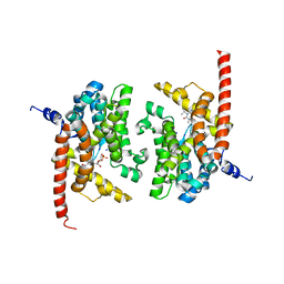
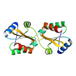
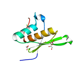
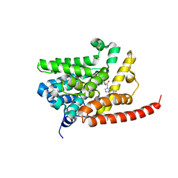 | | human phosphdiesterase 9 substrate complex (ES complex) | | 分子名称: | 3-ISOBUTYL-1-METHYLXANTHINE, CYCLIC GUANOSINE MONOPHOSPHATE, FORMIC ACID, ... | | 著者 | Liu, S, Mansour, M.N, Dillman, K, Perez, J, Danley, D, Menniti, F. | | 登録日 | 2008-07-28 | | 公開日 | 2008-09-16 | | 最終更新日 | 2024-02-21 | | 実験手法 | X-RAY DIFFRACTION (2.7 Å) | | 主引用文献 | Structural basis for the catalytic mechanism of human phosphodiesterase 9.
Proc.Natl.Acad.Sci.Usa, 105, 2008
|
|
3DY8
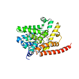 | | Human Phosphodiesterase 9 in complex with product 5'-GMP (E+P complex) | | 分子名称: | 3-ISOBUTYL-1-METHYLXANTHINE, FORMIC ACID, GUANOSINE-5'-MONOPHOSPHATE, ... | | 著者 | Liu, S, Mansour, M.N, Dillman, K, Perez, J, Danley, D, Menniti, F. | | 登録日 | 2008-07-25 | | 公開日 | 2008-09-16 | | 最終更新日 | 2024-02-21 | | 実験手法 | X-RAY DIFFRACTION (2.15 Å) | | 主引用文献 | Structural basis for the catalytic mechanism of human phosphodiesterase 9.
Proc.Natl.Acad.Sci.Usa, 105, 2008
|
|
3DYN
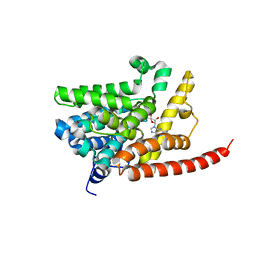 | | human phosphodiestrase 9 in complex with cGMP (Zn inhibited) | | 分子名称: | 3-ISOBUTYL-1-METHYLXANTHINE, CYCLIC GUANOSINE MONOPHOSPHATE, High affinity cGMP-specific 3',5'-cyclic phosphodiesterase 9A, ... | | 著者 | Liu, S, Mansour, M.N, Dillman, K, Perez, J, Danley, D, Menniti, F. | | 登録日 | 2008-07-28 | | 公開日 | 2008-09-16 | | 最終更新日 | 2023-08-30 | | 実験手法 | X-RAY DIFFRACTION (2.1 Å) | | 主引用文献 | Structural basis for the catalytic mechanism of human phosphodiesterase 9.
Proc.Natl.Acad.Sci.Usa, 105, 2008
|
|
3DYS
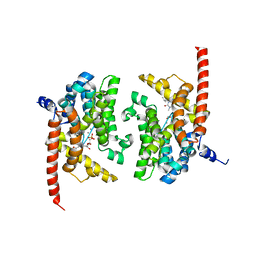 | | human phosphodiestrase-5'GMP complex (EP), produced by soaking with 20mM cGMP+20 mM MnCl2+20 mM MgCl2 for 2 hours, and flash-cooled to liquid nitrogen temperature when substrate was still abudant. | | 分子名称: | 3-ISOBUTYL-1-METHYLXANTHINE, FORMIC ACID, GUANOSINE-5'-MONOPHOSPHATE, ... | | 著者 | Liu, S, Mansour, M.N, Dillman, K, Perez, J, Danley, D, Menniti, F. | | 登録日 | 2008-07-28 | | 公開日 | 2008-09-16 | | 最終更新日 | 2024-02-21 | | 実験手法 | X-RAY DIFFRACTION (2.3 Å) | | 主引用文献 | Structural basis for the catalytic mechanism of human phosphodiesterase 9.
Proc.Natl.Acad.Sci.Usa, 105, 2008
|
|
3DYQ
 | | human phosphodiestrase 9 (inhibited by omitting divalent cation) in complex with cGMP | | 分子名称: | 3-ISOBUTYL-1-METHYLXANTHINE, CYCLIC GUANOSINE MONOPHOSPHATE, High affinity cGMP-specific 3',5'-cyclic phosphodiesterase 9A, ... | | 著者 | Liu, S, Mansour, M.N, Dillman, K, Perez, J, Danley, D, Menniti, F. | | 登録日 | 2008-07-28 | | 公開日 | 2008-09-16 | | 最終更新日 | 2023-08-30 | | 実験手法 | X-RAY DIFFRACTION (2.5 Å) | | 主引用文献 | Structural basis for the catalytic mechanism of human phosphodiesterase 9.
Proc.Natl.Acad.Sci.Usa, 105, 2008
|
|
6STZ
 | |
5G30
 | |
5G31
 | |
5G2Z
 | |
6GD7
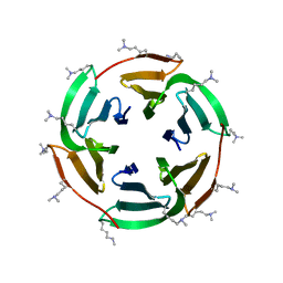
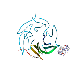
 | | Cytochrome c in complex with Sulfonato-calix[8]arene, H3 form with PEG | | 分子名称: | Cytochrome c iso-1, GLYCEROL, HEME C, ... | | 著者 | Rennie, M.L, Fox, G.C, Crowley, P.B. | | 登録日 | 2018-04-23 | | 公開日 | 2018-08-29 | | 最終更新日 | 2024-01-17 | | 実験手法 | X-RAY DIFFRACTION (1.55 Å) | | 主引用文献 | Auto-regulated Protein Assembly on a Supramolecular Scaffold.
Angew. Chem. Int. Ed. Engl., 57, 2018
|
|
8RQP
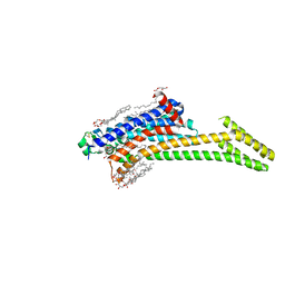
 | | In meso structure of the alginate exporter, AlgE, from Pseudomonas aeruginosa in 7.10 monoacylglycerol | | 分子名称: | 7.10 monoacylglycerol (R-form), 7.10 monoacylglycerol (S-form), Alginate production protein AlgE, ... | | 著者 | Smithers, L, Boland, C, Krawinski, P, Caffrey, M. | | 登録日 | 2024-01-19 | | 公開日 | 2024-04-03 | | 最終更新日 | 2024-04-17 | | 実験手法 | X-RAY DIFFRACTION (1.45 Å) | | 主引用文献 | 7.10 MAG. A Novel Host Monoacylglyceride for In Meso (Lipid Cubic Phase) Crystallization of Membrane Proteins.
Cryst.Growth Des., 24, 2024
|
|
8RQQ
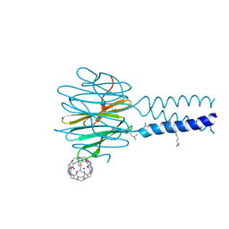
 | | In meso structure of the adenosine A2a G protein-coupled receptor, A2aR, in 7.10 monoacylglycerol | | 分子名称: | 4-{2-[(7-amino-2-furan-2-yl[1,2,4]triazolo[1,5-a][1,3,5]triazin-5-yl)amino]ethyl}phenol, 7.10 monoacylglycerol (R-form), 7.10 monoacylglycerol (S-form), ... | | 著者 | Smithers, L, Krawinski, P, Caffrey, M. | | 登録日 | 2024-01-19 | | 公開日 | 2024-04-03 | | 最終更新日 | 2024-04-17 | | 実験手法 | X-RAY DIFFRACTION (2.37 Å) | | 主引用文献 | 7.10 MAG. A Novel Host Monoacylglyceride for In Meso (Lipid Cubic Phase) Crystallization of Membrane Proteins.
Cryst.Growth Des., 24, 2024
|
|
8RQR
 | | In meso structure of apolipoprotein N-acyltransferase, Lnt, from Escherichia coli in 7.10 monoacylglycerol | | 分子名称: | 7.10 monoacylglycerol (S-form), Apolipoprotein N-acyltransferase, GLYCEROL | | 著者 | Smithers, L, Boland, C, Krawinski, P, Caffrey, M. | | 登録日 | 2024-01-19 | | 公開日 | 2024-04-03 | | 最終更新日 | 2024-04-17 | | 実験手法 | X-RAY DIFFRACTION (2.19 Å) | | 主引用文献 | 7.10 MAG. A Novel Host Monoacylglyceride for In Meso (Lipid Cubic Phase) Crystallization of Membrane Proteins.
Cryst.Growth Des., 24, 2024
|
|
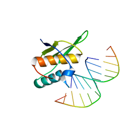
3UKG
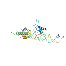 | | Crystal structure of Rap1/DNA complex | | 分子名称: | CALCIUM ION, DNA-binding protein RAP1, telomeric DNA | | 著者 | Matot, B, Le Bihan, Y.-V, Gasparini, S, LeDu, M.H. | | 登録日 | 2011-11-09 | | 公開日 | 2011-12-07 | | 最終更新日 | 2023-09-13 | | 実験手法 | X-RAY DIFFRACTION (2.95 Å) | | 主引用文献 | The orientation of the C-terminal domain of the Saccharomyces cerevisiae Rap1 protein is determined by its binding to DNA.
Nucleic Acids Res., 40, 2012
|
|
3V11
 | |
4DO2
 | |
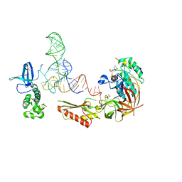
4GPK
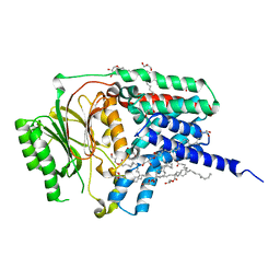
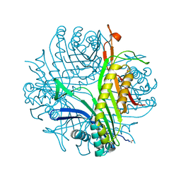
 | | Crystal structure of NprR in complex with its cognate peptide NprX | | 分子名称: | NprR, NprX peptide | | 著者 | Zouhir, S, Guimaraes, B, Perchat, S, Nicaise, M, Lereclus, D, Nessler, S. | | 登録日 | 2012-08-21 | | 公開日 | 2013-07-03 | | 最終更新日 | 2024-02-28 | | 実験手法 | X-RAY DIFFRACTION (3.2 Å) | | 主引用文献 | Peptide-binding dependent conformational changes regulate the transcriptional activity of the quorum-sensor NprR.
Nucleic Acids Res., 41, 2013
|
|
7QO8
 | |
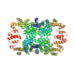
3F2M
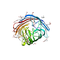
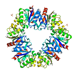
 | | Urate oxidase complexed with 8-azaxanthine at 150 MPa | | 分子名称: | 8-AZAXANTHINE, SODIUM ION, Uricase | | 著者 | Colloc'h, N, Girard, E, Kahn, R, Fourme, R. | | 登録日 | 2008-10-30 | | 公開日 | 2009-11-10 | | 最終更新日 | 2023-11-01 | | 実験手法 | X-RAY DIFFRACTION (1.8 Å) | | 主引用文献 | Structure-function perturbation and dissociation of tetrameric urate oxidase by high hydrostatic pressure.
Biophys.J., 98, 2010
|
|
5OC6
 | |
5OC5
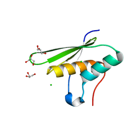 | |
5OC4
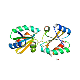
 | | Crystal structure of human tRNA-dihydrouridine(20) synthase dsRBD R361A-R362A mutant | | 分子名称: | CACODYLATE ION, CHLORIDE ION, GLYCEROL, ... | | 著者 | Bou-nader, C, Pecqueur, L, Hamdane, D. | | 登録日 | 2017-06-29 | | 公開日 | 2018-12-26 | | 最終更新日 | 2024-01-17 | | 実験手法 | X-RAY DIFFRACTION (1.71 Å) | | 主引用文献 | Molecular basis for transfer RNA recognition by the double-stranded RNA-binding domain of human dihydrouridine synthase 2.
Nucleic Acids Res., 47, 2019
|
|
7P2H
 | | Dimethylated fusion protein of RSL and mussel adhesion peptide (Mefp) in complex with cucurbit[7]uril, H3 sheet assembly | | 分子名称: | Fucose-binding lectin protein, GLYCEROL, SODIUM ION, ... | | 著者 | Ramberg, K, Engilberge, S, Crowley, P.B. | | 登録日 | 2021-07-06 | | 公開日 | 2021-09-08 | | 最終更新日 | 2024-01-31 | | 実験手法 | X-RAY DIFFRACTION (2.15 Å) | | 主引用文献 | Segregated Protein-Cucurbit[7]uril Crystalline Architectures via Modulatory Peptide Tectons.
Chemistry, 27, 2021
|
|
7P2I
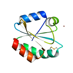
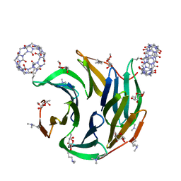 | | Dimethylated fusion protein of RSL and nucleoporin peptide (Nup) in complex with cucurbit[7]uril, F432 cage assembly | | 分子名称: | Fucose-binding lectin protein, cucurbit[7]uril, methyl alpha-L-fucopyranoside | | 著者 | Guagnini, F, Ramberg, K, Crowley, P.B. | | 登録日 | 2021-07-06 | | 公開日 | 2021-09-08 | | 最終更新日 | 2024-01-31 | | 実験手法 | X-RAY DIFFRACTION (1.489 Å) | | 主引用文献 | Segregated Protein-Cucurbit[7]uril Crystalline Architectures via Modulatory Peptide Tectons.
Chemistry, 27, 2021
|
|
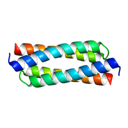
7P2J
 | | Dimethylated fusion protein of RSL and trimeric coiled coil (4dzn) in complex with cucurbit[7]uril, H3 sheet assembly | | 分子名称: | Fucose-binding lectin protein, SODIUM ION, beta-D-mannopyranose, ... | | 著者 | Ramberg, K, Engilberge, S, Crowley, P.B. | | 登録日 | 2021-07-06 | | 公開日 | 2021-09-08 | | 最終更新日 | 2024-01-31 | | 実験手法 | X-RAY DIFFRACTION (1.98 Å) | | 主引用文献 | Segregated Protein-Cucurbit[7]uril Crystalline Architectures via Modulatory Peptide Tectons.
Chemistry, 27, 2021
|
|


