Loading
PDBj
MenuPDBj@FacebookPDBj@TwitterPDBj@YouTubewwPDB FoundationwwPDB
RCSB PDBPDBeBMRBAdv. SearchSearch help
Search by PDB author
3GWC
DownloadVisualize
BU of 3gwc by Molmil
Crystal structure of Mycobacterium tuberculosis thymidylate synthase X bound to FdUMP and FAD
Descriptor: 5-FLUORO-2'-DEOXYURIDINE-5'-MONOPHOSPHATE, FLAVIN-ADENINE DINUCLEOTIDE, GLYCEROL, ...
Authors:Seattle Structural Genomics Center for Infectious Disease (SSGCID)
Deposit date:2009-03-31
Release date:2009-04-28
Last modified:2024-02-21
Method:X-RAY DIFFRACTION (1.9 Å)
Cite:Increasing the structural coverage of tuberculosis drug targets.
Tuberculosis (Edinb), 95, 2015
3H81
DownloadVisualize
BU of 3h81 by Molmil
Crystal structure of enoyl-CoA hydratase from Mycobacterium tuberculosis
Descriptor: CALCIUM ION, GLYCEROL, enoyl-CoA hydratase echA8
Authors:Seattle Structural Genomics Center for Infectious Disease (SSGCID)
Deposit date:2009-04-28
Release date:2009-05-12
Last modified:2023-09-06
Method:X-RAY DIFFRACTION (1.8 Å)
Cite:Increasing the structural coverage of tuberculosis drug targets.
Tuberculosis (Edinb), 95, 2015
3GQT
DownloadVisualize
BU of 3gqt by Molmil
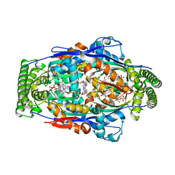
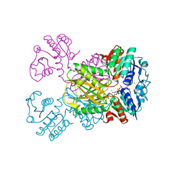
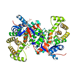
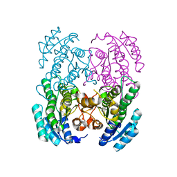
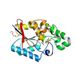
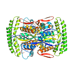
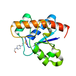
Crystal structure of glutaryl-CoA dehydrogenase from Burkholderia pseudomallei with fragment (1,4-dimethyl-1,2,3,4-tetrahydroquinoxalin-6-yl)methylamine
Descriptor: 1-(1,4-dimethyl-1,2,3,4-tetrahydroquinoxalin-6-yl)methanamine, Glutaryl-CoA dehydrogenase
Authors:Seattle Structural Genomics Center for Infectious Disease (SSGCID)
Deposit date:2009-03-24
Release date:2009-04-28
Last modified:2023-09-06
Method:X-RAY DIFFRACTION (1.99 Å)
Cite:Probing conformational states of glutaryl-CoA dehydrogenase by fragment screening.
Acta Crystallogr.,Sect.F, 67, 2011
3HHJ
DownloadVisualize
BU of 3hhj by Molmil
Crystal structure of mutator mutT from Bartonella henselae
Descriptor: MAGNESIUM ION, Mutator mutT protein
Authors:Seattle Structural Genomics Center for Infectious Disease (SSGCID)
Deposit date:2009-05-15
Release date:2009-05-26
Last modified:2023-09-06
Method:X-RAY DIFFRACTION (2.1 Å)
Cite:Structure of a Nudix hydrolase (MutT) in the Mg(2+)-bound state from Bartonella henselae, the bacterium responsible for cat scratch fever.
Acta Crystallogr.,Sect.F, 67, 2011
3IJP
DownloadVisualize
BU of 3ijp by Molmil
Crystal structure of dihydrodipicolinate reductase from bartonella henselae at 2.0A resolution
Descriptor: CHLORIDE ION, Dihydrodipicolinate reductase, NADP NICOTINAMIDE-ADENINE-DINUCLEOTIDE PHOSPHATE, ...
Authors:Seattle Structural Genomics Center for Infectious Disease (SSGCID)
Deposit date:2009-08-04
Release date:2009-08-11
Last modified:2023-09-06
Method:X-RAY DIFFRACTION (2.3 Å)
Cite:The crystal structure of dihydrodipicolinate reductase from the human-pathogenic bacterium Bartonella henselae strain Houston-1 at 2.3 angstrom resolution.
Acta Crystallogr F Struct Biol Commun, 72, 2016
3IML
DownloadVisualize
BU of 3iml by Molmil
Crystal Structure Of S-Adenosylmethionine Synthetase From Burkholderia Pseudomallei
Descriptor: S-adenosylmethionine synthetase
Authors:Staker, B.L, Seattle Structural Genomics Center for Infectious Disease (SSGCID)
Deposit date:2009-08-10
Release date:2009-09-22
Last modified:2023-09-06
Method:X-RAY DIFFRACTION (2.35 Å)
Cite:Combining functional and structural genomics to sample the essential Burkholderia structome.
Plos One, 8, 2013
3ILY
DownloadVisualize
BU of 3ily by Molmil
Apo crystal structure of protein tyrosine phosphatase from Entamoeba histolytica featuring a disordered active site
Descriptor: Protein tyrosine phosphatase, putative
Authors:Seattle Structural Genomics Center for Infectious Disease (SSGCID)
Deposit date:2009-08-07
Release date:2009-08-25
Last modified:2023-09-06
Method:X-RAY DIFFRACTION (2.2 Å)
Cite:Crystal structure and putative substrate identification for the Entamoeba histolytica low molecular weight tyrosine phosphatase.
Mol.Biochem.Parasitol., 193, 2014
3IDO
DownloadVisualize
BU of 3ido by Molmil
Crystal structure of protein tyrosine phosphatase from Entamoeba histolytica with a phosphotyrosine crude mimic HEPES molecule in the active site
Descriptor: 4-(2-HYDROXYETHYL)-1-PIPERAZINE ETHANESULFONIC ACID, Protein tyrosine phosphatase
Authors:Seattle Structural Genomics Center for Infectious Disease (SSGCID)
Deposit date:2009-07-21
Release date:2009-07-28
Last modified:2023-09-06
Method:X-RAY DIFFRACTION (2.2 Å)
Cite:Crystal structure and putative substrate identification for the Entamoeba histolytica low molecular weight tyrosine phosphatase.
Mol.Biochem.Parasitol., 193, 2014
3HWK
DownloadVisualize
BU of 3hwk by Molmil
Crystal structure of methylcitrate synthase from Mycobacterium tuberculosis
Descriptor: Methylcitrate synthase, SUCCINIC ACID
Authors:Seattle Structural Genomics Center for Infectious Disease (SSGCID)
Deposit date:2009-06-17
Release date:2009-06-30
Last modified:2023-09-06
Method:X-RAY DIFFRACTION (2.3 Å)
Cite:Increasing the structural coverage of tuberculosis drug targets.
Tuberculosis (Edinb), 95, 2015
3ICO
DownloadVisualize
BU of 3ico by Molmil
Crystal structure of 6-phosphogluconolactonase from Mycobacterium tuberculosis
Descriptor: 6-phosphogluconolactonase, SULFATE ION
Authors:Seattle Structural Genomics Center for Infectious Disease (SSGCID)
Deposit date:2009-07-17
Release date:2009-07-28
Last modified:2023-09-06
Method:X-RAY DIFFRACTION (2.15 Å)
Cite:Increasing the structural coverage of tuberculosis drug targets.
Tuberculosis (Edinb), 95, 2015
3GNC
DownloadVisualize
BU of 3gnc by Molmil
Crystal structure of Glutaryl-COA dehydrogenase from Burkholderia Pseudomallei with fragment 6421
Descriptor: 1-(1-methylethyl)-1H-benzimidazole-2-sulfonic acid, 4-(2-HYDROXYETHYL)-1-PIPERAZINE ETHANESULFONIC ACID, Glutaryl-CoA dehydrogenase, ...
Authors:Seattle Structural Genomics Center for Infectious Disease (SSGCID)
Deposit date:2009-03-16
Release date:2009-07-07
Last modified:2023-09-06
Method:X-RAY DIFFRACTION (2.15 Å)
Cite:Probing conformational states of glutaryl-CoA dehydrogenase by fragment screening.
Acta Crystallogr.,Sect.F, 67, 2011
3GTD
DownloadVisualize
BU of 3gtd by Molmil
2.4 Angstrom crystal structure of fumarate hydratase from Rickettsia prowazekii
Descriptor: Fumarate hydratase class II, MALONATE ION, SODIUM ION
Authors:Seattle Structural Genomics Center for Infectious Disease (SSGCID)
Deposit date:2009-03-27
Release date:2009-07-14
Last modified:2023-09-06
Method:X-RAY DIFFRACTION (2.4 Å)
Cite:Structure of fumarate hydratase from Rickettsia prowazekii, the agent of typhus and suspected relative of the mitochondria.
Acta Crystallogr.,Sect.F, 67, 2011
3GVC
DownloadVisualize
BU of 3gvc by Molmil
Crystal structure of probable short-chain dehydrogenase-reductase from Mycobacterium tuberculosis
Descriptor: Probable short-chain type dehydrogenase/reductase
Authors:Seattle Structural Genomics Center for Infectious Disease (SSGCID)
Deposit date:2009-03-30
Release date:2009-04-07
Last modified:2023-09-06
Method:X-RAY DIFFRACTION (2.45 Å)
Cite:Increasing the structural coverage of tuberculosis drug targets.
Tuberculosis (Edinb), 95, 2015
3JVI
DownloadVisualize
BU of 3jvi by Molmil
Product state mimic crystal structure of protein tyrosine phosphatase from Entamoeba histolytica
Descriptor: Protein tyrosine phosphatase, SULFATE ION
Authors:Seattle Structural Genomics Center for Infectious Disease (SSGCID)
Deposit date:2009-09-16
Release date:2009-09-22
Last modified:2023-09-06
Method:X-RAY DIFFRACTION (1.8 Å)
Cite:Crystal structure and putative substrate identification for the Entamoeba histolytica low molecular weight tyrosine phosphatase.
Mol.Biochem.Parasitol., 193, 2014
3GWA
DownloadVisualize
BU of 3gwa by Molmil
1.6 Angstrom crystal structure of 3-oxoacyl-(acyl-carrier-protein) synthase III
Descriptor: 3-oxoacyl-(Acyl-carrier-protein) synthase III
Authors:Seattle Structural Genomics Center for Infectious Disease (SSGCID)
Deposit date:2009-03-31
Release date:2009-04-28
Last modified:2023-09-06
Method:X-RAY DIFFRACTION (1.6 Å)
Cite:Combining functional and structural genomics to sample the essential Burkholderia structome.
Plos One, 8, 2013
3H7F
DownloadVisualize
BU of 3h7f by Molmil
Crystal structure of serine hydroxymethyltransferase from Mycobacterium tuberculosis
Descriptor: MAGNESIUM ION, Serine hydroxymethyltransferase 1
Authors:Seattle Structural Genomics Center for Infectious Disease (SSGCID)
Deposit date:2009-04-24
Release date:2009-05-05
Last modified:2023-11-22
Method:X-RAY DIFFRACTION (1.5 Å)
Cite:Increasing the structural coverage of tuberculosis drug targets.
Tuberculosis (Edinb), 95, 2015
3GP3
DownloadVisualize
BU of 3gp3 by Molmil
Crystal structure of phosphoglyceromutase from Burkholderia pseudomallei with 2-phosphoserine
Descriptor: 2,3-bisphosphoglycerate-dependent phosphoglycerate mutase, PHOSPHITE ION, PHOSPHOSERINE, ...
Authors:Seattle Structural Genomics Center for Infectious Disease (SSGCID)
Deposit date:2009-03-20
Release date:2009-03-31
Last modified:2024-02-21
Method:X-RAY DIFFRACTION (1.5 Å)
Cite:An ensemble of structures of Burkholderia pseudomallei 2,3-bisphosphoglycerate-dependent phosphoglycerate mutase.
Acta Crystallogr.,Sect.F, 67, 2011
3GWE
DownloadVisualize
BU of 3gwe by Molmil
2.1 Angstrom crystal structure of 3-oxoacyl-(acyl-carrier-protein) synthase III
Descriptor: 3-oxoacyl-(Acyl-carrier-protein) synthase III
Authors:Seattle Structural Genomics Center for Infectious Disease (SSGCID)
Deposit date:2009-03-31
Release date:2009-04-28
Last modified:2023-09-06
Method:X-RAY DIFFRACTION (2.1 Å)
Cite:Combining functional and structural genomics to sample the essential Burkholderia structome.
Plos One, 8, 2013
3HE2
DownloadVisualize
BU of 3he2 by Molmil
Crystal structure of enoyl-CoA hydratase from Mycobacterium tuberculosis
Descriptor: TRIETHYLENE GLYCOL, enoyl-CoA hydratase echA6
Authors:Seattle Structural Genomics Center for Infectious Disease (SSGCID)
Deposit date:2009-05-07
Release date:2009-06-09
Last modified:2023-09-06
Method:X-RAY DIFFRACTION (2.3 Å)
Cite:Increasing the structural coverage of tuberculosis drug targets.
Tuberculosis (Edinb), 95, 2015
3K9G
DownloadVisualize
BU of 3k9g by Molmil
CRYSTAL STRUCTURE OF A PLASMID PARTITION PROTEIN FROM BORRELIA BURGDORFERI AT 2.25A RESOLUTION, iodide soak
Descriptor: IODIDE ION, PF-32 protein, SODIUM ION
Authors:Seattle Structural Genomics Center for Infectious Disease (SSGCID)
Deposit date:2009-10-15
Release date:2009-11-03
Last modified:2024-02-21
Method:X-RAY DIFFRACTION (2.25 Å)
Cite:SAD phasing using iodide ions in a high-throughput structural genomics environment.
J Struct Funct Genomics, 12, 2011
3HZG
DownloadVisualize
BU of 3hzg by Molmil
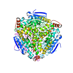
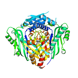
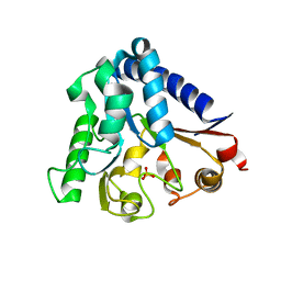
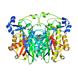
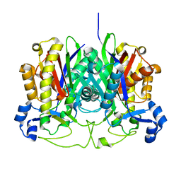
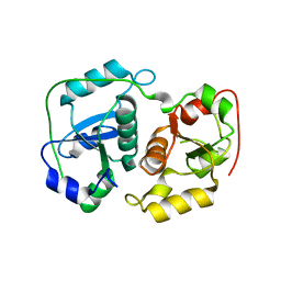
Crystal structure of mycobacterium tuberculosis thymidylate synthase X bound with FAD
Descriptor: FLAVIN-ADENINE DINUCLEOTIDE, GLYCEROL, PHOSPHATE ION, ...
Authors:Staker, B.L, Rathod, P, Hunter, J, Seattle Structural Genomics Center for Infectious Disease (SSGCID)
Deposit date:2009-06-23
Release date:2009-07-07
Last modified:2023-09-06
Method:X-RAY DIFFRACTION (2.45 Å)
Cite:Increasing the structural coverage of tuberculosis drug targets.
Tuberculosis (Edinb), 95, 2015
3I3R
DownloadVisualize
BU of 3i3r by Molmil
X-ray structure dihydrofolate reductase/thymidylate synthase from babesia bovis at 2.35A resolution
Descriptor: CHLORIDE ION, Dihydrofolate reductase/thymidylate synthase
Authors:Seattle Structural Genomics Center for Infectious Disease (SSGCID), Seattle Structural Genomics Center for Infectious Disease (SSGCID)
Deposit date:2009-06-30
Release date:2009-08-18
Last modified:2023-09-06
Method:X-RAY DIFFRACTION (2.35 Å)
Cite:Inhibitor-bound complexes of dihydrofolate reductase-thymidylate synthase from Babesia bovis.
Acta Crystallogr.,Sect.F, 67, 2011
3HWI
DownloadVisualize
BU of 3hwi by Molmil
Crystal structure of probable thiosulfate sulfurtransferase Cysa2 (Rhodanese-like protein) from Mycobacterium tuberculosis
Descriptor: Thiosulfate sulfurtransferase
Authors:Seattle Structural Genomics Center for Infectious Disease (SSGCID)
Deposit date:2009-06-17
Release date:2009-06-30
Last modified:2023-09-06
Method:X-RAY DIFFRACTION (2.29 Å)
Cite:Increasing the structural coverage of tuberculosis drug targets.
Tuberculosis (Edinb), 95, 2015
3K2H
DownloadVisualize
BU of 3k2h by Molmil
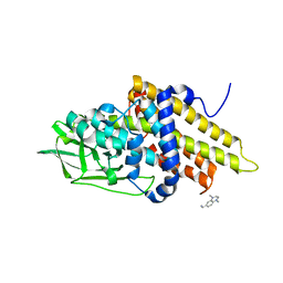
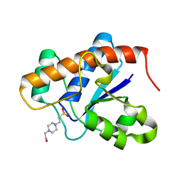
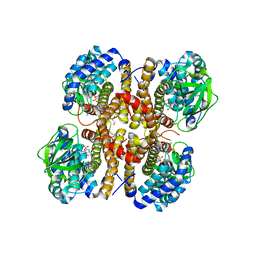
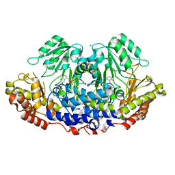
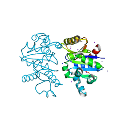
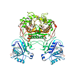
Co-crystal structure of dihydrofolate reductase/thymidylate synthase from Babesia bovis with dUMP, Pemetrexed and NADP
Descriptor: 1,2-ETHANEDIOL, 2'-DEOXYURIDINE 5'-MONOPHOSPHATE, 2-{4-[2-(2-AMINO-4-OXO-4,7-DIHYDRO-3H-PYRROLO[2,3-D]PYRIMIDIN-5-YL)-ETHYL]-BENZOYLAMINO}-PENTANEDIOIC ACID, ...
Authors:Seattle Structural Genomics Center for Infectious Disease (SSGCID)
Deposit date:2009-09-30
Release date:2009-10-13
Last modified:2023-09-06
Method:X-RAY DIFFRACTION (2.2 Å)
Cite:Inhibitor-bound complexes of dihydrofolate reductase-thymidylate synthase from Babesia bovis.
Acta Crystallogr.,Sect.F, 67, 2011
3JS5
DownloadVisualize
BU of 3js5 by Molmil
Crystal structure of protein tyrosine phosphatase from Entamoeba histolytica with Hepes in the active site. High resolution, alternative crystal form with 1 molecule in asymmetric unit
Descriptor: 4-(2-HYDROXYETHYL)-1-PIPERAZINE ETHANESULFONIC ACID, Protein tyrosine phosphatase, SODIUM ION
Authors:Seattle Structural Genomics Center for Infectious Disease (SSGCID)
Deposit date:2009-09-09
Release date:2009-09-22
Last modified:2023-09-06
Method:X-RAY DIFFRACTION (1.94 Å)
Cite:Crystal structure and putative substrate identification for the Entamoeba histolytica low molecular weight tyrosine phosphatase.
Mol.Biochem.Parasitol., 193, 2014

225158

PDB entries from 2024-09-18

PDB statisticsPDBj update infoContact PDBjnumon