1QQT
 
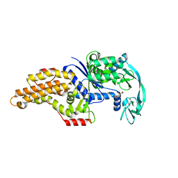
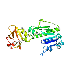
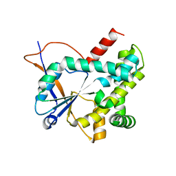
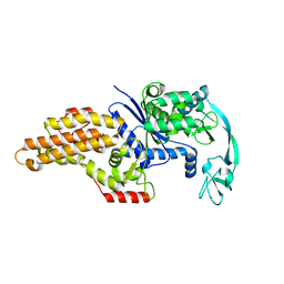 | | METHIONYL-TRNA SYNTHETASE FROM ESCHERICHIA COLI | | 分子名称: | METHIONYL-TRNA SYNTHETASE, ZINC ION | | 著者 | Mechulam, Y, Schmitt, E, Maveyraud, L, Zelwer, C, Nureki, O, Yokoyama, S, Konno, M, Blanquet, S. | | 登録日 | 1999-06-08 | | 公開日 | 2000-01-01 | | 最終更新日 | 2024-02-14 | | 実験手法 | X-RAY DIFFRACTION (2.03 Å) | | 主引用文献 | Crystal structure of Escherichia coli methionyl-tRNA synthetase highlights species-specific features.
J.Mol.Biol., 294, 1999
|
|
3V11
 | |
2QN6
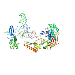
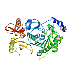
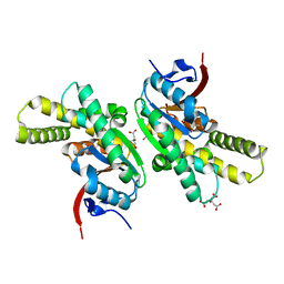
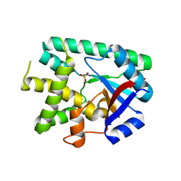
 | | Structure of an archaeal heterotrimeric initiation factor 2 reveals a nucleotide state between the GTP and the GDP states | | 分子名称: | GUANOSINE-5'-DIPHOSPHATE, MAGNESIUM ION, Translation initiation factor 2 alpha subunit, ... | | 著者 | Mechulam, Y, Yatime, L, Blanquet, S, Schmitt, E. | | 登録日 | 2007-07-18 | | 公開日 | 2007-11-06 | | 最終更新日 | 2023-08-30 | | 実験手法 | X-RAY DIFFRACTION (2.15 Å) | | 主引用文献 | Structure of an archaeal heterotrimeric initiation factor 2 reveals a nucleotide state between the GTP and the GDP states.
Proc.Natl.Acad.Sci.Usa, 104, 2007
|
|
5OCD
 | |
6Y4B
 | |
6Y3G
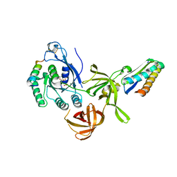
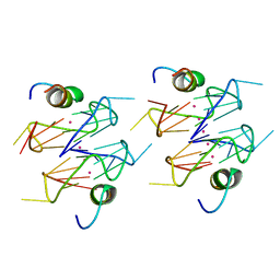
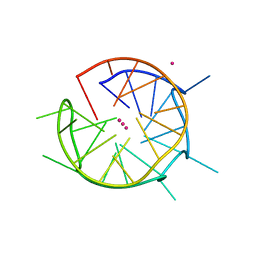
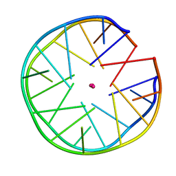
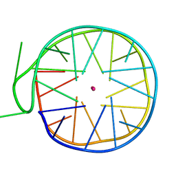
 | | Crystal structure of phenylalanine tRNA from Escherichia coli | | 分子名称: | CALCIUM ION, GLYCEROL, GUANIDINE, ... | | 著者 | Bourgeois, G, Mechulam, Y, Schmitt, E. | | 登録日 | 2020-02-18 | | 公開日 | 2020-12-30 | | 最終更新日 | 2024-01-24 | | 実験手法 | X-RAY DIFFRACTION (3.1 Å) | | 主引用文献 | Structural basis of the interaction between cyclodipeptide synthases and aminoacylated tRNA substrates.
Rna, 26, 2020
|
|
2FMT
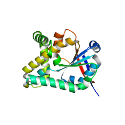
 | | METHIONYL-TRNAFMET FORMYLTRANSFERASE COMPLEXED WITH FORMYL-METHIONYL-TRNAFMET | | 分子名称: | FORMYL-METHIONYL-TRNAFMET2, MAGNESIUM ION, METHIONYL-TRNA FMET FORMYLTRANSFERASE, ... | | 著者 | Schmitt, E, Mechulam, Y, Blanquet, S. | | 登録日 | 1998-07-29 | | 公開日 | 1999-07-29 | | 最終更新日 | 2023-08-02 | | 実験手法 | X-RAY DIFFRACTION (2.8 Å) | | 主引用文献 | Crystal structure of methionyl-tRNAfMet transformylase complexed with the initiator formyl-methionyl-tRNAfMet.
EMBO J., 17, 1998
|
|
8BRV
 | | Escherichia coli methionyl-tRNA synthetase mutant L13M,I297C complexed with beta3-methionine. | | 分子名称: | (3R)-3-amino-5-(methylsulfanyl)pentanoic acid, CITRIC ACID, Methionine--tRNA ligase, ... | | 著者 | Schmitt, E, Mechulam, Y, Nigro, G, Opuu, V, Lazennec-Schurdevin, C, Simonson, T. | | 登録日 | 2022-11-24 | | 公開日 | 2023-08-16 | | 最終更新日 | 2023-11-15 | | 実験手法 | X-RAY DIFFRACTION (1.53 Å) | | 主引用文献 | Redesigning methionyl-tRNA synthetase for beta-methionine activity with adaptive landscape flattening and experiments.
Protein Sci., 32, 2023
|
|
8BRW
 | | Escherichia coli methionyl-tRNA synthetase mutant L13C,I297C | | 分子名称: | Methionine--tRNA ligase, ZINC ION | | 著者 | Schmitt, E, Mechulam, Y, Nigro, G, Opuu, V, Lazennec-Schurdevin, C, Simonson, T. | | 登録日 | 2022-11-24 | | 公開日 | 2023-08-16 | | 最終更新日 | 2023-09-06 | | 実験手法 | X-RAY DIFFRACTION (1.73 Å) | | 主引用文献 | Redesigning methionyl-tRNA synthetase for beta-methionine activity with adaptive landscape flattening and experiments.
Protein Sci., 32, 2023
|
|
8BRX
 | | Escherichia coli methionyl-tRNA synthetase mutant L13C,I297C complexed with beta-3-methionine | | 分子名称: | (3R)-3-amino-5-(methylsulfanyl)pentanoic acid, CITRIC ACID, Methionine--tRNA ligase, ... | | 著者 | Schmitt, E, Mechulam, Y, Nigro, G, Opuu, V, Lazennec-Schurdevin, C, Simonson, T. | | 登録日 | 2022-11-24 | | 公開日 | 2023-08-16 | | 最終更新日 | 2023-11-15 | | 実験手法 | X-RAY DIFFRACTION (1.54 Å) | | 主引用文献 | Redesigning methionyl-tRNA synthetase for beta-methionine activity with adaptive landscape flattening and experiments.
Protein Sci., 32, 2023
|
|
8BRU
 | | Escherichia coli methionyl-tRNA synthetase mutant L13M,I297C | | 分子名称: | CITRIC ACID, Methionine--tRNA ligase, ZINC ION | | 著者 | Schmitt, E, Mechulam, Y, Nigro, G, Opuu, V, Lazennec-Schurdevin, C, Simonson, T. | | 登録日 | 2022-11-24 | | 公開日 | 2023-08-16 | | 最終更新日 | 2023-09-06 | | 実験手法 | X-RAY DIFFRACTION (1.55 Å) | | 主引用文献 | Redesigning methionyl-tRNA synthetase for beta-methionine activity with adaptive landscape flattening and experiments.
Protein Sci., 32, 2023
|
|
3M4X
 | |
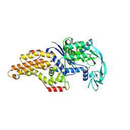
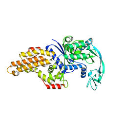
1XTY
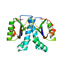 | | Crystal structure of Sulfolobus solfataricus peptidyl-tRNA hydrolase | | 分子名称: | Peptidyl-tRNA hydrolase, SULFATE ION | | 著者 | Fromant, M, Schmitt, E, Mechulam, Y, Lazennec, C, Plateau, P, Blanquet, S. | | 登録日 | 2004-10-25 | | 公開日 | 2005-03-22 | | 最終更新日 | 2024-03-13 | | 実験手法 | X-RAY DIFFRACTION (1.8 Å) | | 主引用文献 | Crystal structure at 1.8 A resolution and identification of active site residues of Sulfolobus solfataricus peptidyl-tRNA hydrolase.
Biochemistry, 44, 2005
|
|
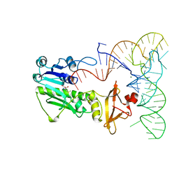
6Q6R
 | | Recognition of different base tetrads by RHAU: X-ray crystal structure of G4 recognition motif bound to the 3-end tetrad of a DNA G-quadruplex | | 分子名称: | ATP-dependent DNA/RNA helicase DHX36, POTASSIUM ION, Parallel stranded DNA G-quadruplex | | 著者 | Heddi, B, Cheong, V.V, Schmitt, E, Mechulam, Y, Phan, A.T. | | 登録日 | 2018-12-11 | | 公開日 | 2019-10-16 | | 最終更新日 | 2024-01-24 | | 実験手法 | X-RAY DIFFRACTION (1.5 Å) | | 主引用文献 | Recognition of different base tetrads by RHAU (DHX36): X-ray crystal structure of the G4 recognition motif bound to the 3'-end tetrad of a DNA G-quadruplex.
J.Struct.Biol., 209, 2020
|
|
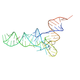
6QJO
 | | DNA containing both right- and left-handed parallel-stranded G-quadruplexes | | 分子名称: | DNA (28-MER), POTASSIUM ION | | 著者 | Winnerdy, F.R, Bakalar, B, Maity, A, Vandana, J.J, Schmitt, E, Mechulam, Y, Phan, A.T. | | 登録日 | 2019-01-24 | | 公開日 | 2019-07-03 | | 最終更新日 | 2024-01-24 | | 実験手法 | X-RAY DIFFRACTION (1.8 Å) | | 主引用文献 | NMR solution and X-ray crystal structures of a DNA molecule containing both right- and left-handed parallel-stranded G-quadruplexes.
Nucleic Acids Res., 47, 2019
|
|
1FMT
 | |
5L4O
 | |
5MLQ
 | | Structure of CDPS from Nocardia brasiliensis | | 分子名称: | CDPS, CITRIC ACID | | 著者 | Bourgeois, G, Seguin, J, Moutiez, M, Babin, M, Belin, P, Mechulam, Y, Gondry, M, Schmitt, E. | | 登録日 | 2016-12-07 | | 公開日 | 2018-05-02 | | 最終更新日 | 2019-05-15 | | 実験手法 | X-RAY DIFFRACTION (3.18 Å) | | 主引用文献 | Structural basis for partition of the cyclodipeptide synthases into two subfamilies.
J.Struct.Biol., 203, 2018
|
|
5MLP
 | | Structure of CDPS from Rickettsiella grylli | | 分子名称: | Uncharacterized protein | | 著者 | Bourgeois, G, Seguin, J, Moutiez, M, Babin, M, Belin, P, Mechulam, Y, Gondry, M, Schmitt, E. | | 登録日 | 2016-12-07 | | 公開日 | 2018-05-02 | | 最終更新日 | 2019-05-15 | | 実験手法 | X-RAY DIFFRACTION (1.99 Å) | | 主引用文献 | Structural basis for partition of the cyclodipeptide synthases into two subfamilies.
J.Struct.Biol., 203, 2018
|
|
6EZ3
 | |
6FQ2
 | | Structure of minimal sequence for left -handed G-quadruplex formation | | 分子名称: | DNA (5'-D(*GP*TP*GP*GP*TP*GP*GP*TP*GP*GP*TP*G)-3'), POTASSIUM ION | | 著者 | Schmitt, E, Mechulam, Y, Phan, A.T, Heddi, B, Bakalar, B. | | 登録日 | 2018-02-13 | | 公開日 | 2018-12-05 | | 最終更新日 | 2024-01-17 | | 実験手法 | X-RAY DIFFRACTION (2.31 Å) | | 主引用文献 | A Minimal Sequence for Left-Handed G-Quadruplex Formation.
Angew. Chem. Int. Ed. Engl., 58, 2019
|
|
5JB3
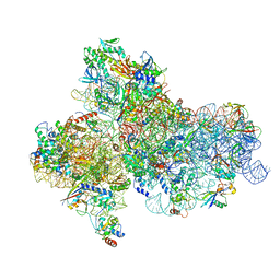
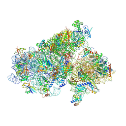 | | Cryo-EM structure of a full archaeal ribosomal translation initiation complex in the P-REMOTE conformation | | 分子名称: | 16S ribosomal RNA, 30S ribosomal protein S10, 30S ribosomal protein S11, ... | | 著者 | Coureux, P.-D, Schmitt, E, Mechulam, Y. | | 登録日 | 2016-04-13 | | 公開日 | 2016-11-30 | | 最終更新日 | 2019-12-11 | | 実験手法 | ELECTRON MICROSCOPY (5.34 Å) | | 主引用文献 | Cryo-EM study of start codon selection during archaeal translation initiation.
Nat Commun, 7, 2016
|
|
5JBH
 | | Cryo-EM structure of a full archaeal ribosomal translation initiation complex in the P-IN conformation | | 分子名称: | 16S ribosomal RNA, 30S ribosomal protein SX, 30S ribosomal protein eL41, ... | | 著者 | Coureux, P.-D, Schmitt, E, Mechulam, Y. | | 登録日 | 2016-04-13 | | 公開日 | 2016-12-07 | | 最終更新日 | 2019-12-11 | | 実験手法 | ELECTRON MICROSCOPY (5.34 Å) | | 主引用文献 | Cryo-EM study of start codon selection during archaeal translation initiation.
Nat Commun, 7, 2016
|
|
6GZ6
 | | Structure of a left-handed G-quadruplex | | 分子名称: | DNA (27-MER), POTASSIUM ION | | 著者 | Bakalar, B, Heddi, B, Schmitt, E, Mechulam, Y, Phan, A.T. | | 登録日 | 2018-07-03 | | 公開日 | 2019-04-24 | | 最終更新日 | 2024-01-17 | | 実験手法 | X-RAY DIFFRACTION (2.006 Å) | | 主引用文献 | A Minimal Sequence for Left-Handed G-Quadruplex Formation.
Angew.Chem.Int.Ed.Engl., 58, 2019
|
|
6JCE
 | | NMR solution and X-ray crystal structures of a DNA containing both right-and left-handed parallel-stranded G-quadruplexes | | 分子名称: | 29-mer DNA | | 著者 | Winnerdy, F.R, Bakalar, B, Maity, A, Vandana, J.J, Mechulam, Y, Schmitt, E, Phan, A.T. | | 登録日 | 2019-01-28 | | 公開日 | 2019-07-10 | | 最終更新日 | 2023-06-14 | | 実験手法 | SOLUTION NMR | | 主引用文献 | NMR solution and X-ray crystal structures of a DNA molecule containing both right- and left-handed parallel-stranded G-quadruplexes.
Nucleic Acids Res., 47, 2019
|
|


