Loading
PDBj
MenuPDBj@FacebookPDBj@TwitterPDBj@YouTubewwPDB FoundationwwPDB
RCSB PDBPDBeBMRBAdv. SearchSearch help
Search by PDB author
2FT8
DownloadVisualize
BU of 2ft8 by Molmil
Structure of Cu(I)azurin, pH8, with the metal-binding loop sequence "CTFPGHSALM" replaced with "CTPHPM"
Descriptor: Azurin, COPPER (I) ION
Authors:Banfield, M.J.
Deposit date:2006-01-24
Release date:2006-04-11
Last modified:2011-07-13
Method:X-RAY DIFFRACTION (1.55 Å)
Cite:Basic requirements for a metal-binding site in a protein: The influence of loop shortening on the cupredoxin azurin.
Proc.Natl.Acad.Sci.Usa, 103, 2006
2FT7
DownloadVisualize
BU of 2ft7 by Molmil
Structure of Cu(I)azurin at pH 6, with the metal-binding loop sequence "CTFPGHSALM" replaced with "CTPHPM"
Descriptor: Azurin, COPPER (I) ION
Authors:Banfield, M.J.
Deposit date:2006-01-24
Release date:2006-04-11
Last modified:2017-10-18
Method:X-RAY DIFFRACTION (1.4 Å)
Cite:Basic requirements for a metal-binding site in a protein: The influence of loop shortening on the cupredoxin azurin.
Proc.Natl.Acad.Sci.Usa, 103, 2006
2FTA
DownloadVisualize
BU of 2fta by Molmil
Structure of Cu(II)azurin with the metal-binding loop sequence "CTFPGHSALM" replaced with "CTPHPFM"
Descriptor: Azurin, COPPER (II) ION, DI(HYDROXYETHYL)ETHER, ...
Authors:Banfield, M.J.
Deposit date:2006-01-24
Release date:2006-04-11
Last modified:2023-08-30
Method:X-RAY DIFFRACTION (1.61 Å)
Cite:Basic requirements for a metal-binding site in a protein: The influence of loop shortening on the cupredoxin azurin.
Proc.Natl.Acad.Sci.Usa, 103, 2006
3LU9
DownloadVisualize
BU of 3lu9 by Molmil
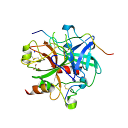
Crystal structure of human thrombin mutant S195A in complex with the extracellular fragment of human PAR1
Descriptor: 2-acetamido-2-deoxy-beta-D-glucopyranose, GLYCEROL, Proteinase-activated receptor 1, ...
Authors:Gandhi, P.S, Chen, Z, Di Cera, E.
Deposit date:2010-02-17
Release date:2010-03-16
Last modified:2023-09-06
Method:X-RAY DIFFRACTION (1.8 Å)
Cite:Crystal structure of thrombin bound to the uncleaved extracellular fragment of PAR1.
J.Biol.Chem., 285, 2010
3QDZ
DownloadVisualize
BU of 3qdz by Molmil
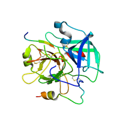
Crystal structure of the human thrombin mutant D102N in complex with the extracellular fragment of human PAR4.
Descriptor: Proteinase-activated receptor 4, Thrombin heavy chain, Thrombin light chain
Authors:Gandhi, P, Chen, Z, Appelbaum, E, Zapata, F, Di Cera, E.
Deposit date:2011-01-19
Release date:2011-06-29
Last modified:2023-09-13
Method:X-RAY DIFFRACTION (2.8 Å)
Cite:Structural basis of thrombin-protease-receptor interactions
IUBMB LIFE, 63, 2011
3R3G
DownloadVisualize
BU of 3r3g by Molmil
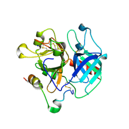
Structure of human thrombin with residues 145-150 of murine thrombin.
Descriptor: 2-acetamido-2-deoxy-beta-D-glucopyranose, SODIUM ION, Thrombin Heavy Chain, ...
Authors:Pozzi, N, Chen, R, Chen, Z, Bah, A, Di Cera, E.
Deposit date:2011-03-15
Release date:2011-05-11
Last modified:2023-09-13
Method:X-RAY DIFFRACTION (1.75 Å)
Cite:Rigidification of the autolysis loop enhances Na(+) binding to thrombin.
Biophys.Chem., 159, 2011
1MAF
DownloadVisualize
BU of 1maf by Molmil
The Active Site Structure of Methylamine Dehydrogenase: Hydrazines Identify C6 as the Reactive Site of the Tryptophan Derived Quinone Cofactor
Descriptor: METHYLAMINE DEHYDROGENASE (HEAVY SUBUNIT), METHYLAMINE DEHYDROGENASE (LIGHT SUBUNIT), NITROGEN MOLECULE
Authors:Huizinga, E.G, Vellieux, F.M.D, Hol, W.G.J.
Deposit date:1992-05-20
Release date:1994-01-31
Last modified:2011-07-13
Method:X-RAY DIFFRACTION (2.6 Å)
Cite:Active site structure of methylamine dehydrogenase: hydrazines identify C6 as the reactive site of the tryptophan-derived quinone cofactor.
Biochemistry, 31, 1992
1MAE
DownloadVisualize
BU of 1mae by Molmil
The Active Site Structure of Methylamine Dehydrogenase: Hydrazines Identify C6 as the Reactive Site of the Tryptophan Derived Quinone Cofactor
Descriptor: METHYLAMINE DEHYDROGENASE (HEAVY SUBUNIT), METHYLAMINE DEHYDROGENASE (LIGHT SUBUNIT), NITROGEN MOLECULE
Authors:Huizinga, E.G, Vellieux, F.M.D, Hol, W.G.J.
Deposit date:1992-05-20
Release date:1994-01-31
Last modified:2011-07-13
Method:X-RAY DIFFRACTION (2.8 Å)
Cite:Active site structure of methylamine dehydrogenase: hydrazines identify C6 as the reactive site of the tryptophan-derived quinone cofactor.
Biochemistry, 31, 1992

218853

PDB entries from 2024-04-24

PDB statisticsPDBj update infoContact PDBjnumon