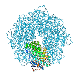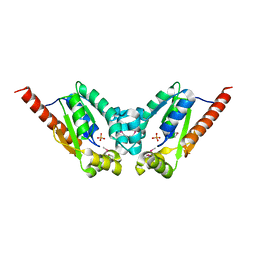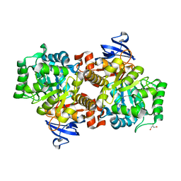
Loading
PDBj
MenuPDBj@FacebookPDBj@TwitterPDBj@YouTubewwPDB FoundationwwPDB
RCSB PDBPDBeBMRBAdv. SearchSearch help
Search by PDB author
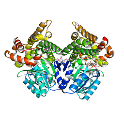
1Y9Q
DownloadVisualize
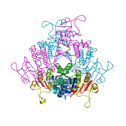
BU of 1y9q by Molmil
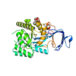
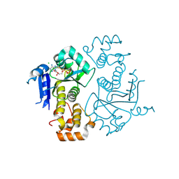
Crystal Structure of HTH_3 family Transcriptional Regulator from Vibrio cholerae
Descriptor: D-METHIONINE, ZINC ION, transcriptional regulator, ...
Authors:Kumaran, D, Swaminathan, S, Burley, S.K, New York SGX Research Center for Structural Genomics (NYSGXRC)
Deposit date:2004-12-16
Release date:2005-01-04
Last modified:2021-02-03
Method:X-RAY DIFFRACTION (1.9 Å)
Cite:Crystal Structure of HTH_3 family Transcriptional Regulator from Vibrio cholerae
To be Published
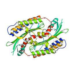
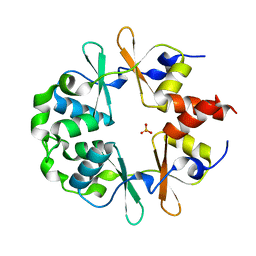
1YAV
DownloadVisualize
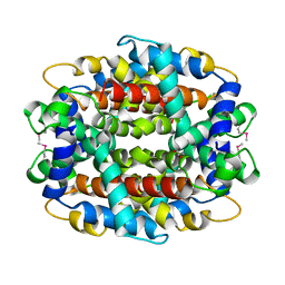
BU of 1yav by Molmil
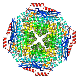
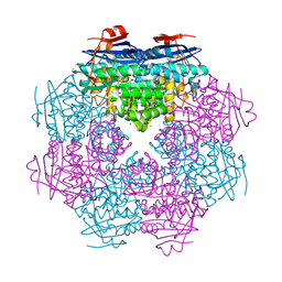
Crystal structure of CBS domain-containing protein ykuL from Bacillus subtilis
Descriptor: SULFATE ION, hypothetical protein BSU14130
Authors:Kumaran, D, Swaminathan, S, Burley, S.K, New York SGX Research Center for Structural Genomics (NYSGXRC)
Deposit date:2004-12-17
Release date:2004-12-28
Last modified:2024-02-14
Method:X-RAY DIFFRACTION (2.1 Å)
Cite:Crystal Structure of a hypothetical protein (ykul) containing CBS domains from Bacillus subtilis.
To be Published
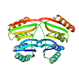
1YBD
DownloadVisualize
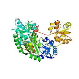
BU of 1ybd by Molmil
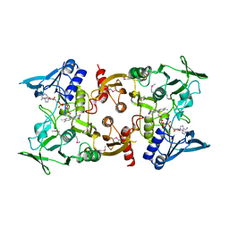
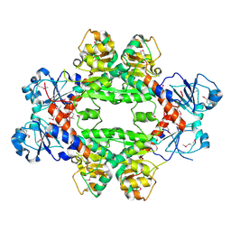
Crystal structure analysis of uridylate kinase from Neisseria meningitidis
Descriptor: FORMIC ACID, GLYCEROL, Uridylate kinase
Authors:Kumaran, D, Swaminathan, S, Burley, S.K, New York SGX Research Center for Structural Genomics (NYSGXRC)
Deposit date:2004-12-20
Release date:2005-02-15
Last modified:2021-02-03
Method:X-RAY DIFFRACTION (2.6 Å)
Cite:Crystal structure analysis of uridylate kinase from Neisseria meningitidis
To be Published
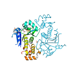
1Y9I
DownloadVisualize
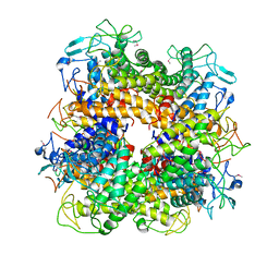
BU of 1y9i by Molmil
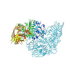
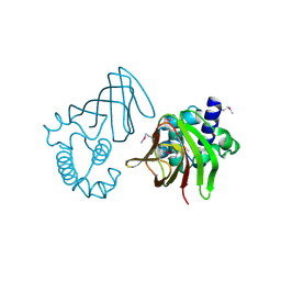
Crystal structure of low temperature requirement C protein from Listeria monocytogenes
Descriptor: CALCIUM ION, GLYCEROL, MAGNESIUM ION, ...
Authors:Kumaran, D, Swaminathan, S, Burley, S.K, New York SGX Research Center for Structural Genomics (NYSGXRC)
Deposit date:2004-12-15
Release date:2004-12-28
Last modified:2021-02-03
Method:X-RAY DIFFRACTION (1.8 Å)
Cite:Crystal structure of phosphatidylglycerophosphatase (PGPase), a putative membrane-bound lipid phosphatase, reveals a novel binuclear metal binding site and two "proton wires".
Proteins, 64, 2006
1YT8
DownloadVisualize
BU of 1yt8 by Molmil
Crystal Structure of Thiosulfate sulfurtransferase from Pseudomonas aeruginosa
Descriptor: GLYCEROL, SULFITE ION, thiosulfate sulfurtransferase
Authors:Kumaran, D, Swaminathan, S, Burley, S.K, New York SGX Research Center for Structural Genomics (NYSGXRC)
Deposit date:2005-02-10
Release date:2005-03-22
Last modified:2021-02-03
Method:X-RAY DIFFRACTION (1.9 Å)
Cite:Crystal Structure of Thiosulfate sulfurtransferase from Pseudomonas aeruginosa
To be Published
2AFA
DownloadVisualize
BU of 2afa by Molmil
Crystal Structure of putative NAG isomerase from Salmonella typhimurium
Descriptor: NAG isomerase
Authors:Kumaran, D, Swaminathan, S, Burley, S.K, New York SGX Research Center for Structural Genomics (NYSGXRC)
Deposit date:2005-07-25
Release date:2005-08-16
Last modified:2021-02-03
Method:X-RAY DIFFRACTION (2.15 Å)
Cite:Crystal Structure of putative NAG isomerase from Salmonella typhimurium
To be Published
2P9B
DownloadVisualize
BU of 2p9b by Molmil
Crystal structure of putative prolidase from Bifidobacterium longum
Descriptor: Possible prolidase
Authors:Kumaran, D, Burley, S.K, Swaminathan, S, New York SGX Research Center for Structural Genomics (NYSGXRC)
Deposit date:2007-03-24
Release date:2007-04-03
Last modified:2021-02-03
Method:X-RAY DIFFRACTION (1.7 Å)
Cite:Crystal structure of putative prolidase from Bifidobacterium longum
To be Published
2PGW
DownloadVisualize
BU of 2pgw by Molmil
Crystal structure of a putative muconate cycloisomerase from Sinorhizobium meliloti 1021
Descriptor: GLYCEROL, Muconate cycloisomerase
Authors:Kumaran, D, Burley, S.K, Swaminathan, S, New York SGX Research Center for Structural Genomics (NYSGXRC)
Deposit date:2007-04-10
Release date:2007-04-24
Last modified:2021-02-03
Method:X-RAY DIFFRACTION (1.95 Å)
Cite:Crystal structure of a putative muconate cycloisomerase from Sinorhizobium meliloti 1021
To be Published
3OC4
DownloadVisualize
BU of 3oc4 by Molmil
Crystal Structure of a pyridine nucleotide-disulfide family oxidoreductase from the Enterococcus faecalis V583
Descriptor: FLAVIN-ADENINE DINUCLEOTIDE, Oxidoreductase, pyridine nucleotide-disulfide family, ...
Authors:Kumaran, D, Baumann, K, Burley, S.K, Swaminathan, S, New York SGX Research Center for Structural Genomics (NYSGXRC)
Deposit date:2010-08-09
Release date:2010-10-06
Last modified:2021-02-10
Method:X-RAY DIFFRACTION (2.6 Å)
Cite:Crystal Structure of a pyridine nucleotide-disulfide family oxidoreductase from the Enterococcus faecalis V583
To be Published
3DEC
DownloadVisualize
BU of 3dec by Molmil
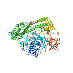
Crystal structure of a glycosyl hydrolases family 2 protein from Bacteroides thetaiotaomicron
Descriptor: Beta-galactosidase, POTASSIUM ION
Authors:Kumaran, D, Bonanno, J, Romero, R, Burley, S.K, Swaminathan, S, New York SGX Research Center for Structural Genomics (NYSGXRC)
Deposit date:2008-06-09
Release date:2008-06-17
Last modified:2023-11-15
Method:X-RAY DIFFRACTION (2.8 Å)
Cite:Crystal Structure of a Glycosyl Hydrolases Family 2 protein from Bacteroides thetaiotaomicron.
To be Published
3DDA
DownloadVisualize
BU of 3dda by Molmil
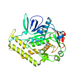
Crystal structure of the catalytic domain of Botulinum neurotoxin serotype a with a snap-25 peptide
Descriptor: Botulinum neurotoxin A light chain, SULFATE ION, Synaptosomal-associated protein 25, ...
Authors:Kumaran, D, Swaminathan, S.
Deposit date:2008-06-05
Release date:2008-09-16
Last modified:2023-08-30
Method:X-RAY DIFFRACTION (1.5 Å)
Cite:Substrate binding mode and its implication on drug design for botulinum neurotoxin A
Plos Pathog., 4, 2008
3DDB
DownloadVisualize
BU of 3ddb by Molmil
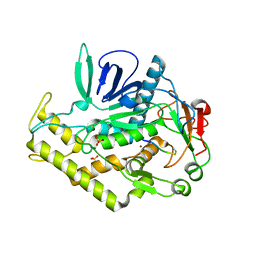
Crystal structure of the catalytic domain of Botulinum neurotoxin serotype a with a substrate analog peptide
Descriptor: Botulinum neurotoxin A light chain, SULFATE ION, Synaptosomal-associated protein 25, ...
Authors:Kumaran, D, Swaminathan, S.
Deposit date:2008-06-05
Release date:2008-09-16
Last modified:2023-08-30
Method:X-RAY DIFFRACTION (1.6 Å)
Cite:Substrate binding mode and its implication on drug design for botulinum neurotoxin A
Plos Pathog., 4, 2008
3EUW
DownloadVisualize
BU of 3euw by Molmil
Crystal Structure of a Myo-inositol dehydrogenase from Corynebacterium glutamicum ATCC 13032
Descriptor: Myo-inositol dehydrogenase, SODIUM ION
Authors:Kumaran, D, Mahmood, A, Burley, S.K, Swaminathan, S, New York SGX Research Center for Structural Genomics (NYSGXRC)
Deposit date:2008-10-11
Release date:2008-10-21
Last modified:2023-12-27
Method:X-RAY DIFFRACTION (2.3 Å)
Cite:Crystal Structure of a Myo-inositol dehydrogenase from Corynebacterium glutamicum ATCC 13032
To be Published
3FFZ
DownloadVisualize
BU of 3ffz by Molmil
Domain organization in Clostridium butulinum neurotoxin type E is unique: Its implication in faster translocation
Descriptor: ACETATE ION, Botulinum neurotoxin type E, SODIUM ION, ...
Authors:Kumaran, D, Eswaramoorthy, S, Swaminathan, S.
Deposit date:2008-12-04
Release date:2008-12-16
Last modified:2023-09-06
Method:X-RAY DIFFRACTION (2.65 Å)
Cite:Domain organization in Clostridium botulinum neurotoxin type E is unique: its implication in faster translocation.
J.Mol.Biol., 386, 2009
3H75
DownloadVisualize
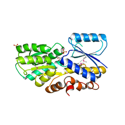
BU of 3h75 by Molmil
Crystal Structure of a Periplasmic Sugar-binding protein from the Pseudomonas fluorescens
Descriptor: GLYCEROL, Periplasmic sugar-binding domain protein, SULFATE ION
Authors:Kumaran, D, Mahmood, A, Burley, S.K, Swaminathan, S, New York SGX Research Center for Structural Genomics (NYSGXRC)
Deposit date:2009-04-24
Release date:2009-05-12
Last modified:2021-02-10
Method:X-RAY DIFFRACTION (1.6 Å)
Cite:Crystal Structure of a Periplasmic Sugar-binding protein from the Pseudomonas fluorescens
To be Published
3SQS
DownloadVisualize
BU of 3sqs by Molmil
Crystal Structure of a putative mandelate racemase/muconate lactonizing protein from Dinoroseobacter shibae DFL 12
Descriptor: ACETATE ION, GLYCEROL, MAGNESIUM ION, ...
Authors:Kumaran, D, Almo, S.C, Swaminathan, S, New York Structural Genomics Research Consortium (NYSGRC)
Deposit date:2011-07-06
Release date:2011-07-27
Last modified:2023-09-13
Method:X-RAY DIFFRACTION (1.9 Å)
Cite:Crystal Structure of a putative mandelate racemase/muconate lactonizing protein from Dinoroseobacter shibae DFL 12
To be Published
3T61
DownloadVisualize
BU of 3t61 by Molmil
Crystal Structure of a gluconokinase from Sinorhizobium meliloti 1021
Descriptor: Gluconokinase, PHOSPHATE ION
Authors:Kumaran, D, Chamala, S, Evans, B, Foti, R, Gizzi, A, Hillerich, B, Kar, A, LaFleur, J, Seidel, R, Villigas, G, Zencheck, W, Almo, S.C, Swaminathan, S, New York Structural Genomics Research Consortium (NYSGRC)
Deposit date:2011-07-28
Release date:2011-08-17
Last modified:2012-03-21
Method:X-RAY DIFFRACTION (2.2 Å)
Cite:Crystal Structure of a gluconokinase from Sinorhizobium meliloti 1021
To be Published
2I9U
DownloadVisualize
BU of 2i9u by Molmil
Crystal Structure of Guanine Deaminase from C. acetobutylicum with bound guanine in the active site
Descriptor: Cytosine/guanine deaminase related protein, FE (III) ION, GLYCEROL, ...
Authors:Kumaran, D, Burley, S.K, Swaminathan, S, New York SGX Research Center for Structural Genomics (NYSGXRC)
Deposit date:2006-09-06
Release date:2006-09-19
Last modified:2024-02-21
Method:X-RAY DIFFRACTION (2.05 Å)
Cite:Crystal Structure of Guanine Deaminase from C. acetobutylicum with bound guanine in the active site
To be Published
1RRM
DownloadVisualize
BU of 1rrm by Molmil
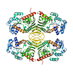
Crystal Structure of Lactaldehyde reductase
Descriptor: ADENOSINE-5-DIPHOSPHORIBOSE, FE (II) ION, Lactaldehyde reductase, ...
Authors:Kumaran, D, Swaminathan, S, Burley, S.K, New York SGX Research Center for Structural Genomics (NYSGXRC)
Deposit date:2003-12-08
Release date:2004-08-17
Last modified:2021-02-03
Method:X-RAY DIFFRACTION (1.6 Å)
Cite:Crystal structure of Lactaldehyde reductase
To be Published
1TXN
DownloadVisualize
BU of 1txn by Molmil
Crystal structure of coproporphyrinogen III oxidase
Descriptor: Coproporphyrinogen III oxidase, GLYCEROL
Authors:Kumaran, D, Swaminathan, S, Burley, S.K, New York SGX Research Center for Structural Genomics (NYSGXRC)
Deposit date:2004-07-05
Release date:2004-11-30
Last modified:2021-02-03
Method:X-RAY DIFFRACTION (1.7 Å)
Cite:Crystal structure of coproporphyrinogen III oxidase
To be Published
1U8S
DownloadVisualize
BU of 1u8s by Molmil
Crystal structure of putative glycine cleavage system transcriptional repressor
Descriptor: glycine cleavage system transcriptional repressor, putative
Authors:Kumaran, D, Swaminathan, S.
Deposit date:2004-08-06
Release date:2004-08-17
Last modified:2024-02-14
Method:X-RAY DIFFRACTION (2.45 Å)
Cite:Crystal structure of putative glycine cleavage system transcriptional repressor
To be Published
1TXZ
DownloadVisualize
BU of 1txz by Molmil
Crystal structure of yeast ymx7, an ADP-ribose-1''-monophosphatase, complexed with ADP-ribose
Descriptor: 1,2-ETHANEDIOL, ADENOSINE-5-DIPHOSPHORIBOSE, Hypothetical 32.1 kDa protein in ADH3-RCA1 intergenic region, ...
Authors:Kumaran, D, Swaminathan, S, Burley, S.K, New York SGX Research Center for Structural Genomics (NYSGXRC)
Deposit date:2004-07-06
Release date:2004-11-30
Last modified:2023-08-23
Method:X-RAY DIFFRACTION (2.05 Å)
Cite:Structure and mechanism of ADP-ribose-1''-monophosphatase (Appr-1''-pase), a ubiquitous cellular processing enzyme.
Protein Sci., 14, 2005
1TY8
DownloadVisualize
BU of 1ty8 by Molmil
Crystal structure of yeast ymx7, an ADP-ribose-1''-monophosphatase, complexed with ADP
Descriptor: ADENOSINE-5'-DIPHOSPHATE, CHLORIDE ION, Hypothetical 32.1 kDa protein in ADH3-RCA1 intergenic region, ...
Authors:Kumaran, D, Swaminathan, S, Burley, S.K, New York SGX Research Center for Structural Genomics (NYSGXRC)
Deposit date:2004-07-07
Release date:2004-11-30
Last modified:2023-08-23
Method:X-RAY DIFFRACTION (2.1 Å)
Cite:CRYSTAL STRUCTURE OF YEAST HYPOTHETICAL PROTEIN YMX7
To be Published
2OG9
DownloadVisualize
BU of 2og9 by Molmil
Crystal Structure of mandelate racemase/muconate lactonizing enzyme from Polaromonas sp. JS666
Descriptor: CALCIUM ION, Mandelate racemase/muconate lactonizing enzyme
Authors:Kumaran, D, Burley, S.K, Swaminathan, S, New York SGX Research Center for Structural Genomics (NYSGXRC)
Deposit date:2007-01-05
Release date:2007-02-27
Last modified:2023-12-27
Method:X-RAY DIFFRACTION (1.9 Å)
Cite:Crystal Structure of mandelate racemase/muconate lactonizing enzyme from Polaromonas sp. JS666
To be Published
4HY3
DownloadVisualize
BU of 4hy3 by Molmil
Crystal structure of a phosphoglycerate oxidoreductase from rhizobium etli
Descriptor: phosphoglycerate oxidoreductase
Authors:Kumaran, D, Chamala, S, Evans, B, Foti, R, Gizzi, A, Hillerich, B, Kar, A, Lafleur, J, Seidel, R, Villigas, G, Zencheck, W, Almo, S.C, Swaminathan, S, New York Structural Genomics Research Consortium (NYSGRC)
Deposit date:2012-11-12
Release date:2012-12-12
Method:X-RAY DIFFRACTION (2.8 Å)
Cite:Crystal structure of a phosphoglycerate oxidoreductase from rhizobium etli
To be Published

219869

PDB entries from 2024-05-15

PDB statisticsPDBj update infoContact PDBjnumon