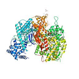
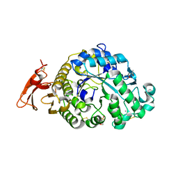
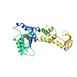
Loading
PDBj
MenuPDBj@FacebookPDBj@TwitterPDBj@YouTubewwPDB FoundationwwPDB
RCSB PDBPDBeBMRBAdv. SearchSearch help
Search by PDB author
6XSJ
DownloadVisualize
BU of 6xsj by Molmil
X-ray structure of a monoclinic form of alpha amylase from Aspergillus at 1.4 A resolution
Descriptor: (4S)-2-METHYL-2,4-PENTANEDIOL, 2-(N-MORPHOLINO)-ETHANESULFONIC ACID, 2-acetamido-2-deoxy-beta-D-glucopyranose, ...
Authors:McPherson, A.
Deposit date:2020-07-15
Release date:2020-10-14
Last modified:2023-10-18
Method:X-RAY DIFFRACTION (1.4 Å)
Cite:Structures of two novel crystal forms of Aspergillus oryzae alpha amylase (taka-amylase).
J.Biosci.Bioeng., 131, 2021
4PNJ
DownloadVisualize
BU of 4pnj by Molmil
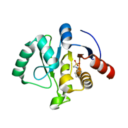
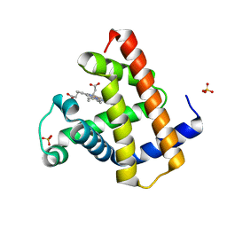
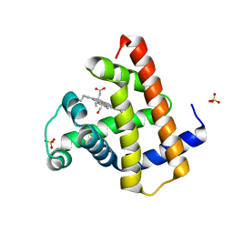
Recombinant Sperm Whale P6 Myoglobin Solved with Single Pulse Free Electron Laser Data
Descriptor: Myoglobin, PROTOPORPHYRIN IX CONTAINING FE, SULFATE ION
Authors:Cohen, A, Gonzalez, A, Lam, W, Lyubimov, A, Sauter, N, Tsai, Y, Uervirojnangkoorn, M, Brunger, A, Soltis, M.
Deposit date:2014-05-23
Release date:2014-11-05
Last modified:2023-09-27
Method:X-RAY DIFFRACTION (1.36 Å)
Cite:Goniometer-based femtosecond crystallography with X-ray free electron lasers.
Proc.Natl.Acad.Sci.USA, 111, 2014
7TWH
DownloadVisualize
BU of 7twh by Molmil
Crystal structure of SARS-CoV-2 NSP3 macrodomain at 293 K (P43 crystal form, 290 kGy)
Descriptor: Non-structural protein 3
Authors:Correy, G.J, Fraser, J.S.
Deposit date:2022-02-07
Release date:2022-02-23
Last modified:2023-10-18
Method:X-RAY DIFFRACTION (1.1 Å)
Cite:The mechanisms of catalysis and ligand binding for the SARS-CoV-2 NSP3 macrodomain from neutron and x-ray diffraction at room temperature.
Sci Adv, 8, 2022
7TWN
DownloadVisualize
BU of 7twn by Molmil
Crystal structure of SARS-CoV-2 NSP3 macrodomain at pH 5 (P43 crystal form)
Descriptor: 2-[N-CYCLOHEXYLAMINO]ETHANE SULFONIC ACID, CITRIC ACID, Non-structural protein 3
Authors:Correy, G.J, Fraser, J.S.
Deposit date:2022-02-07
Release date:2022-02-23
Last modified:2023-10-18
Method:X-RAY DIFFRACTION (0.9 Å)
Cite:The mechanisms of catalysis and ligand binding for the SARS-CoV-2 NSP3 macrodomain from neutron and x-ray diffraction at room temperature.
Sci Adv, 8, 2022
7TWJ
DownloadVisualize
BU of 7twj by Molmil
Crystal structure of SARS-CoV-2 NSP3 macrodomain at pH 4 (P43 crystal form)
Descriptor: CITRIC ACID, Non-structural protein 3
Authors:Correy, G.J, Fraser, J.S.
Deposit date:2022-02-07
Release date:2022-02-23
Last modified:2023-10-18
Method:X-RAY DIFFRACTION (0.9 Å)
Cite:The mechanisms of catalysis and ligand binding for the SARS-CoV-2 NSP3 macrodomain from neutron and x-ray diffraction at room temperature.
Sci Adv, 8, 2022
7TWR
DownloadVisualize
BU of 7twr by Molmil
Crystal structure of SARS-CoV-2 NSP3 macrodomain at pH 8 (P43 crystal form)
Descriptor: ACETATE ION, Non-structural protein 3
Authors:Correy, G.J, Fraser, J.S.
Deposit date:2022-02-07
Release date:2022-02-23
Last modified:2023-10-18
Method:X-RAY DIFFRACTION (0.9 Å)
Cite:The mechanisms of catalysis and ligand binding for the SARS-CoV-2 NSP3 macrodomain from neutron and x-ray diffraction at room temperature.
Sci Adv, 8, 2022
7TWS
DownloadVisualize
BU of 7tws by Molmil
Crystal structure of SARS-CoV-2 NSP3 macrodomain at pH 10 (P43 crystal form)
Descriptor: Non-structural protein 3
Authors:Correy, G.J, Fraser, J.S.
Deposit date:2022-02-07
Release date:2022-02-23
Last modified:2023-10-18
Method:X-RAY DIFFRACTION (0.9 Å)
Cite:The mechanisms of catalysis and ligand binding for the SARS-CoV-2 NSP3 macrodomain from neutron and x-ray diffraction at room temperature.
Sci Adv, 8, 2022
7TWQ
DownloadVisualize
BU of 7twq by Molmil
Crystal structure of SARS-CoV-2 NSP3 macrodomain at pH 9 (P43 crystal form)
Descriptor: Non-structural protein 3
Authors:Correy, G.J, Fraser, J.S.
Deposit date:2022-02-07
Release date:2022-02-23
Last modified:2023-10-18
Method:X-RAY DIFFRACTION (0.9 Å)
Cite:The mechanisms of catalysis and ligand binding for the SARS-CoV-2 NSP3 macrodomain from neutron and x-ray diffraction at room temperature.
Sci Adv, 8, 2022
7TWI
DownloadVisualize
BU of 7twi by Molmil
Crystal structure of SARS-CoV-2 NSP3 macrodomain at 293 K (P43 crystal form, 539 kGy)
Descriptor: Non-structural protein 3
Authors:Correy, G.J, Fraser, J.S.
Deposit date:2022-02-07
Release date:2022-02-23
Last modified:2023-10-18
Method:X-RAY DIFFRACTION (1.1 Å)
Cite:The mechanisms of catalysis and ligand binding for the SARS-CoV-2 NSP3 macrodomain from neutron and x-ray diffraction at room temperature.
Sci Adv, 8, 2022
7TWG
DownloadVisualize
BU of 7twg by Molmil
Crystal structure of SARS-CoV-2 NSP3 macrodomain at 293 K (P43 crystal form, 153 kGy)
Descriptor: Non-structural protein 3
Authors:Correy, G.J, Fraser, J.S.
Deposit date:2022-02-07
Release date:2022-02-23
Last modified:2023-10-18
Method:X-RAY DIFFRACTION (1.1 Å)
Cite:The mechanisms of catalysis and ligand binding for the SARS-CoV-2 NSP3 macrodomain from neutron and x-ray diffraction at room temperature.
Sci Adv, 8, 2022
7TX4
DownloadVisualize
BU of 7tx4 by Molmil
Neutron crystal structure of SARS-CoV-2 NSP3 macrodomain at 293 K (P21 crystal form)
Descriptor: Papain-like protease nsp3
Authors:Correy, G.J, Fraser, J.S, Kovalevsky, A.
Deposit date:2022-02-07
Release date:2022-02-23
Last modified:2023-10-25
Method:NEUTRON DIFFRACTION (1.9 Å), X-RAY DIFFRACTION
Cite:The mechanisms of catalysis and ligand binding for the SARS-CoV-2 NSP3 macrodomain from neutron and x-ray diffraction at room temperature.
Sci Adv, 8, 2022
7TWF
DownloadVisualize
BU of 7twf by Molmil
Crystal structure of SARS-CoV-2 NSP3 macrodomain at 293 K (P43 crystal form, 73 kGy)
Descriptor: Non-structural protein 3
Authors:Correy, G.J, Fraser, J.S.
Deposit date:2022-02-07
Release date:2022-02-23
Last modified:2023-10-18
Method:X-RAY DIFFRACTION (1.1 Å)
Cite:The mechanisms of catalysis and ligand binding for the SARS-CoV-2 NSP3 macrodomain from neutron and x-ray diffraction at room temperature.
Sci Adv, 8, 2022
7TX5
DownloadVisualize
BU of 7tx5 by Molmil
Neutron crystal structure of SARS-CoV-2 NSP3 macrodomain in complex with ADP-ribose at 293 K (C2 crystal form)
Descriptor: ADENOSINE-5-DIPHOSPHORIBOSE, Papain-like protease nsp3
Authors:Correy, G.J, Fraser, J.S, Kovalevsky, A.
Deposit date:2022-02-07
Release date:2022-02-23
Last modified:2023-10-25
Method:NEUTRON DIFFRACTION (1.95 Å), X-RAY DIFFRACTION
Cite:The mechanisms of catalysis and ligand binding for the SARS-CoV-2 NSP3 macrodomain from neutron and x-ray diffraction at room temperature.
Sci Adv, 8, 2022
7TWO
DownloadVisualize
BU of 7two by Molmil
Crystal structure of SARS-CoV-2 NSP3 macrodomain at pH 6 (P43 crystal form)
Descriptor: 2-[N-CYCLOHEXYLAMINO]ETHANE SULFONIC ACID, CITRIC ACID, Non-structural protein 3
Authors:Correy, G.J, Fraser, J.S.
Deposit date:2022-02-07
Release date:2022-02-23
Last modified:2023-10-18
Method:X-RAY DIFFRACTION (0.9 Å)
Cite:The mechanisms of catalysis and ligand binding for the SARS-CoV-2 NSP3 macrodomain from neutron and x-ray diffraction at room temperature.
Sci Adv, 8, 2022
7TWP
DownloadVisualize
BU of 7twp by Molmil
Crystal structure of SARS-CoV-2 NSP3 macrodomain at pH 7 (P43 crystal form)
Descriptor: ACETATE ION, Non-structural protein 3
Authors:Correy, G.J, Fraser, J.S.
Deposit date:2022-02-07
Release date:2022-02-23
Last modified:2023-10-18
Method:X-RAY DIFFRACTION (0.9 Å)
Cite:The mechanisms of catalysis and ligand binding for the SARS-CoV-2 NSP3 macrodomain from neutron and x-ray diffraction at room temperature.
Sci Adv, 8, 2022
7TX3
DownloadVisualize
BU of 7tx3 by Molmil
Neutron crystal structure of SARS-CoV-2 NSP3 macrodomain at 293 K (P43 crystal form)
Descriptor: 2-[N-CYCLOHEXYLAMINO]ETHANE SULFONIC ACID, Non-structural protein 3
Authors:Correy, G.J, Fraser, J.S, Meilleur, F.
Deposit date:2022-02-07
Release date:2022-02-23
Last modified:2023-10-25
Method:NEUTRON DIFFRACTION (1.6 Å), X-RAY DIFFRACTION
Cite:The mechanisms of catalysis and ligand binding for the SARS-CoV-2 NSP3 macrodomain from neutron and x-ray diffraction at room temperature.
Sci Adv, 8, 2022
5W1H
DownloadVisualize
BU of 5w1h by Molmil
Crystal structure of LbaCas13a (C2c2) bound to mature crRNA (24-nt spacer)
Descriptor: GLYCEROL, IODIDE ION, LbaCas13a (C2c2), ...
Authors:Knott, G.J, Doudna, J.A.
Deposit date:2017-06-03
Release date:2017-09-13
Last modified:2024-03-13
Method:X-RAY DIFFRACTION (1.99 Å)
Cite:Guide-bound structures of an RNA-targeting A-cleaving CRISPR-Cas13a enzyme.
Nat. Struct. Mol. Biol., 24, 2017
5W1I
DownloadVisualize
BU of 5w1i by Molmil
Crystal structure of LbaCas13a (C2c2) bound to mature crRNA (20-nt spacer)
Descriptor: IODIDE ION, LbaCas13a (C2c2), mature crRNA
Authors:Knott, G.J, Doudna, J.A.
Deposit date:2017-06-03
Release date:2017-09-13
Last modified:2023-10-04
Method:X-RAY DIFFRACTION (2.2 Å)
Cite:Guide-bound structures of an RNA-targeting A-cleaving CRISPR-Cas13a enzyme.
Nat. Struct. Mol. Biol., 24, 2017
5WLH
DownloadVisualize
BU of 5wlh by Molmil
Crystal structure of LbaCas13a H328A (C2c2) bound to pre-crRNA (24-nt spacer)
Descriptor: GLYCEROL, IODIDE ION, LbaCas13a H328A (C2c2), ...
Authors:Knott, G.J, Doudna, J.A.
Deposit date:2017-07-26
Release date:2017-09-13
Last modified:2023-10-04
Method:X-RAY DIFFRACTION (1.80001032 Å)
Cite:Guide-bound structures of an RNA-targeting A-cleaving CRISPR-Cas13a enzyme.
Nat. Struct. Mol. Biol., 24, 2017
1XWI
DownloadVisualize
BU of 1xwi by Molmil
Crystal Structure of VPS4B
Descriptor: SKD1 protein, SULFATE ION
Authors:Scott, A, Sundquist, W.I, Hill, C.P.
Deposit date:2004-11-01
Release date:2005-10-11
Last modified:2024-02-14
Method:X-RAY DIFFRACTION (2.8 Å)
Cite:Structural and mechanistic studies of VPS4 proteins
Embo J., 24, 2005
8BKH
DownloadVisualize
BU of 8bkh by Molmil
Carboxymyoglobin dark state for comparison with power titration and 23 / 101 mJ/cm2 time series
Descriptor: CARBON MONOXIDE, Myoglobin, PROTOPORPHYRIN IX CONTAINING FE, ...
Authors:Barends, T, Schlichting, I.
Deposit date:2022-11-09
Release date:2024-01-31
Last modified:2024-03-06
Method:X-RAY DIFFRACTION (1.35 Å)
Cite:Influence of pump laser fluence on ultrafast myoglobin structural dynamics.
Nature, 626, 2024
8BKN
DownloadVisualize
BU of 8bkn by Molmil
Carboxymyoglobin dark state for comparison with 5 mJ/cm2 time series
Descriptor: CARBON MONOXIDE, Myoglobin, PROTOPORPHYRIN IX CONTAINING FE, ...
Authors:Barends, T, Schlichting, I.
Deposit date:2022-11-09
Release date:2024-01-31
Last modified:2024-03-06
Method:X-RAY DIFFRACTION (1.29 Å)
Cite:Influence of pump laser fluence on ultrafast myoglobin structural dynamics.
Nature, 626, 2024
8R8I
DownloadVisualize
BU of 8r8i by Molmil
Ensemble-refined carboxymyoglobin photolysis power titration, 56 mJ/cm2
Descriptor: CARBON MONOXIDE, Myoglobin, PROTOPORPHYRIN IX CONTAINING FE, ...
Authors:Barends, T, Schlichting, I.
Deposit date:2023-11-29
Release date:2024-01-31
Last modified:2024-03-06
Method:X-RAY DIFFRACTION (1.4 Å)
Cite:Influence of pump laser fluence on ultrafast myoglobin structural dynamics.
Nature, 626, 2024
8R9D
DownloadVisualize
BU of 8r9d by Molmil
Multicopy-refined carboxymyoglobin photolysis time series at 23 mJ/cm2 laser fluence, 399 fs time delay
Descriptor: CARBON MONOXIDE, Myoglobin, PROTOPORPHYRIN IX CONTAINING FE, ...
Authors:Barends, T, Schlichting, I.
Deposit date:2023-11-30
Release date:2024-01-31
Last modified:2024-03-06
Method:X-RAY DIFFRACTION (1.4 Å)
Cite:Influence of pump laser fluence on ultrafast myoglobin structural dynamics.
Nature, 626, 2024
8R9P
DownloadVisualize
BU of 8r9p by Molmil
Multicopy-refined carboxymyoglobin photolysis time series at 23 mJ/cm2 laser fluence, 1297 fs time delay
Descriptor: CARBON MONOXIDE, Myoglobin, PROTOPORPHYRIN IX CONTAINING FE, ...
Authors:Barends, T, Schlichting, I.
Deposit date:2023-11-30
Release date:2024-01-31
Last modified:2024-03-06
Method:X-RAY DIFFRACTION (1.4 Å)
Cite:Influence of pump laser fluence on ultrafast myoglobin structural dynamics.
Nature, 626, 2024

218853

PDB entries from 2024-04-24

PDB statisticsPDBj update infoContact PDBjnumon