Loading
PDBj
MenuPDBj@FacebookPDBj@X(formerly Twitter)PDBj@BlueSkyPDBj@YouTubewwPDB FoundationwwPDB
RCSB PDBPDBeBMRBAdv. SearchSearch help
Search by PDB author
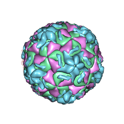
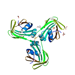
1HRI
DownloadVisualize
BU of 1hri by Molmil
STRUCTURE DETERMINATION OF ANTIVIRAL COMPOUND SCH 38057 COMPLEXED WITH HUMAN RHINOVIRUS 14
Descriptor: 1-[6-(2-CHLORO-4-METHYXYPHENOXY)-HEXYL]-IMIDAZOLE, HUMAN RHINOVIRUS 14 COAT PROTEIN (SUBUNIT VP1), HUMAN RHINOVIRUS 14 COAT PROTEIN (SUBUNIT VP2), ...
Authors:Zhang, A, Nanni, R.G, Oren, D.A, Arnold, E.
Deposit date:1992-10-01
Release date:1993-10-31
Last modified:2024-05-22
Method:X-RAY DIFFRACTION (3 Å)
Cite:Structure determination of antiviral compound SCH 38057 complexed with human rhinovirus 14.
J.Mol.Biol., 230, 1993
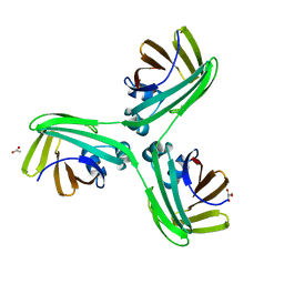
1I9S
DownloadVisualize
BU of 1i9s by Molmil
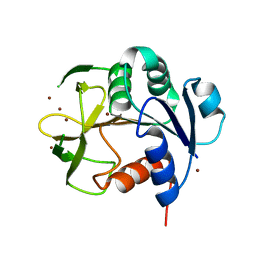
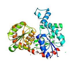
CRYSTAL STRUCTURE OF THE RNA TRIPHOSPHATASE DOMAIN OF MOUSE MRNA CAPPING ENZYME
Descriptor: CACODYLATE ION, ISOPROPYL ALCOHOL, MAGNESIUM ION, ...
Authors:Changela, A, Ho, C.K, Martins, A, Shuman, S, Mondragon, A.
Deposit date:2001-03-20
Release date:2001-05-23
Last modified:2024-02-07
Method:X-RAY DIFFRACTION (1.65 Å)
Cite:Structure and mechanism of the RNA triphosphatase component of mammalian mRNA capping enzyme.
EMBO J., 20, 2001
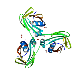
1I9T
DownloadVisualize
BU of 1i9t by Molmil
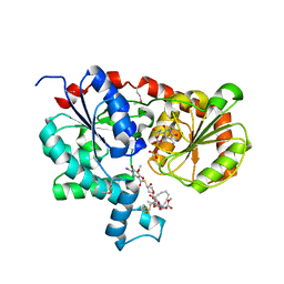
CRYSTAL STRUCTURE OF THE OXIDIZED RNA TRIPHOSPHATASE DOMAIN OF MOUSE MRNA CAPPING ENZYME
Descriptor: CACODYLATE ION, ISOPROPYL ALCOHOL, MAGNESIUM ION, ...
Authors:Changela, A, Ho, C.K, Martins, A, Shuman, S, Mondragon, A.
Deposit date:2001-03-20
Release date:2001-05-23
Last modified:2024-11-13
Method:X-RAY DIFFRACTION (1.7 Å)
Cite:Structure and mechanism of the RNA triphosphatase component of mammalian mRNA capping enzyme.
EMBO J., 20, 2001
1I7D
DownloadVisualize
BU of 1i7d by Molmil
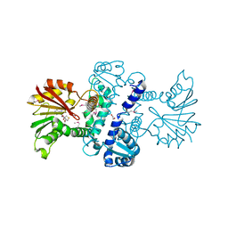
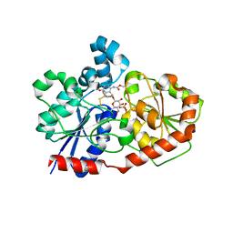
NONCOVALENT COMPLEX OF E.COLI DNA TOPOISOMERASE III WITH AN 8-BASE SINGLE-STRANDED DNA OLIGONUCLEOTIDE
Descriptor: 5'-D(*CP*GP*CP*AP*AP*CP*TP*T)-3', CHLORIDE ION, DNA TOPOISOMERASE III, ...
Authors:Changela, A, DiGate, R.J, Mondragon, A.
Deposit date:2001-03-08
Release date:2001-06-29
Last modified:2023-08-09
Method:X-RAY DIFFRACTION (2.05 Å)
Cite:Crystal structure of a complex of a type IA DNA topoisomerase with a single-stranded DNA molecule.
Nature, 411, 2001
1IAZ
DownloadVisualize
BU of 1iaz by Molmil
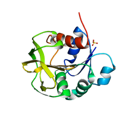
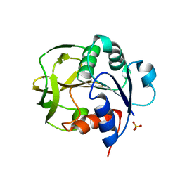
EQUINATOXIN II
Descriptor: EQUINATOXIN II, SULFATE ION
Authors:Athanasiadis, A, Anderluh, G, Macek, P, Turk, D.
Deposit date:2001-03-24
Release date:2001-04-04
Last modified:2024-02-07
Method:X-RAY DIFFRACTION (1.9 Å)
Cite:Crystal structure of the soluble form of equinatoxin II, a pore-forming toxin from the sea anemone Actinia equina.
Structure, 9, 2001
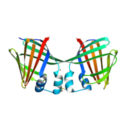
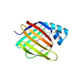
6E6L
DownloadVisualize
BU of 6e6l by Molmil
Crystal structure of the holo retinal-bound domain-swapped dimer Q108K:K40L:T51F:Y60A mutant of human cellular retinol binding protein II
Descriptor: ACETATE ION, RETINAL, Retinol-binding protein 2
Authors:Ghanbarpour, A, Geiger, J.
Deposit date:2018-07-25
Release date:2019-10-16
Last modified:2024-11-06
Method:X-RAY DIFFRACTION (2.08 Å)
Cite:Engineering the hCRBPII Domain-Swapped Dimer into a New Class of Protein Switches.
J.Am.Chem.Soc., 141, 2019
9C88
DownloadVisualize
BU of 9c88 by Molmil
Cryo-EM Structure of a Proteolytic ClpXP AAA+ Machine Translocating a Portion of a Branched-Degron DHFR Substrate
Descriptor: ADENOSINE-5'-DIPHOSPHATE, ADENOSINE-5'-TRIPHOSPHATE, ATP-dependent Clp protease ATP-binding subunit ClpX, ...
Authors:Ghanbarpour, A, Sauer, R.T, Davis, J.H.
Deposit date:2024-06-12
Release date:2024-10-16
Method:ELECTRON MICROSCOPY (2.6 Å)
Cite:Cryo-EM structure of a fully-engaged DHFR-ssrA substrate and the AAA+ ClpXP protease
To Be Published
9C87
DownloadVisualize
BU of 9c87 by Molmil
Cryo-EM Structure of a Proteolytic ClpXP AAA+ Machine Poised to Unfold a Linear-Degron DHFR-ssrA Substrate Bound with MTX
Descriptor: ADENOSINE-5'-DIPHOSPHATE, ADENOSINE-5'-TRIPHOSPHATE, ATP-dependent Clp protease ATP-binding subunit ClpX, ...
Authors:Ghanbarpour, A, Sauer, R.T, Davis, J.H.
Deposit date:2024-06-12
Release date:2024-10-16
Method:ELECTRON MICROSCOPY (3.7 Å)
Cite:Cryo-EM Structure of a Proteolytic ClpXP AAA+ Machine Poised to Unfold a Linear-Degron DHFR-ssrA Substrate Bound with MTX
To Be Published
9CZ1
DownloadVisualize
BU of 9cz1 by Molmil
Cryo-EM structure of a 'hat' portion of FtsH.HflK.HflC complex
Descriptor: Modulator of FtsH protease HflC, Modulator of FtsH protease HflK
Authors:Ghanbarpour, A, Sauer, R.T, Davis, J.H.
Deposit date:2024-08-03
Release date:2024-11-13
Method:ELECTRON MICROSCOPY (3.5 Å)
Cite:Cryo-EM structure of a 'hat' portion of FtsH.HflK.HflC complex
To Be Published
9CZ2
DownloadVisualize
BU of 9cz2 by Molmil
Cryo-EM structure of a nautilus-like HflK/C assembly in complex with FtsH AAA protease
Descriptor: ATP-dependent zinc metalloprotease FtsH, Modulator of FtsH protease HflC, Modulator of FtsH protease HflK
Authors:Ghanbarpour, A, Sauer, R.T, Davis, J.H.
Deposit date:2024-08-03
Release date:2024-11-13
Method:ELECTRON MICROSCOPY (4.4 Å)
Cite:Cryo-EM structure of a nautilus-like HflK/C assembly in complex with FtsH AAA protease
To Be Published
1N6J
DownloadVisualize
BU of 1n6j by Molmil
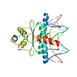
Structural basis of sequence-specific recruitment of histone deacetylases by Myocyte Enhancer Factor-2
Descriptor: 5'-D(*AP*GP*CP*TP*AP*TP*TP*TP*AP*TP*AP*AP*GP*C)-3', 5'-D(*GP*CP*TP*TP*AP*TP*AP*AP*AP*TP*AP*GP*CP*T)-3', Calcineurin-binding protein Cabin 1, ...
Authors:Han, A, Pan, F, Stroud, J.C, Youn, H.D, Liu, J.O, Chen, L.
Deposit date:2002-11-11
Release date:2003-11-11
Last modified:2024-02-14
Method:X-RAY DIFFRACTION (2.2 Å)
Cite:Sequence-specific recruitment of transcriptional co-repressor Cabin1 by myocyte enhancer factor-2
Nature, 422, 2003
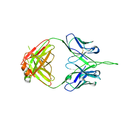
3PIQ
DownloadVisualize
BU of 3piq by Molmil
Crystal structure of human 2909 Fab, a quaternary structure-specific antibody against HIV-1
Descriptor: Human monoclonal antibody 2909 Fab heavy chain, Human monoclonal antibody 2909 Fab light chain
Authors:Changela, A, Gorny, M.K, Zolla-Pazner, S, Kwong, P.D.
Deposit date:2010-11-07
Release date:2011-01-05
Last modified:2024-10-30
Method:X-RAY DIFFRACTION (3.325 Å)
Cite:Crystal Structure of Human Antibody 2909 Reveals Conserved Features of Quaternary Structure-Specific Antibodies That Potently Neutralize HIV-1.
J.Virol., 85, 2011
5ALC
DownloadVisualize
BU of 5alc by Molmil
Ticagrelor antidote candidate Fab 72 in complex with ticagrelor
Descriptor: ANTI-TICAGRELOR FAB 72, HEAVY CHAIN, LIGHT CHAIN, ...
Authors:Buchanan, A, Newton, P, Pehrsson, S, Inghardt, T, Antonsson, T, Svensson, P, Sjogren, T, Oster, L, Janefeldt, A, Sandinge, A, Keyes, F, Austin, M, Spooner, J, Penney, M, Howells, G, Vaughan, T, Nylander, S.
Deposit date:2015-03-07
Release date:2015-04-01
Last modified:2024-11-06
Method:X-RAY DIFFRACTION (1.7 Å)
Cite:Structural and Functional Characterisation of a Specific Antidote for Ticagrelor.
Blood, 125, 2015
5ALB
DownloadVisualize
BU of 5alb by Molmil
Ticagrelor antidote candidate MEDI2452 in complex with ticagrelor
Descriptor: MEDI2452 HEAVY CHAIN, MEDI2452 LIGHT CHAIN, Ticagrelor
Authors:Buchanan, A, Newton, P, Pehrsson, S, Inghardt, T, Antonsson, T, Svensson, P, Sjogren, T, Oster, L, Janefeldt, A, Sandinge, A, Keyes, F, Austin, M, Spooner, J, Penney, M, Howells, G, Vaughan, T, Nylander, S.
Deposit date:2015-03-07
Release date:2015-04-01
Last modified:2024-11-20
Method:X-RAY DIFFRACTION (2.16 Å)
Cite:Structural and Functional Characterisation of a Specific Antidote for Ticagrelor.
Blood, 125, 2015
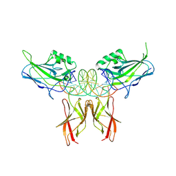
8U9L
DownloadVisualize
BU of 8u9l by Molmil
Crystal Structure of RelA-cRel chimera complex with DNA
Descriptor: DNA (5'-D(P*GP*AP*AP*TP*CP*GP*GP*AP*AP*AP*TP*TP*CP*CP*CP*AP*TP*CP*AP*A)-3'), DNA (5'-D(P*TP*TP*GP*AP*TP*GP*GP*GP*AP*AP*TP*TP*TP*CP*CP*GP*AP*TP*TP*C)-3'), Transcription factor p65,Proto-oncogene c-Rel chimera
Authors:Chang, A, Wu, Y, Li, S.X, Smale, S, Chen, L.
Deposit date:2023-09-19
Release date:2024-10-02
Method:X-RAY DIFFRACTION (3.091 Å)
Cite:Crystal Structure of RelA-cRel chimera complex with DNA
To Be Published
8S1X
DownloadVisualize
BU of 8s1x by Molmil
Crystal structure of Actinonin-bound PDF1 and the computationally designed DBAct553_1 protein binder
Descriptor: ACTINONIN, DBAct553_1, FORMIC ACID, ...
Authors:Marchand, A, Pacesa, M, Correia, B.E.
Deposit date:2024-02-16
Release date:2024-10-30
Method:X-RAY DIFFRACTION (1.88 Å)
Cite:Targeting hybrid neosurface fingerprints for the design of de novo drug-induced protein interactions
To Be Published
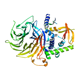
7N40
DownloadVisualize
BU of 7n40 by Molmil
Crystal structure of LIN9-RbAp48-LIN37, a MuvB subcomplex
Descriptor: Histone-binding protein RBBP4, Isoform 2 of Protein lin-9 homolog, Protein lin-37 homolog
Authors:Asthana, A, Ramanan, P, Tripathi, S.M, Rubin, S.M.
Deposit date:2021-06-02
Release date:2022-02-09
Last modified:2023-10-18
Method:X-RAY DIFFRACTION (2.55 Å)
Cite:The MuvB complex binds and stabilizes nucleosomes downstream of the transcription start site of cell-cycle dependent genes.
Nat Commun, 13, 2022
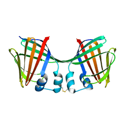
6ON7
DownloadVisualize
BU of 6on7 by Molmil
Crystal Structure of Apo Domain-Swapped Dimer Q108K:T51D:A28C:L36C Mutant of Human Cellular Retinol Binding Protein II
Descriptor: Retinol-binding protein 2
Authors:Ghanbarpour, A, Geiger, J.
Deposit date:2019-04-20
Release date:2019-10-16
Last modified:2024-10-30
Method:X-RAY DIFFRACTION (1.981 Å)
Cite:Engineering the hCRBPII Domain-Swapped Dimer into a New Class of Protein Switches.
J.Am.Chem.Soc., 141, 2019
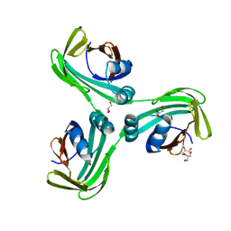
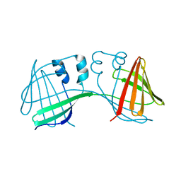
6WP0
DownloadVisualize
BU of 6wp0 by Molmil
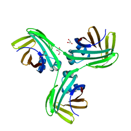
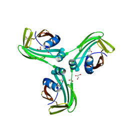
The Crystal Structure of Domain-Swapped Trimer Q108K:T51D variant of HCRBPII
Descriptor: ACETATE ION, GLYCEROL, Retinol-binding protein 2
Authors:Ghanbarpour, A, Geiger, J.
Deposit date:2020-04-26
Release date:2020-08-19
Last modified:2023-10-18
Method:X-RAY DIFFRACTION (2.78 Å)
Cite:Human Cellular Retinol Binding Protein II Forms a Domain-Swapped Trimer Representing a Novel Fold and a New Template for Protein Engineering.
Chembiochem, 21, 2020
1XMK
DownloadVisualize
BU of 1xmk by Molmil
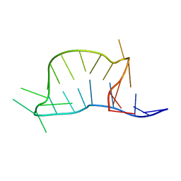
The Crystal structure of the Zb domain from the RNA editing enzyme ADAR1
Descriptor: CADMIUM ION, CHLORIDE ION, Double-stranded RNA-specific adenosine deaminase, ...
Authors:Athanasiadis, A, Placido, D, Maas, S, Brown II, B.A, Lowenhaupt, K, Rich, A.
Deposit date:2004-10-03
Release date:2005-08-02
Last modified:2024-02-14
Method:X-RAY DIFFRACTION (0.97 Å)
Cite:The Crystal Structure of the Z[beta] Domain of the RNA-editing Enzyme ADAR1 Reveals Distinct Conserved Surfaces Among Z-domains.
J.Mol.Biol., 351, 2005
1YN9
DownloadVisualize
BU of 1yn9 by Molmil
Crystal structure of baculovirus RNA 5'-phosphatase complexed with phosphate
Descriptor: PHOSPHATE ION, polynucleotide 5'-phosphatase
Authors:Changela, A, Martins, A, Shuman, S, Mondragon, A.
Deposit date:2005-01-24
Release date:2005-02-22
Last modified:2023-08-23
Method:X-RAY DIFFRACTION (1.5 Å)
Cite:Crystal structure of baculovirus RNA triphosphatase complexed with phosphate
J.Biol.Chem., 280, 2005
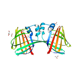
6MCV
DownloadVisualize
BU of 6mcv by Molmil
Crystal Structure of Holo Retinal-Bound Domain-Swapped Dimer of Wild Type Human Cellular Retinol Binding Protein II
Descriptor: RETINAL, Retinol-binding protein 2
Authors:Ghanbarpour, A, Geiger, J.
Deposit date:2018-09-02
Release date:2019-10-16
Last modified:2023-10-11
Method:X-RAY DIFFRACTION (3.3 Å)
Cite:Engineering the hCRBPII Domain-Swapped Dimer into a New Class of Protein Switches.
J.Am.Chem.Soc., 141, 2019
6MQJ
DownloadVisualize
BU of 6mqj by Molmil
Crystal Structure of the All-Trans Retinal bound R111K:Y134F:T54V:R132Q:P39Y:R59Y:L121Q mutant of Human Cellular Retinoic Acid Binding Protein II Irradiated with 532 nm Laser for 10 minutes at 2.1 Angstrom Resolution
Descriptor: Cellular retinoic acid-binding protein 2, RETINAL
Authors:Ghanbarpour, A, Geiger, J.
Deposit date:2018-10-09
Release date:2019-01-09
Last modified:2024-10-30
Method:X-RAY DIFFRACTION (2.125 Å)
Cite:Mimicking Microbial Rhodopsin Isomerization in a Single Crystal.
J. Am. Chem. Soc., 141, 2019
6MOQ
DownloadVisualize
BU of 6moq by Molmil
Crystal Structure of the All-trans Retinal-Bound R111K:Y134F:T54V:R132Q:P39Y:R59Y:L121W Mutant of Human Cellular Retinoic Acid Binding Protein II in the Dark at 2.2 Angstrom Resolution
Descriptor: Cellular retinoic acid-binding protein 2, RETINAL
Authors:Ghanbarpour, A, Geiger, J.
Deposit date:2018-10-04
Release date:2019-01-09
Last modified:2024-11-20
Method:X-RAY DIFFRACTION (2.198 Å)
Cite:Mimicking Microbial Rhodopsin Isomerization in a Single Crystal.
J. Am. Chem. Soc., 141, 2019
3LST
DownloadVisualize
BU of 3lst by Molmil
Crystal Structure of CalO1, Methyltransferase in Calicheamicin Biosynthesis, SAH bound form
Descriptor: 1,2-ETHANEDIOL, CalO1 Methyltransferase, S-ADENOSYL-L-HOMOCYSTEINE
Authors:Chang, A, Singh, S, Bingman, C.A, Thorson, J.S, Phillips Jr, G.N, Center for Eukaryotic Structural Genomics (CESG)
Deposit date:2010-02-12
Release date:2010-03-02
Last modified:2024-10-16
Method:X-RAY DIFFRACTION (2.4 Å)
Cite:Structural characterization of CalO1: a putative orsellinic acid methyltransferase in the calicheamicin-biosynthetic pathway.
Acta Crystallogr.,Sect.D, 67, 2011

227561

PDB entries from 2024-11-20

PDB statisticsPDBj update infoContact PDBjnumon