Loading
PDBj
MenuPDBj@FacebookPDBj@TwitterPDBj@YouTubewwPDB FoundationwwPDB
RCSB PDBPDBeBMRBAdv. SearchSearch help
Search by PDB author
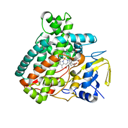
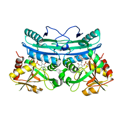
5L1V
DownloadVisualize
BU of 5l1v by Molmil
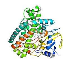
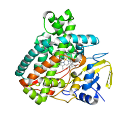
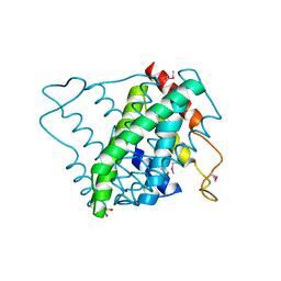
X-ray Structure of M81C mutant of Cytochrome P450 PntM with pentalenolactone F
Descriptor: PROTOPORPHYRIN IX CONTAINING FE, Pentalenolactone synthase, pentalenolactone F
Authors:Duan, L, Jogl, G, Cane, D.E.
Deposit date:2016-07-29
Release date:2016-09-14
Last modified:2023-10-04
Method:X-RAY DIFFRACTION (2.12 Å)
Cite:The Cytochrome P450-Catalyzed Oxidative Rearrangement in the Final Step of Pentalenolactone Biosynthesis: Substrate Structure Determines Mechanism.
J.Am.Chem.Soc., 138, 2016
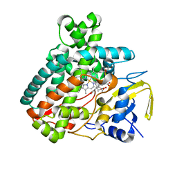
5L1R
DownloadVisualize
BU of 5l1r by Molmil
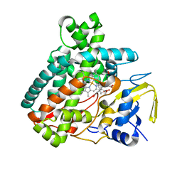
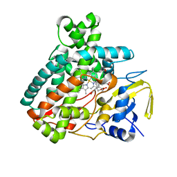
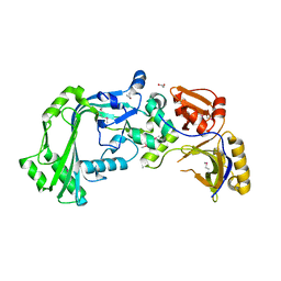
X-ray Structure of the Substrate-free Cytochrome P450 PntM
Descriptor: BICINE, PROTOPORPHYRIN IX CONTAINING FE, Pentalenolactone synthase
Authors:Duan, L, Jogl, G, Cane, D.E.
Deposit date:2016-07-29
Release date:2016-09-14
Last modified:2023-10-04
Method:X-RAY DIFFRACTION (2 Å)
Cite:The Cytochrome P450-Catalyzed Oxidative Rearrangement in the Final Step of Pentalenolactone Biosynthesis: Substrate Structure Determines Mechanism.
J.Am.Chem.Soc., 138, 2016
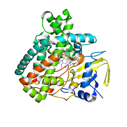
5L1U
DownloadVisualize
BU of 5l1u by Molmil
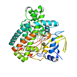
X-ray Structure of M81A mutant of Cytochrome P450 PntM with pentalenolactone F
Descriptor: PROTOPORPHYRIN IX CONTAINING FE, Pentalenolactone synthase, pentalenolactone F
Authors:Duan, L, Jogl, G, Cane, D.E.
Deposit date:2016-07-29
Release date:2016-09-14
Last modified:2023-10-04
Method:X-RAY DIFFRACTION (2.074 Å)
Cite:The Cytochrome P450-Catalyzed Oxidative Rearrangement in the Final Step of Pentalenolactone Biosynthesis: Substrate Structure Determines Mechanism.
J.Am.Chem.Soc., 138, 2016
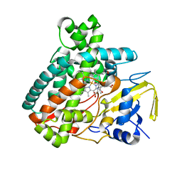
5L1T
DownloadVisualize
BU of 5l1t by Molmil
X-ray Structure of M77S mutant of Cytochrome P450 PntM with pentalenolactone F
Descriptor: PROTOPORPHYRIN IX CONTAINING FE, Pentalenolactone synthase, pentalenolactone F
Authors:Duan, L, Jogl, G, Cane, D.E.
Deposit date:2016-07-29
Release date:2016-09-14
Last modified:2023-10-04
Method:X-RAY DIFFRACTION (2.082 Å)
Cite:The Cytochrome P450-Catalyzed Oxidative Rearrangement in the Final Step of Pentalenolactone Biosynthesis: Substrate Structure Determines Mechanism.
J.Am.Chem.Soc., 138, 2016
6Q1Q
DownloadVisualize
BU of 6q1q by Molmil
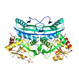
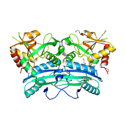
A hypothetical aminotransferase from Mycobacterium tuberculosis, apo form
Descriptor: 1,2-ETHANEDIOL, CITRIC ACID, Probable amino acid aminotransferase
Authors:Duan, L, Sacchettini, J.
Deposit date:2019-08-05
Release date:2020-09-23
Last modified:2023-10-11
Method:X-RAY DIFFRACTION (2.4 Å)
Cite:A hypothetical aminotransferase from Mycobacterium tuberculosis
To Be Published
6Q1S
DownloadVisualize
BU of 6q1s by Molmil
A hypothetical aminotransferase from Mycobacterium tuberculosis, alpha-ketoglutarate and PMP bound form
Descriptor: 2-OXOGLUTARIC ACID, 4'-DEOXY-4'-AMINOPYRIDOXAL-5'-PHOSPHATE, CITRIC ACID, ...
Authors:Duan, L, Sacchettini, J.
Deposit date:2019-08-05
Release date:2020-09-23
Last modified:2023-10-11
Method:X-RAY DIFFRACTION (2.3 Å)
Cite:A hypothetical aminotransferase from Mycobacterium tuberculosis
To Be Published
6Q1R
DownloadVisualize
BU of 6q1r by Molmil
A hypothetical aminotransferase from Mycobacterium tuberculosis, PLP-bound form
Descriptor: CITRIC ACID, GLYCEROL, PYRIDOXAL-5'-PHOSPHATE, ...
Authors:Duan, L, Sacchettini, J.
Deposit date:2019-08-05
Release date:2020-09-23
Last modified:2023-10-11
Method:X-RAY DIFFRACTION (2.702 Å)
Cite:A hypothetical aminotransferase from Mycobacterium tuberculosis
To Be Published
5L1P
DownloadVisualize
BU of 5l1p by Molmil
X-ray Structure of Cytochrome P450 PntM with Pentalenolactone
Descriptor: PROTOPORPHYRIN IX CONTAINING FE, Pentalenolactone synthase, pentalenolactone
Authors:Duan, L, Jogl, G, Cane, D.E.
Deposit date:2016-07-29
Release date:2016-09-14
Last modified:2023-10-04
Method:X-RAY DIFFRACTION (2.28 Å)
Cite:The Cytochrome P450-Catalyzed Oxidative Rearrangement in the Final Step of Pentalenolactone Biosynthesis: Substrate Structure Determines Mechanism.
J.Am.Chem.Soc., 138, 2016
5L1O
DownloadVisualize
BU of 5l1o by Molmil
X-ray Structure of Cytochrome P450 PntM with Pentalenolactone F
Descriptor: PROTOPORPHYRIN IX CONTAINING FE, Pentalenolactone synthase, pentalenolactone F
Authors:Duan, L, Jogl, G, Cane, D.E.
Deposit date:2016-07-29
Release date:2016-09-14
Last modified:2023-10-04
Method:X-RAY DIFFRACTION (2.03 Å)
Cite:The Cytochrome P450-Catalyzed Oxidative Rearrangement in the Final Step of Pentalenolactone Biosynthesis: Substrate Structure Determines Mechanism.
J.Am.Chem.Soc., 138, 2016
5L1Q
DownloadVisualize
BU of 5l1q by Molmil
X-ray Structure of Cytochrome P450 PntM with Dihydropentalenolactone F
Descriptor: Dihydropentalenolactone F, PROTOPORPHYRIN IX CONTAINING FE, Pentalenolactone synthase
Authors:Duan, L, Jogl, G, Cane, D.E.
Deposit date:2016-07-29
Release date:2016-09-14
Last modified:2023-10-04
Method:X-RAY DIFFRACTION (2.03 Å)
Cite:The Cytochrome P450-Catalyzed Oxidative Rearrangement in the Final Step of Pentalenolactone Biosynthesis: Substrate Structure Determines Mechanism.
J.Am.Chem.Soc., 138, 2016
5L1S
DownloadVisualize
BU of 5l1s by Molmil
X-ray Structure of F232L mutant of Cytochrome P450 PntM with pentalenolactone F
Descriptor: PROTOPORPHYRIN IX CONTAINING FE, Pentalenolactone synthase, pentalenolactone F
Authors:Duan, L, Jogl, G, Cane, D.E.
Deposit date:2016-07-29
Release date:2016-09-14
Last modified:2023-10-04
Method:X-RAY DIFFRACTION (2.08 Å)
Cite:The Cytochrome P450-Catalyzed Oxidative Rearrangement in the Final Step of Pentalenolactone Biosynthesis: Substrate Structure Determines Mechanism.
J.Am.Chem.Soc., 138, 2016
5L1W
DownloadVisualize
BU of 5l1w by Molmil
X-ray Structure of 2-Mercaptoethanol modified M81C mutant of Cytochrome P450 PntM with pentalenolactone F
Descriptor: PROTOPORPHYRIN IX CONTAINING FE, Pentalenolactone synthase, pentalenolactone F
Authors:Duan, L, Jogl, G, Cane, D.E.
Deposit date:2016-07-29
Release date:2016-09-14
Last modified:2023-10-04
Method:X-RAY DIFFRACTION (2.06 Å)
Cite:The Cytochrome P450-Catalyzed Oxidative Rearrangement in the Final Step of Pentalenolactone Biosynthesis: Substrate Structure Determines Mechanism.
J.Am.Chem.Soc., 138, 2016
5V93
DownloadVisualize
BU of 5v93 by Molmil
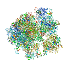
Cryo-EM structure of the 70S ribosome from Mycobacterium tuberculosis bound with Capreomycin
Descriptor: 16S rRNA, 23S rRNA, 30S ribosomal protein S10, ...
Authors:Yang, K, Chang, J.-Y, Cui, Z, Li, X, Meng, R, Duan, L, Thongchol, J, Jakana, J, Huwe, C, Sacchettini, J, Zhang, J.
Deposit date:2017-03-22
Release date:2017-09-20
Last modified:2020-08-12
Method:ELECTRON MICROSCOPY (4 Å)
Cite:Structural insights into species-specific features of the ribosome from the human pathogen Mycobacterium tuberculosis.
Nucleic Acids Res., 45, 2017
5FID
DownloadVisualize
BU of 5fid by Molmil
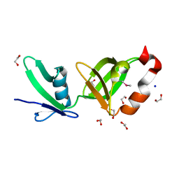
Crystal structure of the protein elicitor MoHrip2 from Magnaporthe oryzae
Descriptor: Elicitor protein Hrip2
Authors:Liu, M, Duan, L, Qiu, D, Liu, X.
Deposit date:2015-12-23
Release date:2016-12-07
Method:X-RAY DIFFRACTION (1.809 Å)
Cite:Crystal Structure Analysis and the Identification of Distinctive Functional Regions of the Protein Elicitor Mohrip2
Front Plant Sci, 7, 2016
8K20
DownloadVisualize
BU of 8k20 by Molmil
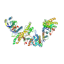
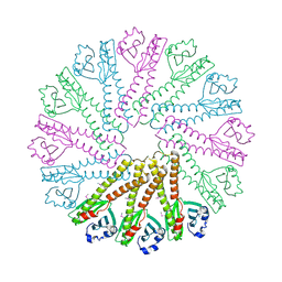
Cryo-EM structure of KEOPS complex from Arabidopsis thaliana
Descriptor: At4g34412, At5g53043, FE (III) ION, ...
Authors:Zheng, X.X, Zhu, L, Duan, L, Zhang, W.H.
Deposit date:2023-07-11
Release date:2024-04-03
Last modified:2024-04-10
Method:ELECTRON MICROSCOPY (3.7 Å)
Cite:Molecular basis of A. thaliana KEOPS complex in biosynthesizing tRNA t6A.
Nucleic Acids Res., 2024
3DCX
DownloadVisualize
BU of 3dcx by Molmil
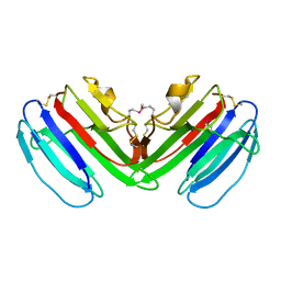
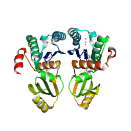
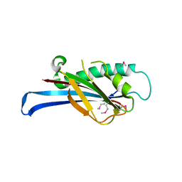
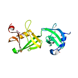
Crystal structure of a duf1696 family protein with a pleckstrin-homology domain (shew_0819) from shewanella loihica pv-4 at 2.00 A resolution
Descriptor: (4S)-2-METHYL-2,4-PENTANEDIOL, CHLORIDE ION, Protein of Unknown Function (DUF1696) with Pleckstrin-homology Domains
Authors:Joint Center for Structural Genomics (JCSG)
Deposit date:2008-06-04
Release date:2008-07-29
Last modified:2023-02-01
Method:X-RAY DIFFRACTION (2 Å)
Cite:Bacterial pleckstrin homology domains: a prokaryotic origin for the PH domain.
J.Mol.Biol., 396, 2010
2GVI
DownloadVisualize
BU of 2gvi by Molmil
Crystal structure of a putative formylmethanofuran dehydrogenase subunit e (ta1109) from thermoplasma acidophilum at 1.87 A resolution
Descriptor: 1,2-ETHANEDIOL, ACETIC ACID, UNKNOWN LIGAND, ...
Authors:Joint Center for Structural Genomics (JCSG)
Deposit date:2006-05-02
Release date:2006-06-13
Last modified:2023-01-25
Method:X-RAY DIFFRACTION (1.87 Å)
Cite:Structures of three members of Pfam PF02663 (FmdE) implicated in microbial methanogenesis reveal a conserved alpha+beta core domain and an auxiliary C-terminal treble-clef zinc finger.
Acta Crystallogr.,Sect.F, 66, 2010
2HAG
DownloadVisualize
BU of 2hag by Molmil
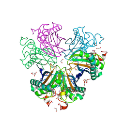
Crystal structure of a putative dyp-type peroxidase protein (so_0740) from shewanella oneidensis at 2.75 A resolution
Descriptor: Melanin biosynthesis protein TyrA, putative
Authors:Joint Center for Structural Genomics (JCSG)
Deposit date:2006-06-12
Release date:2006-08-08
Last modified:2023-01-25
Method:X-RAY DIFFRACTION (2.75 Å)
Cite:Crystal structures of two novel dye-decolorizing peroxidases reveal a beta-barrel fold with a conserved heme-binding motif.
Proteins, 69, 2007
2HUJ
DownloadVisualize
BU of 2huj by Molmil
Crystal structure of a protein of uknown function (NP_471338.1) from Listeria innocua at 1.74 A resolution
Descriptor: Lin2004 protein
Authors:Joint Center for Structural Genomics (JCSG)
Deposit date:2006-07-26
Release date:2006-08-08
Last modified:2023-01-25
Method:X-RAY DIFFRACTION (1.74 Å)
Cite:Crystal structures of MW1337R and lin2004: representatives of a novel protein family that adopt a four-helical bundle fold.
Proteins, 71, 2008
7SIU
DownloadVisualize
BU of 7siu by Molmil
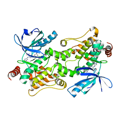
Crystal structure of HPK1 (MAP4K1) complex with inhibitor A-745
Descriptor: 6-[(5R)-5-benzamidocyclohex-1-en-1-yl]-3-[(1-methyl-1H-pyrazol-4-yl)amino]pyrazine-2-carboxamide, Mitogen-activated protein kinase kinase kinase kinase 1, SULFATE ION
Authors:Longenecker, K.L, Korepanova, A, Qiu, W.
Deposit date:2021-10-14
Release date:2022-03-02
Last modified:2023-10-18
Method:X-RAY DIFFRACTION (1.786 Å)
Cite:The HPK1 Inhibitor A-745 Verifies the Potential of Modulating T Cell Kinase Signaling for Immunotherapy.
Acs Chem.Biol., 17, 2022
3NL9
DownloadVisualize
BU of 3nl9 by Molmil
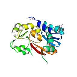
Crystal structure of a putative NTP pyrophosphohydrolase (Exig_1061) from EXIGUOBACTERIUM SP. 255-15 at 1.78 A resolution
Descriptor: 1,2-ETHANEDIOL, putative NTP pyrophosphohydrolase
Authors:Joint Center for Structural Genomics (JCSG)
Deposit date:2010-06-21
Release date:2010-07-21
Last modified:2023-02-01
Method:X-RAY DIFFRACTION (1.78 Å)
Cite:Structure of a putative NTP pyrophosphohydrolase: YP_001813558.1 from Exiguobacterium sibiricum 255-15.
Acta Crystallogr.,Sect.F, 66, 2010
1ZKG
DownloadVisualize
BU of 1zkg by Molmil
Crystal structure of Transcriptional regulator, TETR family (tm1030) from Thermotoga maritima at 2.30 A resolution
Descriptor: UNKNOWN LIGAND, transcriptional regulator, TetR family
Authors:Joint Center for Structural Genomics (JCSG)
Deposit date:2005-05-02
Release date:2005-07-26
Last modified:2023-01-25
Method:X-RAY DIFFRACTION (2.3 Å)
Cite:Crystal structure of TM1030 from Thermotoga maritima at 2.3 A resolution reveals molecular details of its transcription repressor function.
Proteins, 68, 2007
1VR0
DownloadVisualize
BU of 1vr0 by Molmil
Crystal structure of putative 2-phosphosulfolactate phosphatase (15026306) from Clostridium acetobutylicum at 2.6 A resolution
Descriptor: (2R)-3-SULFOLACTIC ACID, MAGNESIUM ION, Probable 2-phosphosulfolactate phosphatase
Authors:Joint Center for Structural Genomics (JCSG)
Deposit date:2005-01-26
Release date:2005-02-15
Last modified:2023-01-25
Method:X-RAY DIFFRACTION (2.49 Å)
Cite:Crystal structure of 2-phosphosulfolactate phosphatase (ComB) from Clostridium acetobutylicum at 2.6 A resolution reveals a new fold with a novel active site.
Proteins, 65, 2006
1VQ3
DownloadVisualize
BU of 1vq3 by Molmil
Crystal structure of Phosphoribosylformylglycinamidine synthase, purS subunit (EC 6.3.5.3) (TM1244) from Thermotoga maritima at 1.90 A resolution
Descriptor: Phosphoribosylformylglycinamidine synthase, purS subunit
Authors:Joint Center for Structural Genomics (JCSG)
Deposit date:2004-12-15
Release date:2004-12-28
Last modified:2023-09-20
Method:X-RAY DIFFRACTION (1.9 Å)
Cite:Crystal structure of phosphoribosylformyl-glycinamidine synthase II, PurS subunit (TM1244) from Thermotoga maritima at 1.90 A resolution.
Proteins, 65, 2006
1VR8
DownloadVisualize
BU of 1vr8 by Molmil
Crystal structure of GTP binding regulator (TM1622) from Thermotoga Maritima at 1.75 A resolution
Descriptor: AZIDE ION, GLYCEROL, GTP binding regulator
Authors:Joint Center for Structural Genomics (JCSG)
Deposit date:2005-02-16
Release date:2005-03-15
Last modified:2023-01-25
Method:X-RAY DIFFRACTION (1.75 Å)
Cite:Crystal structure of an ORFan protein (TM1622) from Thermotoga maritima at 1.75 A resolution reveals a fold similar to the Ran-binding protein Mog1p.
Proteins, 65, 2006


1234>

219869

PDB entries from 2024-05-15

PDB statisticsPDBj update infoContact PDBjnumon