Loading
PDBj
メニューPDBj@FacebookPDBj@TwitterPDBj@YouTubewwPDB FoundationwwPDB
RCSB PDBPDBeBMRBAdv. SearchSearch help
Search by PDB author
3M0R
DownloadVisualize
BU of 3m0r by Molmil
Crystal Structure of the R21D mutant of alpha-spectrin SH3 domain. Crystal obtained in ammonium sulphate at pH 6.
分子名称: SULFATE ION, Spectrin alpha chain, brain
著者Gavira, J.A, Camara-Artigas, A.
登録日2010-03-03
公開日2011-01-19
最終更新日2023-09-06
実験手法X-RAY DIFFRACTION (1.1 Å)
主引用文献Understanding the polymorphic behaviour of a mutant of the alpha-spectrin SH3 domain by means of two 1.1 A structures
Acta Crystallogr.,Sect.D, 2011
3HG6
DownloadVisualize
BU of 3hg6 by Molmil
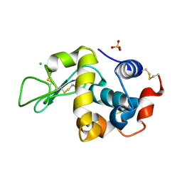
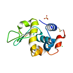
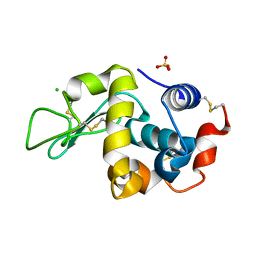
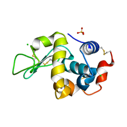
Crystal Structure of the Recombinant Onconase from Rana pipiens
分子名称: GLYCEROL, Onconase, SULFATE ION
著者Camara-Artigas, A, Gavira, J.A, Casares-Atienza, S, Weininger, U, Balbach, J, Garcia-Mira, M.M.
登録日2009-05-13
公開日2010-05-19
最終更新日2023-11-08
実験手法X-RAY DIFFRACTION (1.7 Å)
主引用文献Three-state thermal unfolding of onconase.
Biophys.Chem., 159, 2011
6F9Y
DownloadVisualize
BU of 6f9y by Molmil
Lysozyme crystallized in presence of 10 mM lithium sulphate at pH 4.5
分子名称: CHLORIDE ION, Lysozyme C, SULFATE ION
著者Camara-Artigas, A.
登録日2017-12-15
公開日2018-05-09
最終更新日2024-01-17
実験手法X-RAY DIFFRACTION (1.2 Å)
主引用文献Orthorhombic lysozyme crystallization at acidic pH values driven by phosphate binding.
Acta Crystallogr D Struct Biol, 74, 2018
6F1L
DownloadVisualize
BU of 6f1l by Molmil
Lysozyme crystallized in presence of 100 mM sodium phosphate at pH 4.5
分子名称: CHLORIDE ION, Lysozyme C, PHOSPHATE ION
著者Camara-Artigas, A.
登録日2017-11-22
公開日2018-05-09
最終更新日2024-01-17
実験手法X-RAY DIFFRACTION (1.3 Å)
主引用文献Orthorhombic lysozyme crystallization at acidic pH values driven by phosphate binding.
Acta Crystallogr D Struct Biol, 74, 2018
6F1P
DownloadVisualize
BU of 6f1p by Molmil
Tetragonal Lysozyme crystallized at 298 K and pH 4.5 with phosphate bound
分子名称: CHLORIDE ION, Lysozyme C, PHOSPHATE ION, ...
著者Camara-Artigas, A.
登録日2017-11-22
公開日2018-05-09
最終更新日2024-01-17
実験手法X-RAY DIFFRACTION (1.12 Å)
主引用文献Orthorhombic lysozyme crystallization at acidic pH values driven by phosphate binding.
Acta Crystallogr D Struct Biol, 74, 2018
6F9Z
DownloadVisualize
BU of 6f9z by Molmil
Lysozyme crystallized in presence of 5 mM ammonium sulphate at pH 4.5
分子名称: CHLORIDE ION, Lysozyme C, SULFATE ION
著者Camara-Artigas, A.
登録日2017-12-15
公開日2018-05-09
最終更新日2024-01-17
実験手法X-RAY DIFFRACTION (1.2 Å)
主引用文献Orthorhombic lysozyme crystallization at acidic pH values driven by phosphate binding.
Acta Crystallogr D Struct Biol, 74, 2018
6F1O
DownloadVisualize
BU of 6f1o by Molmil
Orthorhombic Lysozyme crystallized at 298 K and pH 4.5
分子名称: CHLORIDE ION, Lysozyme C, PHOSPHATE ION
著者Camara-Artigas, A.
登録日2017-11-22
公開日2018-05-09
最終更新日2024-01-17
実験手法X-RAY DIFFRACTION (0.96 Å)
主引用文献Orthorhombic lysozyme crystallization at acidic pH values driven by phosphate binding.
Acta Crystallogr D Struct Biol, 74, 2018
6F1R
DownloadVisualize
BU of 6f1r by Molmil
Tetragonal Lysozyme crystallized at 298 K and pH 4.5 with phosphate bound: control experiment
分子名称: CHLORIDE ION, Lysozyme C, SODIUM ION
著者Camara-Artigas, A.
登録日2017-11-22
公開日2018-05-09
最終更新日2024-01-17
実験手法X-RAY DIFFRACTION (1.1 Å)
主引用文献Orthorhombic lysozyme crystallization at acidic pH values driven by phosphate binding.
Acta Crystallogr D Struct Biol, 74, 2018
6F1M
DownloadVisualize
BU of 6f1m by Molmil
Lysozyme crystallized in presence of 100 mM sodium phosphate at pH 4.5: low-humidity form
分子名称: Lysozyme C
著者Camara-Artigas, A.
登録日2017-11-22
公開日2018-05-09
最終更新日2024-01-17
実験手法X-RAY DIFFRACTION (1.48 Å)
主引用文献Orthorhombic lysozyme crystallization at acidic pH values driven by phosphate binding.
Acta Crystallogr D Struct Biol, 74, 2018
6FA0
DownloadVisualize
BU of 6fa0 by Molmil
Lysozyme crystallized in presence of 100 mM ammonium sulphate at pH 4.5
分子名称: CHLORIDE ION, Lysozyme C, SULFATE ION
著者Camara-Artigas, A.
登録日2017-12-15
公開日2018-05-09
最終更新日2024-01-17
実験手法X-RAY DIFFRACTION (1.3 Å)
主引用文献Orthorhombic lysozyme crystallization at acidic pH values driven by phosphate binding.
Acta Crystallogr D Struct Biol, 74, 2018
6F9X
DownloadVisualize
BU of 6f9x by Molmil
Lysozyme crystallized in presence of 100 mM lithium sulphate at pH 4.5
分子名称: CHLORIDE ION, Lysozyme C, SULFATE ION
著者Camara-Artigas, A.
登録日2017-12-15
公開日2018-05-09
最終更新日2024-01-17
実験手法X-RAY DIFFRACTION (1.25 Å)
主引用文献Orthorhombic lysozyme crystallization at acidic pH values driven by phosphate binding.
Acta Crystallogr D Struct Biol, 74, 2018
4OMO
DownloadVisualize
BU of 4omo by Molmil
Crystal structure of the c-Src tyrosine kinase SH3 domain mutant Q128E
分子名称: 4-(2-HYDROXYETHYL)-1-PIPERAZINE ETHANESULFONIC ACID, NICKEL (II) ION, Proto-oncogene tyrosine-protein kinase Src
著者Camara-Artigas, A, Bacarizo, J.
登録日2014-01-27
公開日2014-12-10
最終更新日2023-09-20
実験手法X-RAY DIFFRACTION (1.04 Å)
主引用文献Electrostatic Effects in the Folding of the SH3 Domain of the c-Src Tyrosine Kinase: pH-Dependence in 3D-Domain Swapping and Amyloid Formation.
Plos One, 9, 2014
4OML
DownloadVisualize
BU of 4oml by Molmil
Crystal structure of the intertwined dimer of the c-Src tyrosine kinase SH3 domain mutant Q128R
分子名称: DI(HYDROXYETHYL)ETHER, Proto-oncogene tyrosine-protein kinase Src, TRIETHYLENE GLYCOL
著者Camara-Artigas, A, Bacarizo, J.
登録日2014-01-27
公開日2014-12-10
最終更新日2023-09-20
実験手法X-RAY DIFFRACTION (1.6 Å)
主引用文献Electrostatic Effects in the Folding of the SH3 Domain of the c-Src Tyrosine Kinase: pH-Dependence in 3D-Domain Swapping and Amyloid Formation.
Plos One, 9, 2014
4OMN
DownloadVisualize
BU of 4omn by Molmil
Crystal structure of the intertwined dimer of the c-Src tyrosine kinase SH3 domain mutant Q128E
分子名称: DI(HYDROXYETHYL)ETHER, GLYCEROL, Proto-oncogene tyrosine-protein kinase Src, ...
著者Camara-Artigas, A, Bacarizo, J.
登録日2014-01-27
公開日2014-12-10
最終更新日2023-09-20
実験手法X-RAY DIFFRACTION (1.5 Å)
主引用文献Electrostatic Effects in the Folding of the SH3 Domain of the c-Src Tyrosine Kinase: pH-Dependence in 3D-Domain Swapping and Amyloid Formation.
Plos One, 9, 2014
4OMP
DownloadVisualize
BU of 4omp by Molmil
Crystal structure of the intertwined dimer of the c-Src tyrosine kinase SH3 domain mutant Q128K
分子名称: DI(HYDROXYETHYL)ETHER, Proto-oncogene tyrosine-protein kinase Src, TRIETHYLENE GLYCOL
著者Camara-Artigas, A, Bacarizo, J.
登録日2014-01-27
公開日2014-12-10
最終更新日2023-09-20
実験手法X-RAY DIFFRACTION (2 Å)
主引用文献Electrostatic Effects in the Folding of the SH3 Domain of the c-Src Tyrosine Kinase: pH-Dependence in 3D-Domain Swapping and Amyloid Formation.
Plos One, 9, 2014
4QT7
DownloadVisualize
BU of 4qt7 by Molmil
Crystal structure of the c-Src SH3 domain in complex with a peptide from the Hepatitis C virus NS5A-protein
分子名称: NS5A, Proto-oncogene tyrosine-protein kinase Src
著者Camara-Artigas, A, Bacarizo, J.
登録日2014-07-07
公開日2014-12-24
最終更新日2023-11-08
実験手法X-RAY DIFFRACTION (1.55 Å)
主引用文献Structure of the c-Src-SH3 domain in complex with a proline-rich motif of NS5A protein from the hepatitis C virus.
J.Struct.Biol., 189, 2015
4R61
DownloadVisualize
BU of 4r61 by Molmil
Crystal structure of a rationally designed single-chain protein mimicking a trimeric gp41 N-terminal heptad-repeat region
分子名称: gp41-based construct covNHR3-ABC
著者Camara-Artigas, A, Conejero-Lara, F, Crespillo, S, Casares, S.
登録日2014-08-22
公開日2014-12-10
最終更新日2023-09-20
実験手法X-RAY DIFFRACTION (3.1 Å)
主引用文献Single-chain protein mimetics of the N-terminal heptad-repeat region of gp41 with potential as anti-HIV-1 drugs.
Proc.Natl.Acad.Sci.USA, 111, 2014
4REX
DownloadVisualize
BU of 4rex by Molmil
Crystal structure of the first WW domain of human YAP2 isoform
分子名称: SULFATE ION, Yorkie homolog
著者Camara-Artigas, A.
登録日2014-09-24
公開日2015-08-19
最終更新日2023-09-20
実験手法X-RAY DIFFRACTION (1.6 Å)
主引用文献Crystal structure of the first WW domain of human YAP2 isoform.
J.Struct.Biol., 191, 2015
4HVV
DownloadVisualize
BU of 4hvv by Molmil
Crystal structure of the T98E c-Src-SH3 domain mutant in complex with the high affinity peptide APP12
分子名称: Proto-oncogene tyrosine-protein kinase Src, SULFATE ION, SYNTHETIC PEPTIDE Acetyl-APPLPPRNRP
著者Camara-Artigas, A.
登録日2012-11-07
公開日2013-05-01
最終更新日2023-09-20
実験手法X-RAY DIFFRACTION (1.1 Å)
主引用文献Atomic resolution structures of the c-Src SH3 domain in complex with two high-affinity peptides from classes I and II.
Acta Crystallogr.,Sect.D, 69, 2013
4HVU
DownloadVisualize
BU of 4hvu by Molmil
Crystal structure of the T98D c-Src-SH3 domain mutant in complex with the high affinity peptide APP12
分子名称: ACETYL GROUP, Proto-oncogene tyrosine-protein kinase Src, SULFATE ION, ...
著者Camara-Artigas, A.
登録日2012-11-07
公開日2013-05-01
最終更新日2023-09-20
実験手法X-RAY DIFFRACTION (0.98 Å)
主引用文献Atomic resolution structures of the c-Src SH3 domain in complex with two high-affinity peptides from classes I and II.
Acta Crystallogr.,Sect.D, 69, 2013
4HVW
DownloadVisualize
BU of 4hvw by Molmil
Crystal structure of the T98E c-Src-SH3 domain mutant in complex with the high affinity peptide VSL12
分子名称: ACETYL GROUP, Proto-oncogene tyrosine-protein kinase Src, SULFATE ION, ...
著者Camara-Artigas, A.
登録日2012-11-07
公開日2013-05-01
最終更新日2023-09-20
実験手法X-RAY DIFFRACTION (0.98 Å)
主引用文献Atomic resolution structures of the c-Src SH3 domain in complex with two high-affinity peptides from classes I and II.
Acta Crystallogr.,Sect.D, 69, 2013
4JZ4
DownloadVisualize
BU of 4jz4 by Molmil
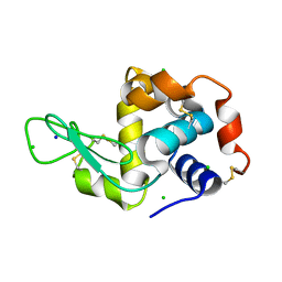
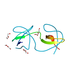
Crystal structure of chicken c-Src-SH3 domain: monomeric form
分子名称: 4-(2-HYDROXYETHYL)-1-PIPERAZINE ETHANESULFONIC ACID, NICKEL (II) ION, Proto-oncogene tyrosine-protein kinase Src
著者Camara-Artigas, A.
登録日2013-04-02
公開日2014-04-23
最終更新日2023-09-20
実験手法X-RAY DIFFRACTION (1.56 Å)
主引用文献Electrostatic Effects in the Folding of the SH3 Domain of the c-Src Tyrosine Kinase: pH-Dependence in 3D-Domain Swapping and Amyloid Formation.
Plos One, 9, 2014
4JZ3
DownloadVisualize
BU of 4jz3 by Molmil
Crystal structure of the chicken c-Src-SH3 domain intertwined dimer
分子名称: DI(HYDROXYETHYL)ETHER, Proto-oncogene tyrosine-protein kinase Src, TRIETHYLENE GLYCOL
著者Camara-Artigas, A.
登録日2013-04-02
公開日2014-04-23
最終更新日2023-09-20
実験手法X-RAY DIFFRACTION (1.852 Å)
主引用文献Electrostatic Effects in the Folding of the SH3 Domain of the c-Src Tyrosine Kinase: pH-Dependence in 3D-Domain Swapping and Amyloid Formation.
Plos One, 9, 2014
2OLP
DownloadVisualize
BU of 2olp by Molmil
Structure and ligand selection of hemoglobin II from Lucina pectinata
分子名称: Hemoglobin II, OXYGEN MOLECULE, PROTOPORPHYRIN IX CONTAINING FE, ...
著者Gavira, J.A, Camara-Artigas, A, de Jesus, W, Lopez-Garriga, J, Garcia-Ruiz, J.M.
登録日2007-01-19
公開日2007-12-18
最終更新日2023-08-30
実験手法X-RAY DIFFRACTION (1.932 Å)
主引用文献Structure and Ligand Selection of Hemoglobin II from Lucina pectinata
J.Biol.Chem., 283, 2008
1Z9J
DownloadVisualize
BU of 1z9j by Molmil
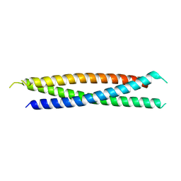
Photosynthetic Reaction Center from Rhodobacter sphaeroides
分子名称: BACTERIOCHLOROPHYLL A, BACTERIOPHEOPHYTIN A, FE (III) ION, ...
著者Thielges, M, Uyeda, G, Camara-Artigas, A, Kalman, L, Williams, J.C, Allen, J.P.
登録日2005-04-02
公開日2005-06-07
最終更新日2023-08-23
実験手法X-RAY DIFFRACTION (4.5 Å)
主引用文献Design of a Redox-Linked Active Metal Site: Manganese Bound to Bacterial Reaction Centers at a Site Resembling That of Photosystem II
Biochemistry, 44, 2005

219869

件を2024-05-15に公開中

PDB statisticsPDBj update infoContact PDBjnumon