Loading
PDBj
MenuPDBj@FacebookPDBj@TwitterPDBj@YouTubewwPDB FoundationwwPDB
RCSB PDBPDBeBMRBAdv. SearchSearch help
Search by PDB author
1Z9K
DownloadVisualize
BU of 1z9k by Molmil
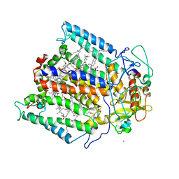
Photosynthetic Reaction Center from Rhodobacter sphaeroides
Descriptor: BACTERIOCHLOROPHYLL A, BACTERIOPHEOPHYTIN A, FE (III) ION, ...
Authors:Camara-Artigas, A, Allen, J.P.
Deposit date:2005-04-02
Release date:2005-06-07
Last modified:2023-08-23
Method:X-RAY DIFFRACTION (4.6 Å)
Cite:Design of a Redox-Linked Active Metal Site: Manganese Bound to Bacterial Reaction Centers at a Site Resembling That of Photosystem II
Biochemistry, 44, 2005
7ZR2
DownloadVisualize
BU of 7zr2 by Molmil
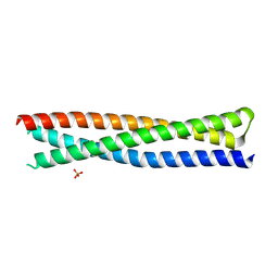
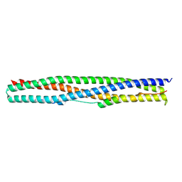
Crystal structure of a chimeric protein mimic of SARS-CoV-2 Spike HR1 in complex with HR2
Descriptor: Spike protein S2', Spike protein S2',Chimeric protein mimic of SARS-CoV-2 Spike HR1
Authors:Camara-Artigas, A, Gavira, J.A, Cano-Munoz, M, Polo-Megias, D, Conejero-Lara, F.
Deposit date:2022-05-03
Release date:2022-11-09
Last modified:2024-01-31
Method:X-RAY DIFFRACTION (1.45 Å)
Cite:Novel chimeric proteins mimicking SARS-CoV-2 spike epitopes with broad inhibitory activity.
Int.J.Biol.Macromol., 222, 2022
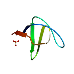
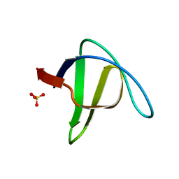
8AH7
DownloadVisualize
BU of 8ah7 by Molmil
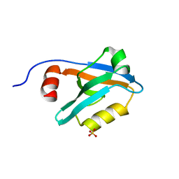
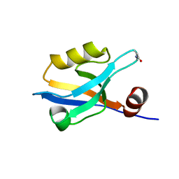
Crystal Structure of the third PDZ domain of PSD-95 protein in the space group P212121 at pH 4.0
Descriptor: SULFATE ION, cDNA FLJ50577, highly similar to Discs large homolog 4
Authors:Camara-Artigas, A, Salinas-Garcia, M.C.
Deposit date:2022-07-20
Release date:2023-03-08
Last modified:2024-02-07
Method:X-RAY DIFFRACTION (1.25 Å)
Cite:pH-Driven Polymorphic Behaviour of the Third PDZ Domain of PSD95: The Role of Electrostatic Interactions
Crystals, 2023
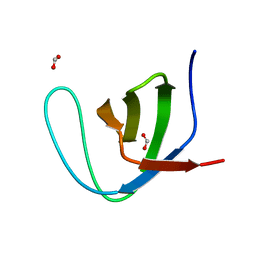
8AH4
DownloadVisualize
BU of 8ah4 by Molmil
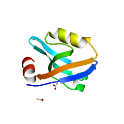
Crystal Structure of the third PDZ domain of PSD-95 protein in the space group P3112 at pH 4.0
Descriptor: ACETATE ION, cDNA FLJ50577, highly similar to Discs large homolog 4
Authors:Camara-Artigas, A, Salinas Garcia, M.C.
Deposit date:2022-07-20
Release date:2023-03-08
Last modified:2024-02-07
Method:X-RAY DIFFRACTION (1.48 Å)
Cite:pH-Driven Polymorphic Behaviour of the Third PDZ Domain of PSD95: The Role of Electrostatic Interactions
Crystals, 2023
8AH6
DownloadVisualize
BU of 8ah6 by Molmil
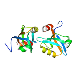
Crystal Structure of the third PDZ domain of PSD-95 protein in the space group P21 at pH 4.0
Descriptor: ACETATE ION, cDNA FLJ50577, highly similar to Discs large homolog 4
Authors:Camara-Artigas, A, Salinas Garcia, M.C.
Deposit date:2022-07-20
Release date:2023-03-08
Last modified:2024-02-07
Method:X-RAY DIFFRACTION (1.63 Å)
Cite:pH-Driven Polymorphic Behaviour of the Third PDZ Domain of PSD95: The Role of Electrostatic Interactions
Crystals, 2023
8AH5
DownloadVisualize
BU of 8ah5 by Molmil
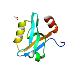
Crystal Structure of the third PDZ domain of PSD-95 protein in the space group P212121 at pH 4.6
Descriptor: ACETATE ION, cDNA FLJ50577, highly similar to Discs large homolog 4
Authors:Camara-Artigas, A, Salinas Garcia, M.C.
Deposit date:2022-07-20
Release date:2023-03-08
Last modified:2024-02-07
Method:X-RAY DIFFRACTION (1.25 Å)
Cite:pH-Driven Polymorphic Behaviour of the Third PDZ Domain of PSD95: The Role of Electrostatic Interactions
Crystals, 2023
8AH8
DownloadVisualize
BU of 8ah8 by Molmil
Crystal Structure of the third PDZ domain of PSD-95 protein in the space group P3121 at pH 3.7
Descriptor: cDNA FLJ50577, highly similar to Discs large homolog 4
Authors:Camara-Artigas, A, Salinas Garcia, M.C.
Deposit date:2022-07-20
Release date:2023-03-08
Last modified:2024-02-07
Method:X-RAY DIFFRACTION (1.5 Å)
Cite:pH-Driven Polymorphic Behaviour of the Third PDZ Domain of PSD95: The Role of Electrostatic Interactions
Crystals, 2023
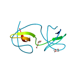
3M0U
DownloadVisualize
BU of 3m0u by Molmil
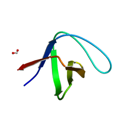
Crystal Structure of the R21D mutant of alpha-spectrin SH3 domain. Hexagonal crystal obtained in sodium formate at pH 6.5.
Descriptor: FORMIC ACID, Spectrin alpha chain, brain
Authors:Gavira, J.A, Camara-Artigas, A.
Deposit date:2010-03-03
Release date:2011-01-19
Last modified:2023-09-06
Method:X-RAY DIFFRACTION (1.1 Å)
Cite:Understanding the polymorphic behaviour of a mutant of the alpha-spectrin SH3 domain by means of two 1.1 A structures
Acta Crystallogr.,Sect.D, 2011
2F0R
DownloadVisualize
BU of 2f0r by Molmil
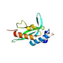
Crystallographic structure of human Tsg101 UEV domain
Descriptor: SULFATE ION, Tumor susceptibility gene 101 protein
Authors:Camara-Artigas, A, Luque, I, Palencia, A, Martinez, J.C, Mateo, P.L.
Deposit date:2005-11-13
Release date:2006-03-28
Last modified:2023-08-23
Method:X-RAY DIFFRACTION (2.26 Å)
Cite:Structure of human TSG101 UEV domain.
Acta Crystallogr.,Sect.D, 62, 2006
3UA6
DownloadVisualize
BU of 3ua6 by Molmil
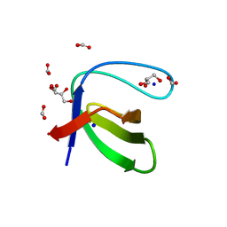
Crystal Structure of the Human Fyn SH3 domain
Descriptor: FORMIC ACID, GLYCEROL, SODIUM ION, ...
Authors:Camara-Artigas, A, Martin-Garcia, J.M.
Deposit date:2011-10-21
Release date:2012-07-25
Last modified:2023-09-13
Method:X-RAY DIFFRACTION (1.85 Å)
Cite:The promiscuous binding of the Fyn SH3 domain to a peptide from the NS5A protein.
Acta Crystallogr.,Sect.D, 68, 2012
3HG6
DownloadVisualize
BU of 3hg6 by Molmil
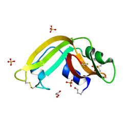
Crystal Structure of the Recombinant Onconase from Rana pipiens
Descriptor: GLYCEROL, Onconase, SULFATE ION
Authors:Camara-Artigas, A, Gavira, J.A, Casares-Atienza, S, Weininger, U, Balbach, J, Garcia-Mira, M.M.
Deposit date:2009-05-13
Release date:2010-05-19
Last modified:2023-11-08
Method:X-RAY DIFFRACTION (1.7 Å)
Cite:Three-state thermal unfolding of onconase.
Biophys.Chem., 159, 2011
6R2G
DownloadVisualize
BU of 6r2g by Molmil
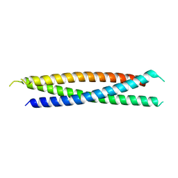
Crystal structure of a single-chain protein mimetic of the gp41 NHR trimer in complex with the synthetic CHR peptide C34
Descriptor: Envelope glycoprotein gp160, PHOSPHATE ION, Single-chain protein mimetics of the N-terminal heptad-repeat region of gp41
Authors:Camara-Artigas, A, Conejero-Lara, F, Jurado, S, Cano-Munoz, M, Morel, B.
Deposit date:2019-03-17
Release date:2019-07-10
Last modified:2024-01-24
Method:X-RAY DIFFRACTION (1.9 Å)
Cite:Structural and Thermodynamic Analysis of HIV-1 Fusion Inhibition Using Small gp41 Mimetic Proteins.
J.Mol.Biol., 431, 2019
4QT7
DownloadVisualize
BU of 4qt7 by Molmil
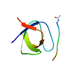
Crystal structure of the c-Src SH3 domain in complex with a peptide from the Hepatitis C virus NS5A-protein
Descriptor: NS5A, Proto-oncogene tyrosine-protein kinase Src
Authors:Camara-Artigas, A, Bacarizo, J.
Deposit date:2014-07-07
Release date:2014-12-24
Last modified:2023-11-08
Method:X-RAY DIFFRACTION (1.55 Å)
Cite:Structure of the c-Src-SH3 domain in complex with a proline-rich motif of NS5A protein from the hepatitis C virus.
J.Struct.Biol., 189, 2015
4R61
DownloadVisualize
BU of 4r61 by Molmil
Crystal structure of a rationally designed single-chain protein mimicking a trimeric gp41 N-terminal heptad-repeat region
Descriptor: gp41-based construct covNHR3-ABC
Authors:Camara-Artigas, A, Conejero-Lara, F, Crespillo, S, Casares, S.
Deposit date:2014-08-22
Release date:2014-12-10
Last modified:2023-09-20
Method:X-RAY DIFFRACTION (3.1 Å)
Cite:Single-chain protein mimetics of the N-terminal heptad-repeat region of gp41 with potential as anti-HIV-1 drugs.
Proc.Natl.Acad.Sci.USA, 111, 2014
4REX
DownloadVisualize
BU of 4rex by Molmil
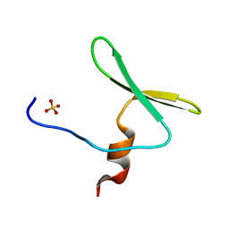
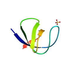
Crystal structure of the first WW domain of human YAP2 isoform
Descriptor: SULFATE ION, Yorkie homolog
Authors:Camara-Artigas, A.
Deposit date:2014-09-24
Release date:2015-08-19
Last modified:2023-09-20
Method:X-RAY DIFFRACTION (1.6 Å)
Cite:Crystal structure of the first WW domain of human YAP2 isoform.
J.Struct.Biol., 191, 2015
4OMO
DownloadVisualize
BU of 4omo by Molmil
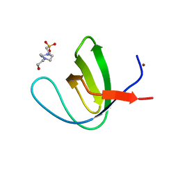
Crystal structure of the c-Src tyrosine kinase SH3 domain mutant Q128E
Descriptor: 4-(2-HYDROXYETHYL)-1-PIPERAZINE ETHANESULFONIC ACID, NICKEL (II) ION, Proto-oncogene tyrosine-protein kinase Src
Authors:Camara-Artigas, A, Bacarizo, J.
Deposit date:2014-01-27
Release date:2014-12-10
Last modified:2023-09-20
Method:X-RAY DIFFRACTION (1.04 Å)
Cite:Electrostatic Effects in the Folding of the SH3 Domain of the c-Src Tyrosine Kinase: pH-Dependence in 3D-Domain Swapping and Amyloid Formation.
Plos One, 9, 2014
4OML
DownloadVisualize
BU of 4oml by Molmil
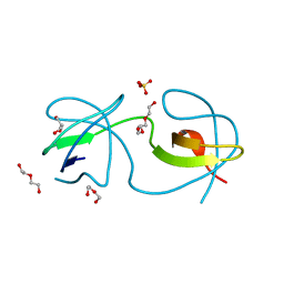
Crystal structure of the intertwined dimer of the c-Src tyrosine kinase SH3 domain mutant Q128R
Descriptor: DI(HYDROXYETHYL)ETHER, Proto-oncogene tyrosine-protein kinase Src, TRIETHYLENE GLYCOL
Authors:Camara-Artigas, A, Bacarizo, J.
Deposit date:2014-01-27
Release date:2014-12-10
Last modified:2023-09-20
Method:X-RAY DIFFRACTION (1.6 Å)
Cite:Electrostatic Effects in the Folding of the SH3 Domain of the c-Src Tyrosine Kinase: pH-Dependence in 3D-Domain Swapping and Amyloid Formation.
Plos One, 9, 2014
4OMN
DownloadVisualize
BU of 4omn by Molmil
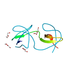
Crystal structure of the intertwined dimer of the c-Src tyrosine kinase SH3 domain mutant Q128E
Descriptor: DI(HYDROXYETHYL)ETHER, GLYCEROL, Proto-oncogene tyrosine-protein kinase Src, ...
Authors:Camara-Artigas, A, Bacarizo, J.
Deposit date:2014-01-27
Release date:2014-12-10
Last modified:2023-09-20
Method:X-RAY DIFFRACTION (1.5 Å)
Cite:Electrostatic Effects in the Folding of the SH3 Domain of the c-Src Tyrosine Kinase: pH-Dependence in 3D-Domain Swapping and Amyloid Formation.
Plos One, 9, 2014
4OMP
DownloadVisualize
BU of 4omp by Molmil
Crystal structure of the intertwined dimer of the c-Src tyrosine kinase SH3 domain mutant Q128K
Descriptor: DI(HYDROXYETHYL)ETHER, Proto-oncogene tyrosine-protein kinase Src, TRIETHYLENE GLYCOL
Authors:Camara-Artigas, A, Bacarizo, J.
Deposit date:2014-01-27
Release date:2014-12-10
Last modified:2023-09-20
Method:X-RAY DIFFRACTION (2 Å)
Cite:Electrostatic Effects in the Folding of the SH3 Domain of the c-Src Tyrosine Kinase: pH-Dependence in 3D-Domain Swapping and Amyloid Formation.
Plos One, 9, 2014
2F2V
DownloadVisualize
BU of 2f2v by Molmil
alpha-spectrin SH3 domain A56G mutant
Descriptor: FORMIC ACID, Spectrin alpha chain, brain
Authors:Camara-Artigas, A, Conejero-Lara, F, Casares, S, Lopez-Mayorga, O, Vega, C.
Deposit date:2005-11-18
Release date:2006-10-31
Last modified:2023-08-23
Method:X-RAY DIFFRACTION (1.85 Å)
Cite:Cooperative propagation of local stability changes from low-stability and high-stability regions in a SH3 domain
Proteins, 67, 2007
2F2W
DownloadVisualize
BU of 2f2w by Molmil
alpha-spectrin SH3 domain R21A mutant
Descriptor: SULFATE ION, Spectrin alpha chain, brain
Authors:Camara-Artigas, A, Conejero-Lara, F, Casares, S, Lopez-Mayorga, O, Vega, C.
Deposit date:2005-11-18
Release date:2006-10-31
Last modified:2023-08-23
Method:X-RAY DIFFRACTION (1.7 Å)
Cite:Cooperative propagation of local stability changes from low-stability and high-stability regions in a SH3 domain
Proteins, 67, 2007
2F2X
DownloadVisualize
BU of 2f2x by Molmil
alpha-spectrin SH3 domain R21G mutant
Descriptor: SULFATE ION, Spectrin alpha chain, brain
Authors:Camara-Artigas, A, Conejero-Lara, F, Casares, S, Lopez-Mayorga, O, Vega, C.
Deposit date:2005-11-18
Release date:2006-10-31
Last modified:2023-08-23
Method:X-RAY DIFFRACTION (1.6 Å)
Cite:Cooperative propagation of local stability changes from low-stability and high-stability regions in a SH3 domain
Proteins, 67, 2007
3M0R
DownloadVisualize
BU of 3m0r by Molmil
Crystal Structure of the R21D mutant of alpha-spectrin SH3 domain. Crystal obtained in ammonium sulphate at pH 6.
Descriptor: SULFATE ION, Spectrin alpha chain, brain
Authors:Gavira, J.A, Camara-Artigas, A.
Deposit date:2010-03-03
Release date:2011-01-19
Last modified:2023-09-06
Method:X-RAY DIFFRACTION (1.1 Å)
Cite:Understanding the polymorphic behaviour of a mutant of the alpha-spectrin SH3 domain by means of two 1.1 A structures
Acta Crystallogr.,Sect.D, 2011
2HDA
DownloadVisualize
BU of 2hda by Molmil
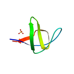
Yes SH3 domain
Descriptor: Proto-oncogene tyrosine-protein kinase Yes, SULFATE ION
Authors:Camara-Artigas, A, Luque, I, Ruiz-Sanz, J, Mateo, P.L, Martin-Garcia, J.M.
Deposit date:2006-06-20
Release date:2007-04-17
Last modified:2023-08-30
Method:X-RAY DIFFRACTION (1.9 Å)
Cite:Crystallographic structure of the SH3 domain of the human c-Yes tyrosine kinase: Loop flexibility and amyloid aggregation.
Febs Lett., 581, 2007
6S7N
DownloadVisualize
BU of 6s7n by Molmil
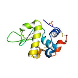
Crystal structure of orthorhombic lysozyme grown at pH 5.5 with a 26% of solvent content
Descriptor: Lysozyme C, SULFATE ION
Authors:Camara-Artigas, A, Plaza-Garrido, M, Salinas-Garcia, M.C.
Deposit date:2019-07-05
Release date:2019-11-20
Last modified:2024-01-24
Method:X-RAY DIFFRACTION (1.2 Å)
Cite:Major conformational changes in the structure of lysozyme obtained from a crystal with a very low solvent content.
Acta Crystallogr.,Sect.F, 75, 2019

218853

PDB entries from 2024-04-24

PDB statisticsPDBj update infoContact PDBjnumon