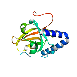
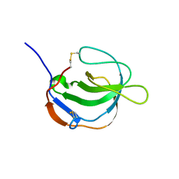
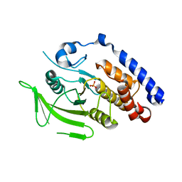
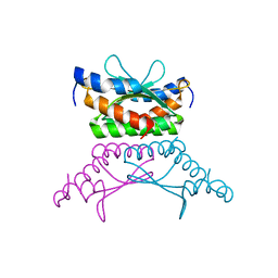
4CMW
 
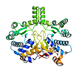 | | Crystal structure of Rv3378c | | 分子名称: | 1,2-ETHANEDIOL, CITRIC ACID, DI(HYDROXYETHYL)ETHER, ... | | 著者 | Layre, E, Lee, H.J, Young, D.C, Martinot, A.J, Buter, J, Minnaard, A.J, Annand, J.W, Fortune, S.M, Snider, B.B, Matsunaga, I, Rubin, E.J, Alber, T, Moody, D.B. | | 登録日 | 2014-01-18 | | 公開日 | 2014-02-19 | | 最終更新日 | 2023-12-20 | | 実験手法 | X-RAY DIFFRACTION (2.209 Å) | | 主引用文献 | Molecular Profiling of Mycobacterium Tuberculosis Identifies Tuberculosinyl Nucleoside Products of the Virulence-Associated Enzyme Rv3378C.
Proc.Natl.Acad.Sci.USA, 111, 2014
|
|
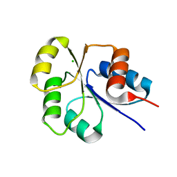
4CMX
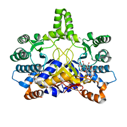 | | Crystal structure of Rv3378c | | 分子名称: | 1,2-ETHANEDIOL, BENZENE HEXACARBOXYLIC ACID, DI(HYDROXYETHYL)ETHER, ... | | 著者 | Layre, E, Lee, H.J, Young, D.C, Martinot, A.J, Buter, J, Minnaard, A.J, Annand, J.W, Fortune, S.M, Snider, B.B, Matsunaga, I, Rubin, E.J, Alber, T, Moody, D.B. | | 登録日 | 2014-01-18 | | 公開日 | 2014-02-19 | | 最終更新日 | 2023-12-20 | | 実験手法 | X-RAY DIFFRACTION (2.36 Å) | | 主引用文献 | Molecular Profiling of Mycobacterium Tuberculosis Identifies Tuberculosinyl Nucleoside Products of the Virulence-Associated Enzyme Rv3378C.
Proc.Natl.Acad.Sci.USA, 111, 2014
|
|
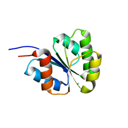
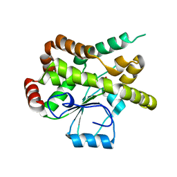
4ESQ
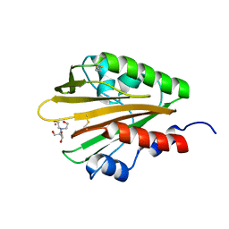 | | Crystal structure of the extracellular domain of PknH from Mycobacterium tuberculosis | | 分子名称: | 2-[BIS-(2-HYDROXY-ETHYL)-AMINO]-2-HYDROXYMETHYL-PROPANE-1,3-DIOL, Serine/threonine protein kinase, TERBIUM(III) ION | | 著者 | Cavazos, A, Prigozhin, D.M, Alber, T. | | 登録日 | 2012-04-23 | | 公開日 | 2012-07-18 | | 最終更新日 | 2017-11-15 | | 実験手法 | X-RAY DIFFRACTION (1.7 Å) | | 主引用文献 | Structure of the sensor domain of Mycobacterium tuberculosis PknH receptor kinase reveals a conserved binding cleft.
J.Mol.Biol., 422, 2012
|
|
4G1H
 | |
4G1J
 | |
2I6F
 | | Receiver domain from Myxococcus xanthus social motility protein FrzS | | 分子名称: | CHLORIDE ION, Response regulator FrzS | | 著者 | Echols, N, Fraser, J, Merlie, J, Zusman, D, Alber, T. | | 登録日 | 2006-08-28 | | 公開日 | 2007-03-13 | | 最終更新日 | 2023-08-30 | | 実験手法 | X-RAY DIFFRACTION (1.9 Å) | | 主引用文献 | An atypical receiver domain controls the dynamic polar localization of the Myxococcus xanthus social motility protein FrzS.
Mol.Microbiol., 65, 2007
|
|
2NT4
 | | Receiver domain from Myxococcus xanthus social motility protein FrzS (H92F mutant) | | 分子名称: | CHLORIDE ION, Response regulator homolog | | 著者 | Echols, N, Fraser, J, Weisfield, S, Merlie, J, Zusman, D, Alber, T. | | 登録日 | 2006-11-06 | | 公開日 | 2007-03-13 | | 最終更新日 | 2023-08-30 | | 実験手法 | X-RAY DIFFRACTION (1.02 Å) | | 主引用文献 | An atypical receiver domain controls the dynamic polar localization of the Myxococcus xanthus social motility protein FrzS.
Mol.Microbiol., 65, 2007
|
|
2NT3
 | | Receiver domain from Myxococcus xanthus social motility protein FrzS (Y102A Mutant) | | 分子名称: | Response regulator homolog | | 著者 | Fraser, J.S, Echols, N, Merlie, J.P, Zusman, D.R, Alber, T. | | 登録日 | 2006-11-06 | | 公開日 | 2007-03-13 | | 最終更新日 | 2023-08-30 | | 実験手法 | X-RAY DIFFRACTION (1.3 Å) | | 主引用文献 | An atypical receiver domain controls the dynamic polar localization of the Myxococcus xanthus social motility protein FrzS.
Mol.Microbiol., 65, 2007
|
|
3KA4
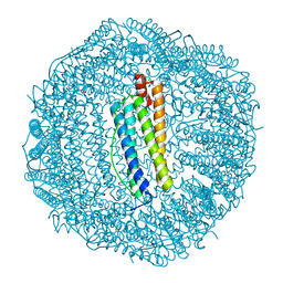 | | Frog M-ferritin with cobalt | | 分子名称: | CHLORIDE ION, COBALT (II) ION, Ferritin, ... | | 著者 | Tosha, T, Ng, H.L, Theil, E, Alber, T, Bhattasali, O. | | 登録日 | 2009-10-18 | | 公開日 | 2010-10-06 | | 最終更新日 | 2023-09-06 | | 実験手法 | X-RAY DIFFRACTION (1.4 Å) | | 主引用文献 | Moving Metal Ions through Ferritin-Protein Nanocages from Three-Fold Pores to Catalytic Sites.
J.Am.Chem.Soc., 132, 2010
|
|
3KA6
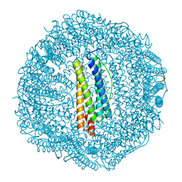 | | Frog M-ferritin, EED mutant, with cobalt | | 分子名称: | CHLORIDE ION, COBALT (II) ION, Ferritin, ... | | 著者 | Tosha, T, Ng, H.L, Theil, E, Alber, T, Bhattasali, O. | | 登録日 | 2009-10-18 | | 公開日 | 2010-10-06 | | 最終更新日 | 2023-09-06 | | 実験手法 | X-RAY DIFFRACTION (1.4 Å) | | 主引用文献 | Moving Metal Ions through Ferritin-Protein Nanocages from Three-Fold Pores to Catalytic Sites.
J.Am.Chem.Soc., 132, 2010
|
|
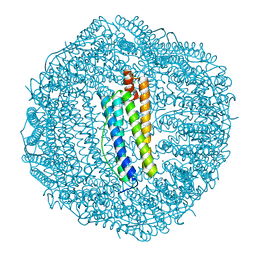
3KA8
 | | Frog M-ferritin, EQH mutant, with cobalt | | 分子名称: | CHLORIDE ION, COBALT (II) ION, Ferritin, ... | | 著者 | Tosha, T, Ng, H.L, Theil, E, Alber, T, Bhattasali, O. | | 登録日 | 2009-10-19 | | 公開日 | 2010-10-06 | | 最終更新日 | 2023-09-06 | | 実験手法 | X-RAY DIFFRACTION (1.35 Å) | | 主引用文献 | Moving Metal Ions through Ferritin-Protein Nanocages from Three-Fold Pores to Catalytic Sites.
J.Am.Chem.Soc., 132, 2010
|
|
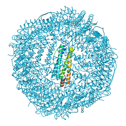
3KA3
 | | Frog M-ferritin with magnesium | | 分子名称: | CHLORIDE ION, Ferritin, middle subunit, ... | | 著者 | Tosha, T, Ng, H.L, Theil, E, Alber, T, Bhattasali, O. | | 登録日 | 2009-10-18 | | 公開日 | 2010-10-06 | | 最終更新日 | 2024-02-21 | | 実験手法 | X-RAY DIFFRACTION (1.4 Å) | | 主引用文献 | Moving Metal Ions through Ferritin-Protein Nanocages from Three-Fold Pores to Catalytic Sites.
J.Am.Chem.Soc., 132, 2010
|
|
3KA9
 | | Frog M-ferritin, EEH mutant, with cobalt | | 分子名称: | CHLORIDE ION, COBALT (II) ION, Ferritin, ... | | 著者 | Tosha, T, Ng, H.L, Theil, E, Alber, T, Bhattasali, O. | | 登録日 | 2009-10-19 | | 公開日 | 2010-10-06 | | 最終更新日 | 2023-09-06 | | 実験手法 | X-RAY DIFFRACTION (1.45 Å) | | 主引用文献 | Frog M-ferritin, EEH mutant, with cobalt
To be Published
|
|
1I1J
 | | STRUCTURE OF MELANOMA INHIBITORY ACTIVITY PROTEIN: A MEMBER OF A NEW FAMILY OF SECRETED PROTEINS | | 分子名称: | MELANOMA DERIVED GROWTH REGULATORY PROTEIN | | 著者 | Lougheed, J.C, Holton, J.M, Alber, T, Bazan, J.F, Handel, T.M. | | 登録日 | 2001-02-02 | | 公開日 | 2001-05-16 | | 最終更新日 | 2011-07-13 | | 実験手法 | X-RAY DIFFRACTION (1.39 Å) | | 主引用文献 | Structure of melanoma inhibitory activity protein, a member of a recently identified family of secreted proteins.
Proc.Natl.Acad.Sci.USA, 98, 2001
|
|
3SE1
 | | Frog M-ferritin with magnesium, R72D mutant | | 分子名称: | CHLORIDE ION, Ferritin, middle subunit, ... | | 著者 | Tosha, T, Ng, H.L, Alber, T, Theil, E.C. | | 登録日 | 2011-06-10 | | 公開日 | 2012-02-29 | | 最終更新日 | 2024-02-28 | | 実験手法 | X-RAY DIFFRACTION (1.65 Å) | | 主引用文献 | Frog M-ferritin with magnesium, R72D mutant
TO BE PUBLISHED
|
|
3SHX
 | | Frog M-ferritin with magnesium, L134P mutant | | 分子名称: | CHLORIDE ION, Ferritin, middle subunit, ... | | 著者 | Tosha, T, Ng, H.L, Alber, T, Theil, E.C. | | 登録日 | 2011-06-17 | | 公開日 | 2012-02-29 | | 最終更新日 | 2024-02-28 | | 実験手法 | X-RAY DIFFRACTION (1.35 Å) | | 主引用文献 | Frog M-ferritin with magnesium, L134P mutant
To be Published
|
|
3SH6
 | | Frog M-ferritin, D122R mutant, with magnesium | | 分子名称: | CHLORIDE ION, Ferritin, middle subunit, ... | | 著者 | Tosha, T, Ng, H.L, Alber, T, Theil, E.C. | | 登録日 | 2011-06-16 | | 公開日 | 2012-02-29 | | 最終更新日 | 2024-02-28 | | 実験手法 | X-RAY DIFFRACTION (1.4 Å) | | 主引用文献 | Frog M-ferritin, D122R mutant, with magnesium
To be Published
|
|
3M4U
 | |
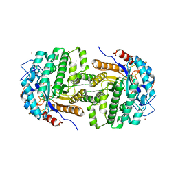
3CSU
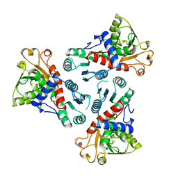 | | CATALYTIC TRIMER OF ESCHERICHIA COLI ASPARTATE TRANSCARBAMOYLASE | | 分子名称: | CALCIUM ION, PROTEIN (ASPARTATE CARBAMOYLTRANSFERASE) | | 著者 | Beernink, P.T, Endrizzi, J.A, Alber, T, Schachman, H.K. | | 登録日 | 1999-04-22 | | 公開日 | 1999-05-11 | | 最終更新日 | 2023-08-30 | | 実験手法 | X-RAY DIFFRACTION (1.88 Å) | | 主引用文献 | Assessment of the allosteric mechanism of aspartate transcarbamoylase based on the crystalline structure of the unregulated catalytic subunit.
Proc.Natl.Acad.Sci.USA, 96, 1999
|
|
3F61
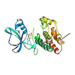 | |
3GOF
 | | Calmodulin bound to peptide from macrophage nitric oxide synthase | | 分子名称: | CALCIUM ION, Calmodulin, Nitric oxide synthase, ... | | 著者 | Ng, H.L, Greenstein, A, Marletta, M, Wand, A.J, Alber, T. | | 登録日 | 2009-03-19 | | 公開日 | 2010-03-31 | | 最終更新日 | 2024-02-21 | | 実験手法 | X-RAY DIFFRACTION (1.45 Å) | | 主引用文献 | Structural diversity in calmodulin recognition of nitric oxide synthases
To be Published
|
|
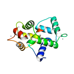
1RU0
 | | Crystal structure of DCoH2, a paralog of DCoH, the Dimerization Cofactor of HNF-1 | | 分子名称: | DcoH-like protein DCoHm | | 著者 | Rose, R.B, Pullen, K.E, Bayle, J.H, Crabtree, G.R, Alber, T. | | 登録日 | 2003-12-10 | | 公開日 | 2004-10-12 | | 最終更新日 | 2023-08-23 | | 実験手法 | X-RAY DIFFRACTION (1.6 Å) | | 主引用文献 | Biochemical and structural basis for partially redundant enzymatic and transcriptional functions of DCoH and DCoH2
Biochemistry, 43, 2004
|
|
1TGG
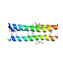 | | RH3 DESIGNED RIGHT-HANDED COILED COIL TRIMER | | 分子名称: | CHLORIDE ION, NICKEL (II) ION, right-handed coiled coil trimer | | 著者 | Plecs, J.J, Harbury, P.B, Kim, P.S, Alber, T. | | 登録日 | 2004-05-28 | | 公開日 | 2004-10-12 | | 最終更新日 | 2011-07-13 | | 実験手法 | X-RAY DIFFRACTION (2 Å) | | 主引用文献 | Structural test of the parameterized-backbone method for protein design
J.Mol.Biol., 342, 2004
|
|
1YWF
 | |
2XHY
 | | Crystal Structure of E.coli BglA | | 分子名称: | 6-PHOSPHO-BETA-GLUCOSIDASE BGLA, BROMIDE ION, SULFATE ION | | 著者 | Totir, M, Zubieta, C, Echols, N, May, A.P, Gee, C.L, nanao, M, alber, T. | | 登録日 | 2010-06-24 | | 公開日 | 2011-07-06 | | 最終更新日 | 2023-12-20 | | 実験手法 | X-RAY DIFFRACTION (2.3 Å) | | 主引用文献 | Macro-to-Micro Structural Proteomics: Native Source Proteins for High-Throughput Crystallization.
Plos One, 7, 2012
|
|


