Loading
PDBj
MenuPDBj@FacebookPDBj@TwitterPDBj@YouTubewwPDB FoundationwwPDB
RCSB PDBPDBeBMRBAdv. SearchSearch help
Search by PDB author
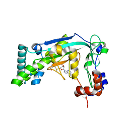
8RXR
DownloadVisualize
BU of 8rxr by Molmil
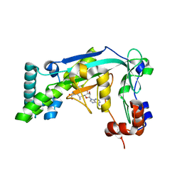
Crystal structure of VPS34 in complex with inhibitor SB02024
Descriptor: 4-[(3R)-3-methylmorpholin-4-yl]-2-[(2R)-2-(trifluoromethyl)piperidin-1-yl]-3H-pyridin-6-one, DI(HYDROXYETHYL)ETHER, DIMETHYL SULFOXIDE, ...
Authors:Tresaugues, L, Yu, Y, Bogdan, M, Parpal, S, Silvander, C, Lindstrom, J, Simeon, J, Timson, M.J, Al-Hashimi, H, Smith, B.D, Flynn, D.L, Viklund, J, Martinsson, J, De Milito, A, Andersson, M.
Deposit date:2024-02-07
Release date:2024-03-20
Last modified:2024-08-21
Method:X-RAY DIFFRACTION (2.06 Å)
Cite:Combining VPS34 inhibitors with STING agonists enhances type I interferon signaling and anti-tumor efficacy.
Mol Oncol, 18, 2024
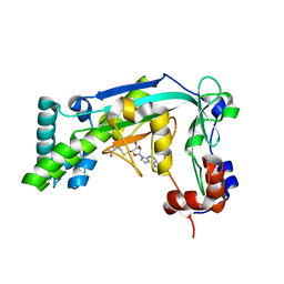
6UEQ
DownloadVisualize
BU of 6ueq by Molmil
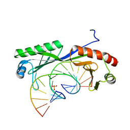
Structure of TBP bound to C-C mismatch containing TATA site
Descriptor: DNA (5'-D(*GP*CP*TP*AP*TP*AP*AP*AP*AP*CP*GP*GP*CP*A)-3'), DNA (5'-D(*TP*GP*CP*CP*CP*TP*TP*TP*TP*AP*TP*AP*GP*C)-3'), SULFATE ION, ...
Authors:Schumacher, M.A, Al-Hashimi, H.
Deposit date:2019-09-22
Release date:2020-09-02
Last modified:2023-10-11
Method:X-RAY DIFFRACTION (2.4 Å)
Cite:DNA mismatches reveal conformational penalties in protein-DNA recognition.
Nature, 587, 2020
6UEP
DownloadVisualize
BU of 6uep by Molmil
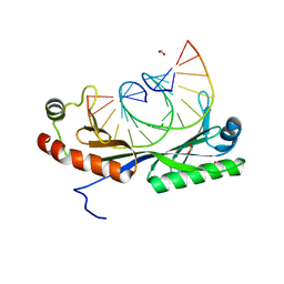
Structure of A. thaliana TBP bound to a DNA site with a C-C mismatch
Descriptor: DNA (5'-D(*GP*CP*TP*AP*TP*AP*AP*AP*CP*GP*GP*GP*CP*A)-3'), DNA (5'-D(*TP*GP*CP*CP*CP*CP*TP*TP*TP*AP*TP*AP*GP*C)-3'), FORMIC ACID, ...
Authors:Schumacher, M.A, Al-hashimi, H.
Deposit date:2019-09-22
Release date:2020-09-02
Last modified:2024-03-13
Method:X-RAY DIFFRACTION (2.05 Å)
Cite:DNA mismatches reveal conformational penalties in protein-DNA recognition.
Nature, 587, 2020
6UER
DownloadVisualize
BU of 6uer by Molmil
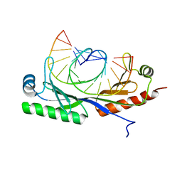
Crystal form 2: Structure of TBP bound to C-C mismatch at pH 7
Descriptor: DNA (5'-D(*GP*CP*TP*AP*TP*AP*AP*AP*CP*GP*GP*GP*CP*A)-3'), DNA (5'-D(*TP*GP*CP*CP*CP*CP*TP*TP*TP*AP*TP*AP*GP*C)-3'), TATA-box-binding protein 1
Authors:Schumacher, M.A, Al-Hashimi, H.
Deposit date:2019-09-22
Release date:2020-09-02
Last modified:2023-10-11
Method:X-RAY DIFFRACTION (2.5 Å)
Cite:DNA mismatches reveal conformational penalties in protein-DNA recognition.
Nature, 587, 2020
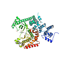
6BDY
DownloadVisualize
BU of 6bdy by Molmil
Crystal Structure of the MetH Reactivation Domain bound to Sinefungin
Descriptor: Methionine synthase, SINEFUNGIN
Authors:Fick, R.J, Vander Lee, L.P, Trievel, R.C.
Deposit date:2017-10-24
Release date:2018-05-23
Last modified:2023-10-04
Method:X-RAY DIFFRACTION (1.512 Å)
Cite:Water-Mediated Carbon-Oxygen Hydrogen Bonding Facilitates S-Adenosylmethionine Recognition in the Reactivation Domain of Cobalamin-Dependent Methionine Synthase.
Biochemistry, 57, 2018
6BM5
DownloadVisualize
BU of 6bm5 by Molmil
Crystal Structure of the MetH Reactivation Domain bound to AdoMet
Descriptor: Methionine synthase, S-ADENOSYLMETHIONINE
Authors:Fick, R.J, Vander Lee, L.P, Trievel, R.C.
Deposit date:2017-11-13
Release date:2018-05-23
Last modified:2023-10-04
Method:X-RAY DIFFRACTION (1.5 Å)
Cite:Water-Mediated Carbon-Oxygen Hydrogen Bonding Facilitates S-Adenosylmethionine Recognition in the Reactivation Domain of Cobalamin-Dependent Methionine Synthase.
Biochemistry, 57, 2018
6BM6
DownloadVisualize
BU of 6bm6 by Molmil
Crystal Structure of the MetH Reactivation Domain bound to AdoHcy
Descriptor: Methionine synthase, S-ADENOSYL-L-HOMOCYSTEINE
Authors:Fick, R.J, Vander Lee, L.P, Trievel, R.C.
Deposit date:2017-11-13
Release date:2018-05-23
Last modified:2023-10-04
Method:X-RAY DIFFRACTION (1.504 Å)
Cite:Water-Mediated Carbon-Oxygen Hydrogen Bonding Facilitates S-Adenosylmethionine Recognition in the Reactivation Domain of Cobalamin-Dependent Methionine Synthase.
Biochemistry, 57, 2018

226707

PDB entries from 2024-10-30

PDB statisticsPDBj update infoContact PDBjnumon