Loading
PDBj
MenuPDBj@FacebookPDBj@X(formerly Twitter)PDBj@BlueSkyPDBj@YouTubewwPDB FoundationwwPDBDonate
RCSB PDBPDBeBMRBAdv. SearchSearch help
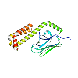
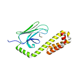
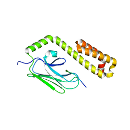
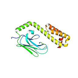
Search by PDB author
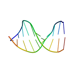
122D
DownloadVisualize
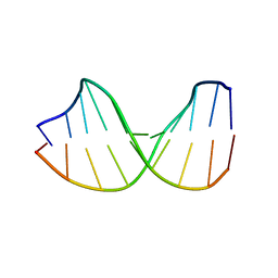
BU of 122d by Molmil
DNA HELIX STRUCTURE AND REFINEMENT ALGORITHM: COMPARISON OF MODELS FOR D(CCAGGCM==5==CTGG) DERIVED FROM NUCLSQ, TNT, AND X-PLOR
Descriptor: DNA (5'-D(*CP*CP*AP*GP*GP*CP*(5CM)P*TP*GP*G)-3')
Authors:Hahn, M, Heinemann, U.
Deposit date:1993-05-03
Release date:1993-10-15
Last modified:2024-02-07
Method:X-RAY DIFFRACTION (1.7 Å)
Cite:DNA helix structure and refinement algorithm: comparison of models for d(CCAGGCm5CTGG) derived from NUCLSQ, TNT and X-PLOR.
Acta Crystallogr.,Sect.D, 49, 1993
123D
DownloadVisualize
BU of 123d by Molmil
DNA HELIX STRUCTURE AND REFINEMENT ALGORITHM: COMPARISON OF MODELS FOR D(CCAGGCM==5==CTGG) DERIVED FROM NUCLSQ, TNT, AND X-PLOR
Descriptor: DNA (5'-D(*CP*CP*AP*GP*GP*CP*(5CM)P*TP*GP*G)-3')
Authors:Hahn, M, Heinemann, U.
Deposit date:1993-05-03
Release date:1993-10-15
Last modified:2024-02-07
Method:X-RAY DIFFRACTION (1.7 Å)
Cite:DNA helix structure and refinement algorithm: comparison of models for d(CCAGGCm5CTGG) derived from NUCLSQ, TNT and X-PLOR.
Acta Crystallogr.,Sect.D, 49, 1993
1E4X
DownloadVisualize
BU of 1e4x by Molmil
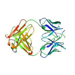
crossreactive binding of a circularized peptide to an anti-TGFalpha antibody Fab-fragment
Descriptor: CYCLIC PEPTIDE, TAB2
Authors:Hahn, M, Winkler, D, Misselwitz, R, Wessner, H, Welfle, K, Zahn, G, Schneider-Mergener, J, Hoehne, W.
Deposit date:2000-07-12
Release date:2001-07-12
Last modified:2024-10-23
Method:X-RAY DIFFRACTION (1.9 Å)
Cite:Cross-Reactive Binding of Cyclic Peptides to an Anti-Tgf Alpha Antibody Fab Fragment: An X-Ray Structural and Thermodynamic Analysis
J.Mol.Biol., 314, 2001
1E4W
DownloadVisualize
BU of 1e4w by Molmil
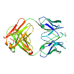
crossreactive binding of a circularized peptide to an anti-TGFalpha antibody Fab-fragment
Descriptor: CHLORIDE ION, CYCLIC PEPTIDE, NICKEL (II) ION, ...
Authors:Hahn, M, Winkler, D, Misselwitz, R, Wessner, H, Welfle, K, Zahn, G, Schneider-Mergener, J, Hoehne, W.
Deposit date:2000-07-12
Release date:2001-07-12
Last modified:2024-10-16
Method:X-RAY DIFFRACTION (1.95 Å)
Cite:Cross-Reactive Binding of Cyclic Peptides to an Anti-Tgf Alpha Antibody Fab Fragment: An X-Ray Structural and Thermodynamic Analysis
J.Mol.Biol., 314, 2001
5DL8
DownloadVisualize
BU of 5dl8 by Molmil
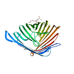
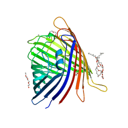
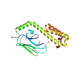
Crystal structure of Acinetobacter baumannii OccAB4
Descriptor: (HYDROXYETHYLOXY)TRI(ETHYLOXY)OCTANE, Benzoate transport porin BenP, CALCIUM ION
Authors:Zahn, M, Basle, A, van den Berg, B.
Deposit date:2015-09-04
Release date:2016-02-03
Last modified:2024-01-10
Method:X-RAY DIFFRACTION (2.2 Å)
Cite:Structural Insights into Outer Membrane Permeability of Acinetobacter baumannii.
Structure, 24, 2016
5DL5
DownloadVisualize
BU of 5dl5 by Molmil
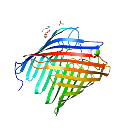
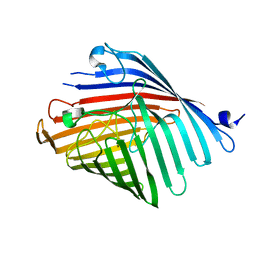
Crystal structure of Acinetobacter baumannii OccAB1
Descriptor: (HYDROXYETHYLOXY)TRI(ETHYLOXY)OCTANE, Membrane protein, PHOSPHATE ION
Authors:Zahn, M, Basle, A, van den Berg, B.
Deposit date:2015-09-04
Release date:2016-02-03
Last modified:2024-01-10
Method:X-RAY DIFFRACTION (2.05 Å)
Cite:Structural Insights into Outer Membrane Permeability of Acinetobacter baumannii.
Structure, 24, 2016
5DL7
DownloadVisualize
BU of 5dl7 by Molmil
Crystal structure of Acinetobacter baumannii OccAB3
Descriptor: (HYDROXYETHYLOXY)TRI(ETHYLOXY)OCTANE, CALCIUM ION, Porin
Authors:Zahn, M, Basle, A, van den Berg, B.
Deposit date:2015-09-04
Release date:2016-02-03
Last modified:2024-01-10
Method:X-RAY DIFFRACTION (1.75 Å)
Cite:Structural Insights into Outer Membrane Permeability of Acinetobacter baumannii.
Structure, 24, 2016
5DL6
DownloadVisualize
BU of 5dl6 by Molmil
Crystal structure of Acinetobacter baumannii OccAB2
Descriptor: Putative porin for vanillate trafficking (VanP)
Authors:Zahn, M, Basle, A, van den Berg, B.
Deposit date:2015-09-04
Release date:2016-02-03
Last modified:2024-01-10
Method:X-RAY DIFFRACTION (2.9 Å)
Cite:Structural Insights into Outer Membrane Permeability of Acinetobacter baumannii.
Structure, 24, 2016
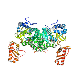
7PT2
DownloadVisualize
BU of 7pt2 by Molmil
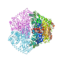
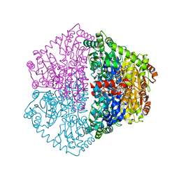
Actinobacterial 2-hydroxyacyl-CoA lyase (AcHACL) mutant E493Q structure in complex with substrate 2-HIB-CoA and inactive cofactor 3-deaza-ThDP
Descriptor: 2-hydroxyacyl-CoA lyase, 2-{4-[(4-AMINO-2-METHYLPYRIMIDIN-5-YL)METHYL]-3-METHYLTHIOPHEN-2-YL}ETHYL TRIHYDROGEN DIPHOSPHATE, MAGNESIUM ION, ...
Authors:Zahn, M, Rohwerder, T.
Deposit date:2021-09-25
Release date:2022-02-02
Last modified:2024-06-19
Method:X-RAY DIFFRACTION (1.762 Å)
Cite:Mechanistic details of the actinobacterial lyase-catalyzed degradation reaction of 2-hydroxyisobutyryl-CoA.
J.Biol.Chem., 298, 2022
7PT1
DownloadVisualize
BU of 7pt1 by Molmil
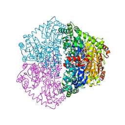
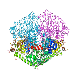
Actinobacterial 2-hydroxyacyl-CoA lyase (AcHACL) structure in complex with substrate 2-HIB-CoA and inactive cofactor 3-deaza-ThDP
Descriptor: 2-hydroxyacyl-CoA lyase, 2-{4-[(4-AMINO-2-METHYLPYRIMIDIN-5-YL)METHYL]-3-METHYLTHIOPHEN-2-YL}ETHYL TRIHYDROGEN DIPHOSPHATE, MAGNESIUM ION, ...
Authors:Zahn, M, Rohwerder, T.
Deposit date:2021-09-25
Release date:2022-02-02
Last modified:2024-01-31
Method:X-RAY DIFFRACTION (1.553 Å)
Cite:Mechanistic details of the actinobacterial lyase-catalyzed degradation reaction of 2-hydroxyisobutyryl-CoA.
J.Biol.Chem., 298, 2022
7PT3
DownloadVisualize
BU of 7pt3 by Molmil
Actinobacterial 2-hydroxyacyl-CoA lyase (AcHACL) mutant E493A structure in complex with substrate 2-HIB-CoA and inactive cofactor 3-deaza-ThDP
Descriptor: 2-hydroxyacyl-CoA lyase, 2-{4-[(4-AMINO-2-METHYLPYRIMIDIN-5-YL)METHYL]-3-METHYLTHIOPHEN-2-YL}ETHYL TRIHYDROGEN DIPHOSPHATE, MAGNESIUM ION, ...
Authors:Zahn, M, Rohwerder, T.
Deposit date:2021-09-25
Release date:2022-02-02
Last modified:2024-06-19
Method:X-RAY DIFFRACTION (1.625 Å)
Cite:Mechanistic details of the actinobacterial lyase-catalyzed degradation reaction of 2-hydroxyisobutyryl-CoA.
J.Biol.Chem., 298, 2022
7PT4
DownloadVisualize
BU of 7pt4 by Molmil
Actinobacterial 2-hydroxyacyl-CoA lyase (AcHACL) structure in complex with a covalently bound reaction intermediate as well as products formyl-CoA and acetone
Descriptor: 2-hydroxyacyl-CoA lyase, 3-[(4-AMINO-2-METHYLPYRIMIDIN-5-YL)METHYL]-2-{(1R,11R,15S,17R)-19-[(2R,3S,4R,5R)-5-(6-AMINO-9H-PURIN-9-YL)-4-HYDROXY-3-(PHOSPHONOOXY)TETRAHYDROFURAN-2-YL]-1,11,15,17-TETRAHYDROXY-12,12-DIMETHYL-15,17-DIOXIDO-6,10-DIOXO-14,16,18-TRIOXA-2-THIA-5,9-DIAZA-15,17-DIPHOSPHANONADEC-1-YL}-5-(2-{[(R)-HYDROXY(PHOSPHONOOXY)PHOSPHORYL]OXY}ETHYL)-4-METHYL-1,3-THIAZOL-3-IUM, ACETONE, ...
Authors:Zahn, M, Rohwerder, T.
Deposit date:2021-09-25
Release date:2022-02-02
Last modified:2024-01-31
Method:X-RAY DIFFRACTION (1.64 Å)
Cite:Mechanistic details of the actinobacterial lyase-catalyzed degradation reaction of 2-hydroxyisobutyryl-CoA.
J.Biol.Chem., 298, 2022
1CPN
DownloadVisualize
BU of 1cpn by Molmil
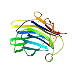
NATIVE-LIKE IN VIVO FOLDING OF A CIRCULARLY PERMUTED JELLYROLL PROTEIN SHOWN BY CRYSTAL STRUCTURE ANALYSIS
Descriptor: CALCIUM ION, CIRCULARLY PERMUTED
Authors:Hahn, M, Heinemann, U.
Deposit date:1994-03-11
Release date:1994-06-22
Last modified:2024-11-13
Method:X-RAY DIFFRACTION (1.8 Å)
Cite:Native-like in vivo folding of a circularly permuted jellyroll protein shown by crystal structure analysis.
Proc.Natl.Acad.Sci.USA, 91, 1994
4F00
DownloadVisualize
BU of 4f00 by Molmil
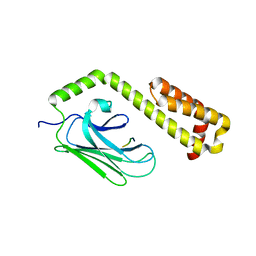
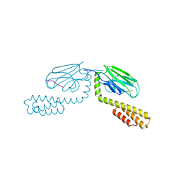
Crystal structure of the substrate binding domain of E.coli DnaK in complex with an apidaecin fragment from the bumblebee (residues 3 to 11)
Descriptor: Apidaecin, Chaperone protein DnaK
Authors:Zahn, M, Straeter, N.
Deposit date:2012-05-03
Release date:2013-04-17
Last modified:2024-02-28
Method:X-RAY DIFFRACTION (1.95 Å)
Cite:Structural Studies on the Forward and Reverse Binding Modes of Peptides to the Chaperone DnaK.
J.Mol.Biol., 425, 2013
4EZV
DownloadVisualize
BU of 4ezv by Molmil
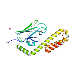
Crystal structure of the substrate binding domain of E.coli DnaK in complex with PR-bombesin in space group P21212
Descriptor: Chaperone protein DnaK, Proline rich bombesin-related protein
Authors:Zahn, M, Straeter, N.
Deposit date:2012-05-03
Release date:2013-04-24
Last modified:2024-02-28
Method:X-RAY DIFFRACTION (2.1 Å)
Cite:Structural studies of DnaK in complex with proline rich antimicrobial peptides reveal two different peptide binding modes
To be Published
4EZQ
DownloadVisualize
BU of 4ezq by Molmil
Crystal structure of the substrate binding domain of E.coli DnaK in complex with the C-terminal part of pyrrhocoricin (residues 12 to 20)
Descriptor: Chaperone protein DnaK, Pyrrhocoricin
Authors:Zahn, M, Straeter, N.
Deposit date:2012-05-03
Release date:2013-04-17
Last modified:2024-02-28
Method:X-RAY DIFFRACTION (2 Å)
Cite:Structural Studies on the Forward and Reverse Binding Modes of Peptides to the Chaperone DnaK.
J.Mol.Biol., 425, 2013
4EZU
DownloadVisualize
BU of 4ezu by Molmil
Crystal structure of the substrate binding domain of E.coli DnaK in complex with PR-bombesin in space group I222
Descriptor: Chaperone protein DnaK, Proline rich bombesin-related protein
Authors:Zahn, M, Straeter, N.
Deposit date:2012-05-03
Release date:2013-04-24
Last modified:2024-02-28
Method:X-RAY DIFFRACTION (1.9 Å)
Cite:Structural studies of DnaK in complex with proline rich antimicrobial peptides reveal two different peptide binding modes
To be Published
4EZS
DownloadVisualize
BU of 4ezs by Molmil
Crystal structure of the substrate binding domain of E.coli DnaK in complex with metchnikowin (residues 20 to 26)
Descriptor: Chaperone protein DnaK, Metchnikowin, SULFATE ION
Authors:Zahn, M, Straeter, N.
Deposit date:2012-05-03
Release date:2013-04-24
Last modified:2024-02-28
Method:X-RAY DIFFRACTION (1.9 Å)
Cite:Structural studies of DnaK in complex with proline rich antimicrobial peptides reveal two different peptide binding modes
To be Published
4EZY
DownloadVisualize
BU of 4ezy by Molmil
Crystal structure of the substrate binding domain of E.coli DnaK in complex with the designer peptide NRLILTG
Descriptor: Chaperone protein DnaK, SULFATE ION, synthetic peptide NRLILTG
Authors:Zahn, M, Straeter, N.
Deposit date:2012-05-03
Release date:2013-04-17
Last modified:2024-02-28
Method:X-RAY DIFFRACTION (1.85 Å)
Cite:Structural Studies on the Forward and Reverse Binding Modes of Peptides to the Chaperone DnaK.
J.Mol.Biol., 425, 2013
4EZN
DownloadVisualize
BU of 4ezn by Molmil
Crystal structure of the substrate binding domain of E.coli DnaK in complex with pyrrhocoricin
Descriptor: Chaperone protein DnaK, Pyrrhocoricin
Authors:Zahn, M, Straeter, N.
Deposit date:2012-05-03
Release date:2013-04-17
Last modified:2024-02-28
Method:X-RAY DIFFRACTION (1.8 Å)
Cite:Structural Studies on the Forward and Reverse Binding Modes of Peptides to the Chaperone DnaK.
J.Mol.Biol., 425, 2013
4EZX
DownloadVisualize
BU of 4ezx by Molmil
Crystal structure of the substrate binding domain of E.coli DnaK in complex with the designer peptide NRLMLTG
Descriptor: Chaperone protein DnaK, SULFATE ION, synthetic peptide NRLMLTG
Authors:Zahn, M, Straeter, N.
Deposit date:2012-05-03
Release date:2013-04-17
Last modified:2024-02-28
Method:X-RAY DIFFRACTION (1.7 Å)
Cite:Structural Studies on the Forward and Reverse Binding Modes of Peptides to the Chaperone DnaK.
J.Mol.Biol., 425, 2013
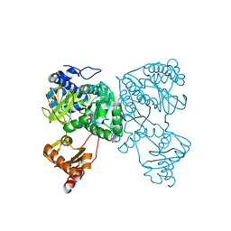
6HDW
DownloadVisualize
BU of 6hdw by Molmil
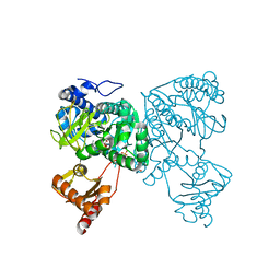
Crystal structure of 2-Hydroxyisobutyryl-CoA Ligase (HCL) in the postadenylation state in complex with 2-HIB-AMP
Descriptor: 2-hydroxyisobutyryl-CoA synthetase, SULFATE ION, [[(2~{R},3~{S},4~{R},5~{R})-5-(6-aminopurin-9-yl)-3,4-bis(oxidanyl)oxolan-2-yl]methoxy-oxidanyl-phosphoryl] 2-methyl-2-oxidanyl-propanoate
Authors:Zahn, M, Rohwerder, T, Strater, N.
Deposit date:2018-08-20
Release date:2019-08-28
Last modified:2024-01-17
Method:X-RAY DIFFRACTION (2.3 Å)
Cite:Structures of 2-Hydroxyisobutyric Acid-CoA Ligase Reveal Determinants of Substrate Specificity and Describe a Multi-Conformational Catalytic Cycle.
J.Mol.Biol., 431, 2019
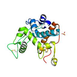
1DXJ
DownloadVisualize
BU of 1dxj by Molmil
Structure of the chitinase from jack bean
Descriptor: CLASS II CHITINASE, SULFATE ION
Authors:Hahn, M, Hennig, M, Schlesier, B, Hohne, W.
Deposit date:2000-01-10
Release date:2000-08-29
Last modified:2024-10-16
Method:X-RAY DIFFRACTION (1.8 Å)
Cite:Structure of Jack Bean Chitinase
Acta Crystallogr.,Sect.D, 56, 2000
6HDY
DownloadVisualize
BU of 6hdy by Molmil
Crystal structure of 2-Hydroxyisobutyryl-CoA Ligase (HCL) in the postadenylation state in complex with S3-HB-AMP
Descriptor: (3S)-3-HYDROXYBUTANOIC ACID, 2-hydroxyisobutyryl-CoA synthetase, SULFATE ION, ...
Authors:Zahn, M, Rohwerder, T, Strater, N.
Deposit date:2018-08-20
Release date:2019-08-28
Last modified:2024-01-17
Method:X-RAY DIFFRACTION (2.2 Å)
Cite:Structures of 2-Hydroxyisobutyric Acid-CoA Ligase Reveal Determinants of Substrate Specificity and Describe a Multi-Conformational Catalytic Cycle.
J.Mol.Biol., 431, 2019
6HE2
DownloadVisualize
BU of 6he2 by Molmil
Crystal structure of an open conformation of 2-Hydroxyisobutyryl-CoA Ligase (HCL) in complex with 2-HIB-AMP and CoA
Descriptor: 2-hydroxyisobutyryl-CoA synthetase, ADENOSINE MONOPHOSPHATE, COENZYME A, ...
Authors:Zahn, M, Rohwerder, T, Strater, N.
Deposit date:2018-08-20
Release date:2019-08-28
Last modified:2024-01-17
Method:X-RAY DIFFRACTION (2.3 Å)
Cite:Structures of 2-Hydroxyisobutyric Acid-CoA Ligase Reveal Determinants of Substrate Specificity and Describe a Multi-Conformational Catalytic Cycle.
J.Mol.Biol., 431, 2019

237423

PDB entries from 2025-06-11

PDB statisticsPDBj update infoContact PDBjnumon