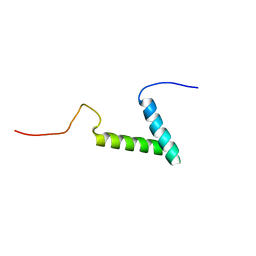
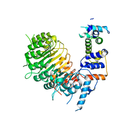
7MY4
 
 | |
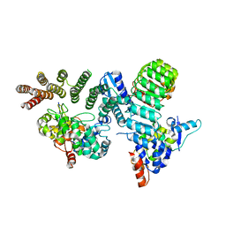
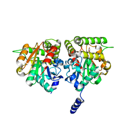
1YKD
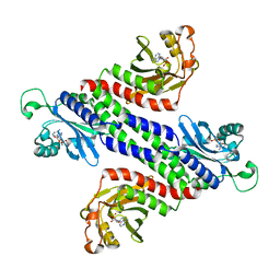 | | Crystal Structure of the Tandem GAF Domains from a Cyanobacterial Adenylyl Cyclase: Novel Modes of Ligand-Binding and Dimerization | | 分子名称: | ADENOSINE-3',5'-CYCLIC-MONOPHOSPHATE, adenylate cyclase | | 著者 | Martinez, S.E, Bruder, S, Schultz, A, Zheng, N, Schultz, J.E, Beavo, J.A, Linder, J.U. | | 登録日 | 2005-01-17 | | 公開日 | 2005-02-22 | | 最終更新日 | 2024-02-14 | | 実験手法 | X-RAY DIFFRACTION (1.9 Å) | | 主引用文献 | Crystal structure of the tandem GAF domains from a cyanobacterial adenylyl cyclase: Modes of ligand binding and dimerization
Proc.Natl.Acad.Sci.USA, 102, 2005
|
|
2AZE
 | | Structure of the Rb C-terminal domain bound to an E2F1-DP1 heterodimer | | 分子名称: | Retinoblastoma-associated protein, Transcription factor Dp-1, Transcription factor E2F1 | | 著者 | Rubin, S.M, Gall, A.L, Zheng, N, Pavletich, N.P. | | 登録日 | 2005-09-10 | | 公開日 | 2006-01-31 | | 最終更新日 | 2024-02-14 | | 実験手法 | X-RAY DIFFRACTION (2.55 Å) | | 主引用文献 | Structure of the Rb C-terminal domain bound to E2F1-DP1: a mechanism for phosphorylation-induced E2F release.
Cell(Cambridge,Mass.), 123, 2005
|
|
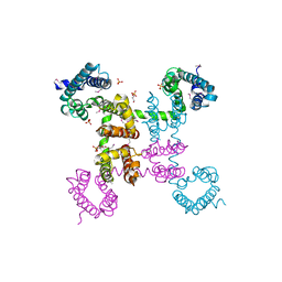
2B5L
 | | Crystal Structure of DDB1 In Complex with Simian Virus 5 V Protein | | 分子名称: | Nonstructural protein V, ZINC ION, damage-specific DNA binding protein 1 | | 著者 | Li, T, Chen, X, Garbutt, K.C, Zhou, P, Zheng, N. | | 登録日 | 2005-09-28 | | 公開日 | 2006-02-28 | | 最終更新日 | 2023-08-23 | | 実験手法 | X-RAY DIFFRACTION (2.85 Å) | | 主引用文献 | Structure of DDB1 in complex with a paramyxovirus V protein: viral hijack of a propeller cluster in ubiquitin ligase.
Cell(Cambridge,Mass.), 124, 2006
|
|
3C6N
 | | Small molecule agonists and antagonists of F-box protein-substrate interactions in auxin perception and signaling | | 分子名称: | (2S)-8-[(tert-butoxycarbonyl)amino]-2-(1H-indol-3-yl)octanoic acid, INOSITOL HEXAKISPHOSPHATE, SKP1-like protein 1A, ... | | 著者 | Tan, X, Zheng, N, Hayashi, K. | | 登録日 | 2008-02-04 | | 公開日 | 2008-04-22 | | 最終更新日 | 2023-08-30 | | 実験手法 | X-RAY DIFFRACTION (2.6 Å) | | 主引用文献 | Small-molecule agonists and antagonists of F-box protein-substrate interactions in auxin perception and signaling.
Proc.Natl.Acad.Sci.Usa, 105, 2008
|
|
6O60
 | | Crystal structure of GGTase3-FBXL2-SKP1 complex | | 分子名称: | F-box/LRR-repeat protein 2, Geranylgeranyl transferase type-2 subunit beta, Protein prenyltransferase alpha subunit repeat-containing protein 1, ... | | 著者 | Wang, H, Zheng, N. | | 登録日 | 2019-03-04 | | 公開日 | 2019-06-26 | | 最終更新日 | 2023-10-11 | | 実験手法 | X-RAY DIFFRACTION (2.503 Å) | | 主引用文献 | GGTase3 is a newly identified geranylgeranyltransferase targeting a ubiquitin ligase.
Nat.Struct.Mol.Biol., 26, 2019
|
|
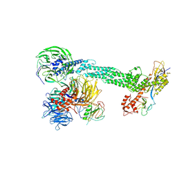
4EKW
 | | Crystal structure of the NavAb voltage-gated sodium channel (wild-type, 3.2 A) | | 分子名称: | 1,2-DIMYRISTOYL-SN-GLYCERO-3-PHOSPHOCHOLINE, Ion transport protein, PHOSPHATE ION | | 著者 | Payandeh, J, Gamal El-Din, T.M, Scheuer, T, Zheng, N, Catterall, W.A. | | 登録日 | 2012-04-10 | | 公開日 | 2012-05-16 | | 最終更新日 | 2023-12-06 | | 実験手法 | X-RAY DIFFRACTION (3.21 Å) | | 主引用文献 | Crystal structure of a voltage-gated sodium channel in two potentially inactivated states.
Nature, 486, 2012
|
|
2B5N
 | | Crystal Structure of the DDB1 BPB Domain | | 分子名称: | ISOPROPYL ALCOHOL, damage-specific DNA binding protein 1 | | 著者 | Li, T, Chen, X, Garbutt, K.C, Zhou, P, Zheng, N. | | 登録日 | 2005-09-28 | | 公開日 | 2006-02-28 | | 最終更新日 | 2024-02-14 | | 実験手法 | X-RAY DIFFRACTION (2.8 Å) | | 主引用文献 | Structure of DDB1 in complex with a paramyxovirus V protein: viral hijack of a propeller cluster in ubiquitin ligase.
Cell(Cambridge,Mass.), 124, 2006
|
|
2B5M
 | | Crystal Structure of DDB1 | | 分子名称: | damage-specific DNA binding protein 1 | | 著者 | Li, T, Chen, X, Garbutt, K.C, Zhou, P, Zheng, N. | | 登録日 | 2005-09-28 | | 公開日 | 2006-02-28 | | 最終更新日 | 2011-07-13 | | 実験手法 | X-RAY DIFFRACTION (2.92 Å) | | 主引用文献 | Structure of DDB1 in complex with a paramyxovirus V protein: viral hijack of a propeller cluster in ubiquitin ligase.
Cell(Cambridge,Mass.), 124, 2006
|
|
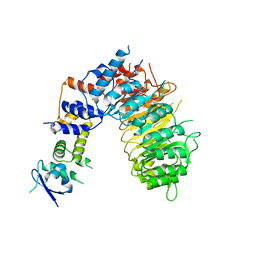
2HYE
 | | Crystal Structure of the DDB1-Cul4A-Rbx1-SV5V Complex | | 分子名称: | Cullin-4A, DNA damage-binding protein 1, Nonstructural protein V, ... | | 著者 | Angers, S, Li, T, Yi, X, MacCoss, M.J, Moon, R.T, Zheng, N. | | 登録日 | 2006-08-05 | | 公開日 | 2006-10-03 | | 最終更新日 | 2017-10-18 | | 実験手法 | X-RAY DIFFRACTION (3.1 Å) | | 主引用文献 | Molecular architecture and assembly of the DDB1-CUL4A ubiquitin ligase machinery.
Nature, 443, 2006
|
|
6BRP
 | | F-box protein form 2 | | 分子名称: | F-box/LRR-repeat MAX2 homolog, SKP1-like protein 1A | | 著者 | Shabek, N, Zheng, N, Mao, H, Hinds, T.R, Ticchiarelli, F, Leyser, O. | | 登録日 | 2017-11-30 | | 公開日 | 2018-11-21 | | 最終更新日 | 2024-03-13 | | 実験手法 | X-RAY DIFFRACTION (2.39 Å) | | 主引用文献 | Structural plasticity of D3-D14 ubiquitin ligase in strigolactone signalling.
Nature, 563, 2018
|
|
6BRQ
 | | Crystal structure of rice ASK1-D3 ubiquitin ligase complex crystal form 3 | | 分子名称: | F-box/LRR-repeat MAX2 homolog, SKP1-like protein 1A | | 著者 | Shabek, N, Zheng, N, Mao, H, Hinds, T.R, Ticchiarelli, F, Leyser, O. | | 登録日 | 2017-11-30 | | 公開日 | 2018-11-21 | | 最終更新日 | 2024-03-13 | | 実験手法 | X-RAY DIFFRACTION (2.99 Å) | | 主引用文献 | Structural plasticity of D3-D14 ubiquitin ligase in strigolactone signalling.
Nature, 563, 2018
|
|
6BRT
 | | F-box protein CTH with hydrolase | | 分子名称: | (5R)-5-hydroxy-3-methylfuran-2(5H)-one, D3-CTH-D14-D-ring | | 著者 | Shabek, N, Zheng, N, Mao, H, Hinds, T.R, Ticchiarelli, F, Leyser, O. | | 登録日 | 2017-12-01 | | 公開日 | 2018-11-21 | | 最終更新日 | 2024-03-13 | | 実験手法 | X-RAY DIFFRACTION (2.393 Å) | | 主引用文献 | Structural plasticity of D3-D14 ubiquitin ligase in strigolactone signalling.
Nature, 563, 2018
|
|
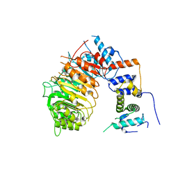
6BRO
 | |
6C1P
 | | HypoPP mutant | | 分子名称: | 1,2-DIMYRISTOYL-SN-GLYCERO-3-PHOSPHOCHOLINE, CHAPSO, Ion transport protein, ... | | 著者 | Catterall, W.A, Zheng, N, Jiang, D, Gamal El-Din, T.M. | | 登録日 | 2018-01-05 | | 公開日 | 2018-05-16 | | 最終更新日 | 2023-10-04 | | 実験手法 | X-RAY DIFFRACTION (2.9 Å) | | 主引用文献 | Structural basis for gating pore current in periodic paralysis.
Nature, 557, 2018
|
|
3I7P
 | | Crystal Structure of DDB1 in Complex with the H-Box Motif of WDR40A | | 分子名称: | DNA damage-binding protein 1, WD repeat-containing protein 40A | | 著者 | Li, T, Robert, E.I, Breugel, P.C.V, Strubin, M, Zheng, N. | | 登録日 | 2009-07-08 | | 公開日 | 2009-12-08 | | 最終更新日 | 2023-09-06 | | 実験手法 | X-RAY DIFFRACTION (3 Å) | | 主引用文献 | A promiscuous alpha-helical motif anchors viral hijackers and substrate receptors to the CUL4-DDB1 ubiquitin ligase machinery.
Nat.Struct.Mol.Biol., 17, 2010
|
|
3I7O
 | | Crystal Structure of DDB1 in Complex with the H-Box Motif of IQWD1 | | 分子名称: | DNA damage-binding protein 1, IQ motif and WD repeat-containing protein 1 | | 著者 | Li, T, Robert, E.I, Breugel, P.C.V, Strubin, M, Zheng, N. | | 登録日 | 2009-07-08 | | 公開日 | 2009-12-08 | | 最終更新日 | 2023-09-06 | | 実験手法 | X-RAY DIFFRACTION (2.8 Å) | | 主引用文献 | A promiscuous alpha-helical motif anchors viral hijackers and substrate receptors to the CUL4-DDB1 ubiquitin ligase machinery.
Nat.Struct.Mol.Biol., 17, 2010
|
|
3I7N
 | | Crystal Structure of DDB1 in Complex with the H-Box Motif of WDTC1 | | 分子名称: | DNA damage-binding protein 1, WD and tetratricopeptide repeats protein 1 | | 著者 | Li, T, Robert, E.I, Breugel, P.C.V, Strubin, M, Zheng, N. | | 登録日 | 2009-07-08 | | 公開日 | 2009-12-08 | | 最終更新日 | 2023-09-06 | | 実験手法 | X-RAY DIFFRACTION (2.8 Å) | | 主引用文献 | A promiscuous alpha-helical motif anchors viral hijackers and substrate receptors to the CUL4-DDB1 ubiquitin ligase machinery.
Nat.Struct.Mol.Biol., 17, 2010
|
|
3I7H
 | | Crystal Structure of DDB1 in Complex with the H-Box Motif of HBX | | 分子名称: | DNA damage-binding protein 1, X protein | | 著者 | Li, T, Robert, E.I, Breugel, P.C.V, Strubin, M, Zheng, N. | | 登録日 | 2009-07-08 | | 公開日 | 2009-12-08 | | 最終更新日 | 2023-09-06 | | 実験手法 | X-RAY DIFFRACTION (2.9 Å) | | 主引用文献 | A promiscuous alpha-helical motif anchors viral hijackers and substrate receptors to the CUL4-DDB1 ubiquitin ligase machinery.
Nat.Struct.Mol.Biol., 17, 2010
|
|
3I7L
 | | Crystal Structure of DDB1 in Complex with the H-Box Motif of DDB2 | | 分子名称: | DNA damage-binding protein 1, DNA damage-binding protein 2 | | 著者 | Li, T, Robert, E.I, Breugel, P.C.V, Strubin, M, Zheng, N. | | 登録日 | 2009-07-08 | | 公開日 | 2009-12-08 | | 最終更新日 | 2023-09-06 | | 実験手法 | X-RAY DIFFRACTION (2.8 Å) | | 主引用文献 | A promiscuous alpha-helical motif anchors viral hijackers and substrate receptors to the CUL4-DDB1 ubiquitin ligase machinery.
Nat.Struct.Mol.Biol., 17, 2010
|
|
3I7K
 | | Crystal Structure of DDB1 in Complex with the H-Box Motif of WHX | | 分子名称: | DNA damage-binding protein 1, X protein | | 著者 | Li, T, Robert, E.I, Breugel, P.C.V, Strubin, M, Zheng, N. | | 登録日 | 2009-07-08 | | 公開日 | 2009-12-08 | | 最終更新日 | 2023-09-06 | | 実験手法 | X-RAY DIFFRACTION (2.8 Å) | | 主引用文献 | A promiscuous alpha-helical motif anchors viral hijackers and substrate receptors to the CUL4-DDB1 ubiquitin ligase machinery.
Nat.Struct.Mol.Biol., 17, 2010
|
|
3I8E
 | | Crystal Structure of DDB1 in Complex with the H-Box Motif of WDR42A | | 分子名称: | DNA damage-binding protein 1, WD repeat-containing protein 42A | | 著者 | Li, T, Robert, E.I, Breugel, P.C.V, Strubin, M, Zheng, N. | | 登録日 | 2009-07-09 | | 公開日 | 2009-12-08 | | 最終更新日 | 2023-09-06 | | 実験手法 | X-RAY DIFFRACTION (3.4 Å) | | 主引用文献 | A promiscuous alpha-helical motif anchors viral hijackers and substrate receptors to the CUL4-DDB1 ubiquitin ligase machinery.
Nat.Struct.Mol.Biol., 17, 2010
|
|
3I89
 | | Crystal Structure of DDB1 in Complex with the H-Box Motif of WDR22 | | 分子名称: | DNA damage-binding protein 1, WD repeat-containing protein 22 | | 著者 | Li, T, Robert, E.I, Breugel, P.C.V, Strubin, M, Zheng, N. | | 登録日 | 2009-07-09 | | 公開日 | 2009-12-08 | | 最終更新日 | 2023-09-06 | | 実験手法 | X-RAY DIFFRACTION (3 Å) | | 主引用文献 | A promiscuous alpha-helical motif anchors viral hijackers and substrate receptors to the CUL4-DDB1 ubiquitin ligase machinery.
Nat.Struct.Mol.Biol., 17, 2010
|
|
3I8C
 | | Crystal Structure of DDB1 in Complex with the H-Box Motif of WDR21A | | 分子名称: | DNA damage-binding protein 1, WD repeat-containing protein 21A | | 著者 | Li, T, Robert, E.I, Breugel, P.C.V, Strubin, M, Zheng, N. | | 登録日 | 2009-07-09 | | 公開日 | 2009-12-08 | | 最終更新日 | 2023-09-06 | | 実験手法 | X-RAY DIFFRACTION (2.8 Å) | | 主引用文献 | A promiscuous alpha-helical motif anchors viral hijackers and substrate receptors to the CUL4-DDB1 ubiquitin ligase machinery.
Nat.Struct.Mol.Biol., 17, 2010
|
|
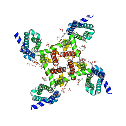
3M99
 | | Structure of the Ubp8-Sgf11-Sgf73-Sus1 SAGA DUB module | | 分子名称: | Protein SUS1, SAGA-associated factor 11, SAGA-associated factor 73, ... | | 著者 | Kohler, A, Zimmerman, E, Schneider, M, Hurt, E, Zheng, N. | | 登録日 | 2010-03-21 | | 公開日 | 2010-05-05 | | 最終更新日 | 2023-09-06 | | 実験手法 | X-RAY DIFFRACTION (2.7 Å) | | 主引用文献 | Structural basis for assembly and activation of the heterotetrameric SAGA histone H2B deubiquitinase module.
Cell(Cambridge,Mass.), 141, 2010
|
|


