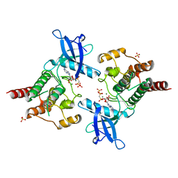
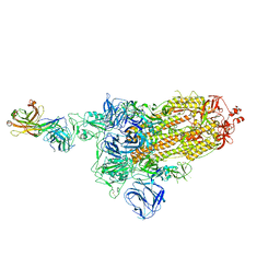
7MET
 
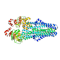 | | A. baumannii MsbA in complex with TBT1 decoupler | | 分子名称: | 2-(4-chlorobenzamido)-4,5,6,7-tetrahydro-1-benzothiophene-3-carboxylic acid, ATP-dependent lipid A-core flippase | | 著者 | Thelot, F, Liao, M. | | 登録日 | 2021-04-07 | | 公開日 | 2021-10-06 | | 最終更新日 | 2021-11-10 | | 実験手法 | ELECTRON MICROSCOPY (3.97 Å) | | 主引用文献 | Distinct allosteric mechanisms of first-generation MsbA inhibitors.
Science, 374, 2021
|
|
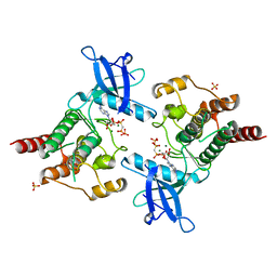
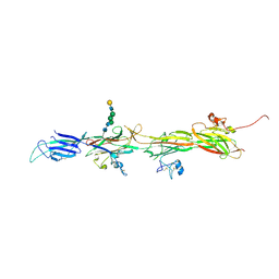
7MEW
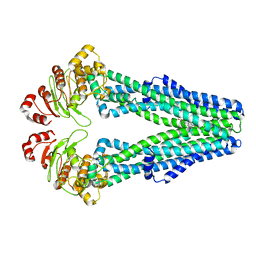 | | E. coli MsbA in complex with G247 | | 分子名称: | (2E)-3-{1-cyclopropyl-7-[(1S)-1-(3,6-dichloro-2-fluorophenyl)ethoxy]naphthalen-2-yl}prop-2-enoic acid, ATP-dependent lipid A-core flippase | | 著者 | Thelot, F, Liao, M. | | 登録日 | 2021-04-08 | | 公開日 | 2021-10-06 | | 最終更新日 | 2021-11-10 | | 実験手法 | ELECTRON MICROSCOPY (3.9 Å) | | 主引用文献 | Distinct allosteric mechanisms of first-generation MsbA inhibitors.
Science, 374, 2021
|
|
6JLU
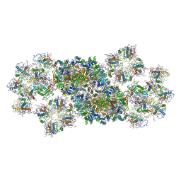 | | Structure of PSII-FCP supercomplex from a centric diatom Chaetoceros gracilis at 3.02 angstrom resolution | | 分子名称: | (3S,3'R,5R,6S,7cis)-7',8'-didehydro-5,6-dihydro-5,6-epoxy-beta,beta-carotene-3,3'-diol, (3S,3'S,5R,5'R,6S,6'R,8'R)-3,5'-dihydroxy-8-oxo-6',7'-didehydro-5,5',6,6',7,8-hexahydro-5,6-epoxy-beta,beta-caroten-3'-yl acetate, 1,2-DI-O-ACYL-3-O-[6-DEOXY-6-SULFO-ALPHA-D-GLUCOPYRANOSYL]-SN-GLYCEROL, ... | | 著者 | Pi, X, Zhao, S, Wang, W, Kuang, T, Sui, S, Shen, J. | | 登録日 | 2019-03-06 | | 公開日 | 2019-07-31 | | 最終更新日 | 2019-11-06 | | 実験手法 | ELECTRON MICROSCOPY (3.02 Å) | | 主引用文献 | The pigment-protein network of a diatom photosystem II-light-harvesting antenna supercomplex.
Science, 365, 2019
|
|
7WVQ
 | |
7WVP
 | |
3O8V
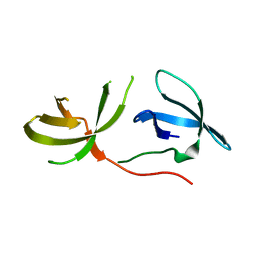 | | Crystal Structure of the Tudor Domains from FXR1 | | 分子名称: | Fragile X mental retardation syndrome-related protein 1 | | 著者 | Lam, R, Guo, Y.H, Adams-Cioaba, M, Bian, C.B, Mackenzie, F, Bountra, C, Weigelt, J, Arrowsmith, C.H, Edwards, A.M, Bochkarev, A, Min, J, Structural Genomics Consortium (SGC) | | 登録日 | 2010-08-03 | | 公開日 | 2010-08-18 | | 最終更新日 | 2024-02-21 | | 実験手法 | X-RAY DIFFRACTION (2.5 Å) | | 主引用文献 | Structural Studies of the Tandem Tudor Domains of Fragile X Mental Retardation Related Proteins FXR1 and FXR2.
Plos One, 5, 2010
|
|
2PSQ
 | |
2PVY
 | |
2PY3
 | |
2PZP
 | |
2Q0B
 | |
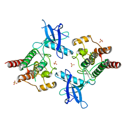
2QS8
 | | Crystal structure of a Xaa-Pro dipeptidase with bound methionine in the active site | | 分子名称: | MAGNESIUM ION, METHIONINE, Xaa-Pro Dipeptidase | | 著者 | Kumaran, D, Burley, S.K, Swaminathan, S, New York SGX Research Center for Structural Genomics (NYSGXRC) | | 登録日 | 2007-07-30 | | 公開日 | 2007-08-21 | | 最終更新日 | 2021-02-03 | | 実験手法 | X-RAY DIFFRACTION (2.33 Å) | | 主引用文献 | Functional annotation of two new carboxypeptidases from the amidohydrolase superfamily of enzymes.
Biochemistry, 48, 2009
|
|
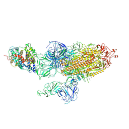
7DD2
 | |
7DK4
 | |
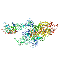
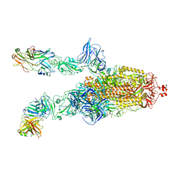
7DDN
 | | SARS-Cov2 S protein at open state | | 分子名称: | Spike glycoprotein | | 著者 | Cong, Y, Liu, C.X. | | 登録日 | 2020-10-29 | | 公開日 | 2020-11-25 | | 最終更新日 | 2021-01-27 | | 実験手法 | ELECTRON MICROSCOPY (6.3 Å) | | 主引用文献 | Development and structural basis of a two-MAb cocktail for treating SARS-CoV-2 infections.
Nat Commun, 12, 2021
|
|
7DDD
 | | SARS-Cov2 S protein at close state | | 分子名称: | Spike glycoprotein | | 著者 | Cong, Y, Liu, C.X. | | 登録日 | 2020-10-28 | | 公開日 | 2020-11-25 | | 最終更新日 | 2021-01-27 | | 実験手法 | ELECTRON MICROSCOPY (3 Å) | | 主引用文献 | Development and structural basis of a two-MAb cocktail for treating SARS-CoV-2 infections.
Nat Commun, 12, 2021
|
|
7DK7
 | |
7DCX
 | |
7DCC
 | |
7DK6
 | |
7DD8
 | |
7DK5
 | |
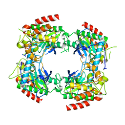
6M1H
 | | CryoEM structure of human PAC1 receptor in complex with maxadilan | | 分子名称: | Guanine nucleotide-binding protein G(I)/G(S)/G(O) subunit gamma-2, Guanine nucleotide-binding protein G(I)/G(S)/G(T) subunit beta-1, Guanine nucleotide-binding protein G(s) subunit alpha isoforms short, ... | | 著者 | Song, X, Wang, J, Zhang, D, Wang, H.W, Ma, Y. | | 登録日 | 2020-02-26 | | 公開日 | 2020-03-11 | | 最終更新日 | 2020-05-27 | | 実験手法 | ELECTRON MICROSCOPY (3.6 Å) | | 主引用文献 | Cryo-EM structures of PAC1 receptor reveal ligand binding mechanism.
Cell Res., 30, 2020
|
|
6M1I
 | | CryoEM structure of human PAC1 receptor in complex with PACAP38 | | 分子名称: | Guanine nucleotide-binding protein G(I)/G(S)/G(O) subunit gamma-2, Guanine nucleotide-binding protein G(I)/G(S)/G(T) subunit beta-1, Guanine nucleotide-binding protein G(s) subunit alpha isoforms short, ... | | 著者 | Song, X, Wang, J, Zhang, D, Wang, H.W, Ma, Y. | | 登録日 | 2020-02-26 | | 公開日 | 2020-03-11 | | 最終更新日 | 2020-05-27 | | 実験手法 | ELECTRON MICROSCOPY (3.5 Å) | | 主引用文献 | Cryo-EM structures of PAC1 receptor reveal ligand binding mechanism.
Cell Res., 30, 2020
|
|
8BQU
 | | Molecular basis of ZP3/ZP1 heteropolymerization: crystal structure of a native vertebrate egg coat filament | | 分子名称: | Choriogenin H, Zona pellucida sperm-binding protein 3, beta-D-galactopyranose-(1-4)-2-acetamido-2-deoxy-beta-D-glucopyranose-(1-2)-alpha-D-mannopyranose-(1-3)-[alpha-D-mannopyranose-(1-6)]beta-D-mannopyranose-(1-4)-2-acetamido-2-deoxy-beta-D-glucopyranose-(1-4)-2-acetamido-2-deoxy-beta-D-glucopyranose | | 著者 | Bokhove, M, de Sanctis, D, Yasumasu, S, Jovine, L. | | 登録日 | 2022-11-21 | | 公開日 | 2024-03-13 | | 最終更新日 | 2024-03-27 | | 実験手法 | X-RAY DIFFRACTION (2.7 Å) | | 主引用文献 | ZP2 cleavage blocks polyspermy by modulating the architecture of the egg coat.
Cell, 187, 2024
|
|


