1VKB
 
 | |
1VKY
 | |
1VR0
 | |
1VLR
 | |
1VQ3
 | |
1VKM
 | |
1VJ1
 | |
1VQR
 | |
1VR8
 | |
1VPZ
 | |
1VK3
 | |
1VKH
 | |
1VL4
 | |
1VJO
 | |
1VR3
 | |
1VRM
 | |
1VQ0
 | |
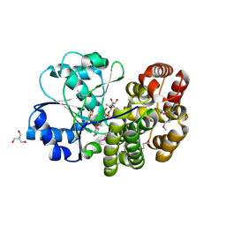
4MKC
 | | Crystal Structure of Anaplastic Lymphoma Kinase Complexed with LDK378 | | Descriptor: | 5-chloro-N~2~-[5-methyl-4-(piperidin-4-yl)-2-(propan-2-yloxy)phenyl]-N~4~-[2-(propan-2-ylsulfonyl)phenyl]pyrimidine-2,4-diamine, ALK tyrosine kinase receptor, GLYCEROL | | Authors: | Lee, C.C, Spraggon, G. | | Deposit date: | 2013-09-04 | | Release date: | 2014-04-09 | | Last modified: | 2023-09-20 | | Method: | X-RAY DIFFRACTION (2.01 Å) | | Cite: | The ALK Inhibitor Ceritinib Overcomes Crizotinib Resistance in Non-Small Cell Lung Cancer.
Cancer Discov, 4, 2014
|
|
3L9P
 | |
3LCS
 | |
4Z55
 | |
1O58
 | |
1O5U
 | |
1O4W
 | |
1O2D
 | |


