Loading
PDBj
メニューPDBj@FacebookPDBj@TwitterPDBj@YouTubewwPDB FoundationwwPDB
RCSB PDBPDBeBMRBAdv. SearchSearch help
Search by PDB author
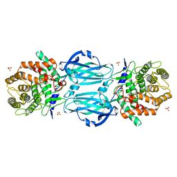
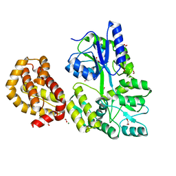
4WMS
DownloadVisualize
BU of 4wms by Molmil
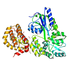
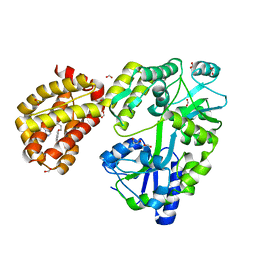
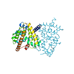
STRUCTURE OF APO MBP-MCL1 AT 1.9A
分子名称: 1,2-ETHANEDIOL, FORMIC ACID, MAGNESIUM ION, ...
著者Clifton, M.C, Dranow, D.M.
登録日2014-10-09
公開日2015-05-06
最終更新日2023-09-27
実験手法X-RAY DIFFRACTION (1.9 Å)
主引用文献A Maltose-Binding Protein Fusion Construct Yields a Robust Crystallography Platform for MCL1.
Plos One, 10, 2015
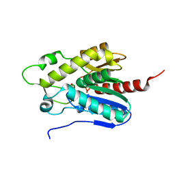
7CCM
DownloadVisualize
BU of 7ccm by Molmil
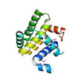
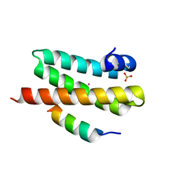
Crystal structure of selenomethionine-derived human BFK
分子名称: Bcl-2-like protein 15
著者Oh, E.K, Jang, D.M, Han, B.W.
登録日2020-06-17
公開日2021-06-23
実験手法X-RAY DIFFRACTION (2.45 Å)
主引用文献Structural analyses on human BFK, a Novel BCL-2 Family Member.
To Be Published
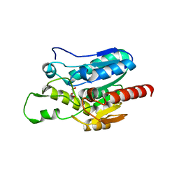
4WXU
DownloadVisualize
BU of 4wxu by Molmil
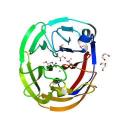
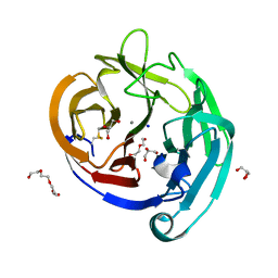
Crystal Structure of the Selenomthionine Incorporated Myocilin Olfactomedin Domain E396D Variant.
分子名称: CALCIUM ION, GLYCEROL, HEXAETHYLENE GLYCOL, ...
著者Donegan, R.K, Freeman, D.M, Lieberman, R.L.
登録日2014-11-14
公開日2015-04-01
実験手法X-RAY DIFFRACTION (2.092 Å)
主引用文献Structural basis for misfolding in myocilin-associated glaucoma.
Hum.Mol.Genet., 24, 2015
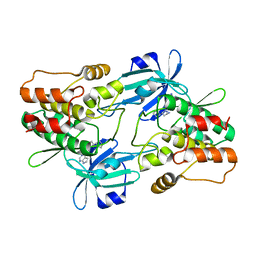
7CCL
DownloadVisualize
BU of 7ccl by Molmil
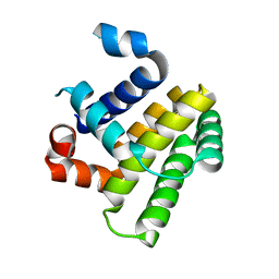
Crystal structure of human BFK
分子名称: Bcl-2-like protein 15
著者Jang, D.M, Oh, E.K, Han, B.W.
登録日2020-06-17
公開日2021-06-23
最終更新日2023-11-29
実験手法X-RAY DIFFRACTION (2.7 Å)
主引用文献Structural analyses on human BFK, a Novel BCL-2 Family Member.
To Be Published
4WMX
DownloadVisualize
BU of 4wmx by Molmil
The structure of MBP-MCL1 bound to ligand 6 at 2.0A
分子名称: 4-ethenyl-2-[(phenylsulfonyl)amino]benzoic acid, FORMIC ACID, MAGNESIUM ION, ...
著者Clifton, M.C, Dranow, D.M.
登録日2014-10-09
公開日2015-05-06
最終更新日2023-09-27
実験手法X-RAY DIFFRACTION (2 Å)
主引用文献A Maltose-Binding Protein Fusion Construct Yields a Robust Crystallography Platform for MCL1.
Plos One, 10, 2015
4WZX
DownloadVisualize
BU of 4wzx by Molmil
ULK3 regulates cytokinetic abscission by phosphorylating ESCRT-III proteins
分子名称: COBALT (II) ION, IST1 homolog, SULFATE ION, ...
著者Caballe, A, Wenzel, D.M, Agromayor, M, Alam, S.L, Skalicky, J.J, Kloc, M, Carlton, J.G, Labrador, L, Sundquist, W.I, Martin-Serrano, J.
登録日2014-11-20
公開日2015-06-03
最終更新日2024-02-28
実験手法X-RAY DIFFRACTION (1.3859 Å)
主引用文献ULK3 regulates cytokinetic abscission by phosphorylating ESCRT-III proteins.
Elife, 4, 2015
4WR2
DownloadVisualize
BU of 4wr2 by Molmil
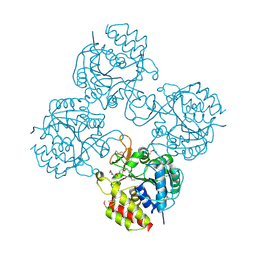
Crystal structure of a putative pyrimidine-specific ribonucleoside hydrolase (RihA) Protein from Shewanella loihica PV-4 (SHEW_0697, Target PSI-029635) with divalent cation and PEG 400 bound at the active site
分子名称: CALCIUM ION, PENTAETHYLENE GLYCOL, Pyrimidine-specific ribonucleoside hydrolase RihA
著者Himmel, D.M, Bhosle, R, Toro, R, Hillerich, B, Gizzi, A, Garforth, S, Kar, A, Chan, M.K, Lafluer, J, Patel, H, Matikainen, B, Chamala, S, Lim, S, Celikgil, A, Villegas, G, Evans, B, Love, J, Fiser, A, Seidel, R.D, Bonanno, J.B, Almo, S.C, New York Structural Genomics Research Consortium (NYSGRC)
登録日2014-10-22
公開日2014-11-12
最終更新日2023-12-27
実験手法X-RAY DIFFRACTION (1.7 Å)
主引用文献Crystal structure of a putative pyrimidine-specific ribonucleoside hydrolase (RihA) Protein from Shewanella loihica PV-4 (SHEW_0697, Target PSI-029635) with divalent cation and PEG 400 bound at the active site
To be published
4WUD
DownloadVisualize
BU of 4wud by Molmil
N-terminal 43 kDa fragment of the E. coli DNA gyrase B subunit grown from no salt condition
分子名称: CHLORIDE ION, DNA gyrase subunit B, MAGNESIUM ION, ...
著者Hearnshaw, S.J, Chung, T.T, Stevenson, C.E.M, Maxwell, A, Lawson, D.M.
登録日2014-10-31
公開日2015-04-08
最終更新日2024-01-10
実験手法X-RAY DIFFRACTION (1.95 Å)
主引用文献The role of monovalent cations in the ATPase reaction of DNA gyrase
Acta Crystallogr.,Sect.D, 71, 2015
4WMT
DownloadVisualize
BU of 4wmt by Molmil
STRUCTURE OF MBP-MCL1 BOUND TO ligand 1 AT 2.35A
分子名称: 1,2-ETHANEDIOL, 7-[2-(1H-imidazol-1-yl)-4-methylpyridin-3-yl]-3-[3-(naphthalen-1-yloxy)propyl]-1-[2-oxo-2-(piperazin-1-yl)ethyl]-1H-indole-2-carboxylic acid, FORMIC ACID, ...
著者Clifton, M.C, Dranow, D.M.
登録日2014-10-09
公開日2015-05-06
最終更新日2023-09-27
実験手法X-RAY DIFFRACTION (2.35 Å)
主引用文献A Maltose-Binding Protein Fusion Construct Yields a Robust Crystallography Platform for MCL1.
Plos One, 10, 2015
4WXS
DownloadVisualize
BU of 4wxs by Molmil
Crystal Structure of the E396D SNP Variant of the Myocilin Olfactomedin Domain
分子名称: CALCIUM ION, GLYCEROL, HEXAETHYLENE GLYCOL, ...
著者Donegan, R.K, Freeman, D.M, Lieberman, R.L.
登録日2014-11-14
公開日2015-04-01
実験手法X-RAY DIFFRACTION (1.9 Å)
主引用文献Structural basis for misfolding in myocilin-associated glaucoma.
Hum.Mol.Genet., 24, 2015
1OKK
DownloadVisualize
BU of 1okk by Molmil
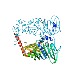
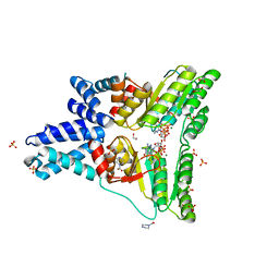
HOMO-HETERODIMERIC COMPLEX OF THE SRP GTPASES
分子名称: 1,2-ETHANEDIOL, CELL DIVISION PROTEIN FTSY, MAGNESIUM ION, ...
著者Focia, P.J, Freymann, D.M.
登録日2003-07-26
公開日2004-01-19
最終更新日2023-12-13
実験手法X-RAY DIFFRACTION (2.05 Å)
主引用文献Heterodimeric Gtpase Core of the Srp Targeting Complex
Science, 303, 2004
4XHD
DownloadVisualize
BU of 4xhd by Molmil
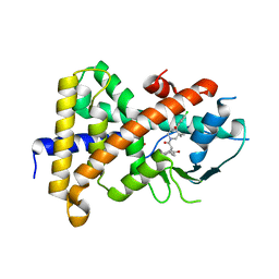
STRUCTURE OF HUMAN PREGNANE X RECEPTOR LIGAND BINDING DOMAIN WITH COMPOUND-1
分子名称: GLYCEROL, N-{(2R)-1-[(4S)-4-(4-chlorophenyl)-4-hydroxy-3,3-dimethylpiperidin-1-yl]-3-methyl-1-oxobutan-2-yl}-2-cyclopropylacetamide, Nuclear receptor subfamily 1 group I member 2
著者Khan, J.A, Camac, D.M.
登録日2015-01-05
公開日2015-01-28
最終更新日2024-02-28
実験手法X-RAY DIFFRACTION (2.4 Å)
主引用文献Developing Adnectins That Target SRC Co-Activator Binding to PXR: A Structural Approach toward Understanding Promiscuity of PXR.
J.Mol.Biol., 427, 2015
4Y2D
DownloadVisualize
BU of 4y2d by Molmil
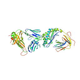
Crystal structure of the mCD1d/7DW8-5/iNKTCR ternary complex
分子名称: 11-(4-fluorophenyl)-N-[(2S,3S,4R)-1-(alpha-D-galactopyranosyloxy)-3,4-dihydroxyoctadecan-2-yl]undecanamide, 2-acetamido-2-deoxy-beta-D-glucopyranose, 2-acetamido-2-deoxy-beta-D-glucopyranose-(1-4)-2-acetamido-2-deoxy-beta-D-glucopyranose, ...
著者Zajonc, D.M, Yu, E.D.
登録日2015-02-09
公開日2015-05-27
最終更新日2023-09-27
実験手法X-RAY DIFFRACTION (3.05 Å)
主引用文献Structural modifications of alphaGalCer in both lipid and carbohydrate moiety influence activation of murine and human iNKT cells
To Be Published
7CXE
DownloadVisualize
BU of 7cxe by Molmil
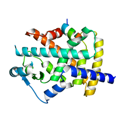
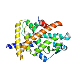
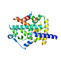
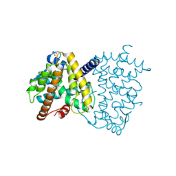
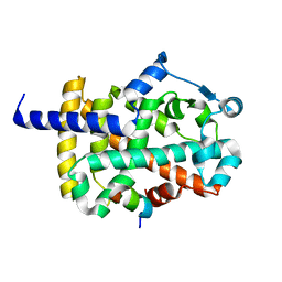
The ligand-free structure of human PPARgamma LBD R280C mutant
分子名称: Peroxisome proliferator-activated receptor gamma
著者Jang, D.M, Han, B.W.
登録日2020-09-01
公開日2021-09-01
最終更新日2023-11-29
実験手法X-RAY DIFFRACTION (2.5 Å)
主引用文献The ligand-free structure of human PPARgamma LBD
To Be Published
7CXJ
DownloadVisualize
BU of 7cxj by Molmil
The ligand-free structure of human PPARgamma LBD R288C mutant in the presence of the SRC-1 coactivator peptide
分子名称: 16-mer peptide from Nuclear receptor coactivator 1, Peroxisome proliferator-activated receptor gamma
著者Jang, D.M, Han, B.W.
登録日2020-09-01
公開日2021-09-01
最終更新日2023-11-29
実験手法X-RAY DIFFRACTION (2.65 Å)
主引用文献The ligand-free structure of human PPARgamma LBD
To Be Published
7CXL
DownloadVisualize
BU of 7cxl by Molmil
The ligand-free structure of human PPARgamma LBD S289C mutant in the presence of the SRC-1 coactivator peptide
分子名称: 16-mer peptide from Nuclear receptor coactivator 1, MALONIC ACID, Peroxisome proliferator-activated receptor gamma
著者Jang, D.M, Han, B.W.
登録日2020-09-01
公開日2021-09-01
最終更新日2023-11-29
実験手法X-RAY DIFFRACTION (2.7 Å)
主引用文献The ligand-free structure of human PPARgamma LBD
To Be Published
7CXI
DownloadVisualize
BU of 7cxi by Molmil
The ligand-free structure of human PPARgamma LBD F287Y mutant in the presence of the SRC-1 coactivator peptide
分子名称: 16-mer peptide from Nuclear receptor coactivator 1, MALONATE ION, Peroxisome proliferator-activated receptor gamma
著者Jang, D.M, Han, B.W.
登録日2020-09-01
公開日2021-09-01
最終更新日2023-11-29
実験手法X-RAY DIFFRACTION (2.3 Å)
主引用文献The ligand-free structure of human PPARgamma LBD
To Be Published
7CXF
DownloadVisualize
BU of 7cxf by Molmil
The ligand-free structure of human PPARgamma LBD C285Y mutant in the presence of the SRC-1 coactivator peptide
分子名称: 16-mer peptide from Nuclear receptor coactivator 1, MALONIC ACID, Peroxisome proliferator-activated receptor gamma
著者Jang, D.M, Han, B.W.
登録日2020-09-01
公開日2021-09-01
最終更新日2023-11-29
実験手法X-RAY DIFFRACTION (2.35 Å)
主引用文献The ligand-free structure of human PPARgamma LBD
To Be Published
7CXK
DownloadVisualize
BU of 7cxk by Molmil
The ligand-free structure of human PPARgamma LBD R288H mutant in the presence of the SRC-1 coactivator peptide
分子名称: 16-mer peptide from Nuclear receptor coactivator 1, MALONATE ION, Peroxisome proliferator-activated receptor gamma
著者Jang, D.M, Han, B.W.
登録日2020-09-01
公開日2021-09-01
最終更新日2023-11-29
実験手法X-RAY DIFFRACTION (2.2 Å)
主引用文献The ligand-free structure of human PPARgamma LBD
To Be Published
7CXG
DownloadVisualize
BU of 7cxg by Molmil
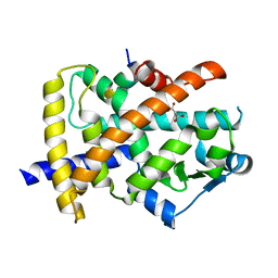
The ligand-free structure of human PPARgamma LBD Q286E mutant
分子名称: GLYCEROL, Peroxisome proliferator-activated receptor gamma
著者Jang, D.M, Han, B.W.
登録日2020-09-01
公開日2021-09-01
最終更新日2023-11-29
実験手法X-RAY DIFFRACTION (1.88 Å)
主引用文献The ligand-free structure of human PPARgamma LBD
To Be Published
7CXH
DownloadVisualize
BU of 7cxh by Molmil
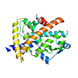
The ligand-free structure of human PPARgamma LBD Q286E mutant in the presence of the SRC-1 coactivator peptide
分子名称: 16-mer peptide from Nuclear receptor coactivator 1, Peroxisome proliferator-activated receptor gamma
著者Jang, D.M, Han, B.W.
登録日2020-09-01
公開日2021-09-01
最終更新日2023-11-29
実験手法X-RAY DIFFRACTION (2.3 Å)
主引用文献The ligand-free structure of human PPARgamma LBD
To Be Published
4XHC
DownloadVisualize
BU of 4xhc by Molmil
rhamnosidase from Klebsiella oxytoca with rhamnose bound
分子名称: Alpha-L-rhamnosidase, SULFATE ION, alpha-L-rhamnopyranose
著者O'Neill, E.O, Stevenson, C.E.M, Patterson, M.J, Rejzek, M, Chauvin, A, Lawson, D.M, Field, R.A.
登録日2015-01-05
公開日2015-04-15
最終更新日2024-05-08
実験手法X-RAY DIFFRACTION (2.7 Å)
主引用文献Crystal structure of a novel two domain GH78 family alpha-rhamnosidase from Klebsiella oxytoca with rhamnose bound.
Proteins, 83, 2015
7E04
DownloadVisualize
BU of 7e04 by Molmil
Crystal structure of Bomgl, a monoacylglycerol lipase from marine Bacillus sp.
分子名称: GLYCEROL, Lipase
著者Wang, Y.H, Wang, J, Lan, D.M.
登録日2021-01-27
公開日2022-02-02
最終更新日2023-11-29
実験手法X-RAY DIFFRACTION (1.25 Å)
主引用文献Crystal structure of Bomgl, a monoacylglycerol lipase from marine Bacillus
To Be Published
7E0N
DownloadVisualize
BU of 7e0n by Molmil
Crystal structure of Monoacylglycerol Lipase chimera
分子名称: GLYCEROL, Thermostable monoacylglycerol lipase,Lipase
著者Lan, D.M.
登録日2021-01-28
公開日2022-03-16
最終更新日2023-11-29
実験手法X-RAY DIFFRACTION (1.85 Å)
主引用文献Crystal structure of Monoacylglycerol Lipase chimera
To Be Published
7F3G
DownloadVisualize
BU of 7f3g by Molmil
Crystal structure of DCLK1 kinase domain in complex with ruxolitinib
分子名称: (3R)-3-cyclopentyl-3-[4-(7H-pyrrolo[2,3-d]pyrimidin-4-yl)-1H-pyrazol-1-yl]propanenitrile, Isoform 4 of Serine/threonine-protein kinase DCLK1
著者Lim, H.J, Jang, D.M, Kim, H.S.
登録日2021-06-16
公開日2021-08-18
最終更新日2023-11-29
実験手法X-RAY DIFFRACTION (2.1 Å)
主引用文献Structural Basis of Inhibition of DCLK1 by Ruxolitinib.
Int J Mol Sci, 22, 2021

220472

件を2024-05-29に公開中

PDB statisticsPDBj update infoContact PDBjnumon