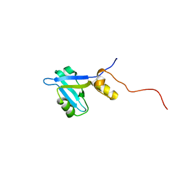
Loading
PDBj
MenuPDBj@FacebookPDBj@TwitterPDBj@YouTubewwPDB FoundationwwPDB
RCSB PDBPDBeBMRBAdv. SearchSearch help
Search by PDB author
2KJD
DownloadVisualize
BU of 2kjd by Molmil
Solution structure of extended PDZ2 domain from NHERF1 (150-270)
Descriptor: Sodium/hydrogen exchange regulatory cofactor NHE-RF1
Authors:Bhattacharya, S, Cowburn, D, Bu, Z.
Deposit date:2009-05-27
Release date:2009-12-29
Last modified:2022-03-16
Method:SOLUTION NMR
Cite:A dynamic intramolecular conformational switch autoregulates the scaffolding protein NHERF1
To be Published
2KRG
DownloadVisualize
BU of 2krg by Molmil
Solution Structure of human sodium/ hydrogen exchange regulatory factor 1(150-358)
Descriptor: Na(+)/H(+) exchange regulatory cofactor NHE-RF1
Authors:Bhattacharya, S, Dai, Z, Li, J, Baxter, S, Callaway, D.J.E, Cowburn, D, Bu, Z.
Deposit date:2009-12-17
Release date:2009-12-29
Last modified:2023-07-26
Method:SOLUTION NMR
Cite:A conformational switch in the scaffolding protein NHERF1 controls autoinhibition and complex formation.
J.Biol.Chem., 285, 2010
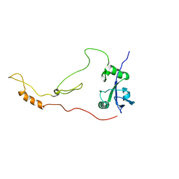
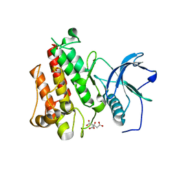
4HDI
DownloadVisualize
BU of 4hdi by Molmil
Crystal Structure of 3E5 IgG3 FAB from mus musculus
Descriptor: Ig heavy chain V region RF, Ig gamma-3 chain C region, Kappa light chain variable region, ...
Authors:Janda, A, Eryilmaz, E, Kim, J, Cordero, R.J.B, Cowburn, D, Casadevall, A.
Deposit date:2012-10-02
Release date:2013-04-17
Last modified:2023-09-20
Method:X-RAY DIFFRACTION (2.449 Å)
Cite:Global structures of IgG isotypes expressing identical variable regions.
Mol.Immunol., 56, 2013
1AYC
DownloadVisualize
BU of 1ayc by Molmil
CRYSTAL STRUCTURES OF PEPTIDE COMPLEXES OF THE AMINO-TERMINAL SH2 DOMAIN OF THE SYP TYROSINE PHOSPHATASE
Descriptor: PEPTIDE PDGFR-740, PROTEIN-TYROSINE PHOSPHATASE SYP (N-TERMINAL SH2 DOMAIN)
Authors:Lee, C.-H, Kuriyan, J.
Deposit date:1994-05-15
Release date:1994-08-31
Last modified:2011-07-13
Method:X-RAY DIFFRACTION (2.3 Å)
Cite:Crystal structures of peptide complexes of the amino-terminal SH2 domain of the Syp tyrosine phosphatase.
Structure, 2, 1994
1AYA
DownloadVisualize
BU of 1aya by Molmil
CRYSTAL STRUCTURES OF PEPTIDE COMPLEXES OF THE AMINO-TERMINAL SH2 DOMAIN OF THE SYP TYROSINE PHOSPHATASE
Descriptor: PEPTIDE PDGFR-1009, PROTEIN-TYROSINE PHOSPHATASE SYP (N-TERMINAL SH2 DOMAIN)
Authors:Lee, C.-H, Kuriyan, J.
Deposit date:1994-05-15
Release date:1994-08-31
Last modified:2011-07-13
Method:X-RAY DIFFRACTION (2.05 Å)
Cite:Crystal structures of peptide complexes of the amino-terminal SH2 domain of the Syp tyrosine phosphatase.
Structure, 2, 1994
1AYB
DownloadVisualize
BU of 1ayb by Molmil
CRYSTAL STRUCTURES OF PEPTIDE COMPLEXES OF THE AMINO-TERMINAL SH2 DOMAIN OF THE SYP TYROSINE PHOSPHATASE
Descriptor: PEPTIDE IRS-1-895, PROTEIN-TYROSINE PHOSPHATASE SYP (N-TERMINAL SH2 DOMAIN)
Authors:Lee, C.-H, Kuriyan, J.
Deposit date:1994-05-15
Release date:1994-08-31
Last modified:2011-07-13
Method:X-RAY DIFFRACTION (3 Å)
Cite:Crystal structures of peptide complexes of the amino-terminal SH2 domain of the Syp tyrosine phosphatase.
Structure, 2, 1994
1AYD
DownloadVisualize
BU of 1ayd by Molmil
CRYSTAL STRUCTURES OF PEPTIDE COMPLEXES OF THE AMINO-TERMINAL SH2 DOMAIN OF THE SYP TYROSINE PHOSPHATASE
Descriptor: PROTEIN-TYROSINE PHOSPHATASE SYP (N-TERMINAL SH2 DOMAIN)
Authors:Lee, C.-H, Kuriyan, J.
Deposit date:1994-05-15
Release date:1994-08-31
Last modified:2024-02-07
Method:X-RAY DIFFRACTION (2.2 Å)
Cite:Crystal structures of peptide complexes of the amino-terminal SH2 domain of the Syp tyrosine phosphatase.
Structure, 2, 1994
1SHB
DownloadVisualize
BU of 1shb by Molmil
CRYSTAL STRUCTURE OF THE PHOSPHOTYROSINE RECOGNITION DOMAIN SH2 OF V-SRC COMPLEXED WITH TYROSINE-PHOSPHORYLATED PEPTIDES
Descriptor: PHOSPHOPEPTIDE B, V-SRC SH2 DOMAIN
Authors:Waksman, G, Kuriyan, J.
Deposit date:1992-08-18
Release date:1993-10-31
Last modified:2011-07-13
Method:X-RAY DIFFRACTION (2 Å)
Cite:Crystal structure of the phosphotyrosine recognition domain SH2 of v-src complexed with tyrosine-phosphorylated peptides.
Nature, 358, 1992
1SHA
DownloadVisualize
BU of 1sha by Molmil
CRYSTAL STRUCTURE OF THE PHOSPHOTYROSINE RECOGNITION DOMAIN SH2 OF V-SRC COMPLEXED WITH TYROSINE-PHOSPHORYLATED PEPTIDES
Descriptor: PHOSPHOPEPTIDE A, V-SRC SH2 DOMAIN
Authors:Waksman, G, Kuriyan, J.
Deposit date:1992-08-18
Release date:1993-10-31
Last modified:2011-07-13
Method:X-RAY DIFFRACTION (1.5 Å)
Cite:Crystal structure of the phosphotyrosine recognition domain SH2 of v-src complexed with tyrosine-phosphorylated peptides.
Nature, 358, 1992
1SPR
DownloadVisualize
BU of 1spr by Molmil
BINDING OF A HIGH AFFINITY PHOSPHOTYROSYL PEPTIDE TO THE SRC SH2 DOMAIN: CRYSTAL STRUCTURES OF THE COMPLEXED AND PEPTIDE-FREE FORMS
Descriptor: PHOSPHATE ION, SRC TYROSINE KINASE SH2 DOMAIN
Authors:Waksman, G, Kuriyan, J.
Deposit date:1993-03-05
Release date:1994-05-31
Last modified:2024-02-14
Method:X-RAY DIFFRACTION (2.5 Å)
Cite:Binding of a high affinity phosphotyrosyl peptide to the Src SH2 domain: crystal structures of the complexed and peptide-free forms.
Cell(Cambridge,Mass.), 72, 1993
1SPS
DownloadVisualize
BU of 1sps by Molmil
BINDING OF A HIGH AFFINITY PHOSPHOTYROSYL PEPTIDE TO THE SRC SH2 DOMAIN: CRYSTAL STRUCTURES OF THE COMPLEXED AND PEPTIDE-FREE FORMS
Descriptor: PEPTIDE YEEI, SRC SH2 DOMAIN
Authors:Waksman, G, Kuriyan, J.
Deposit date:1993-03-05
Release date:1994-05-31
Last modified:2011-07-13
Method:X-RAY DIFFRACTION (2.7 Å)
Cite:Binding of a high affinity phosphotyrosyl peptide to the Src SH2 domain: crystal structures of the complexed and peptide-free forms.
Cell(Cambridge,Mass.), 72, 1993
1CKB
DownloadVisualize
BU of 1ckb by Molmil
STRUCTURAL BASIS FOR THE SPECIFIC INTERACTION OF LYSINE-CONTAINING PROLINE-RICH PEPTIDES WITH THE N-TERMINAL SH3 DOMAIN OF C-CRK
Descriptor: C-CRK N-TERMINAL SH3 DOMAIN, SOS PEPTIDE (PRO-PRO-PRO-VAL-PRO-PRO-ARG-ARG-ARG-ARG)
Authors:Wu, X, Kuriyan, J.
Deposit date:1995-01-24
Release date:1995-05-08
Last modified:2024-02-07
Method:X-RAY DIFFRACTION (1.9 Å)
Cite:Structural basis for the specific interaction of lysine-containing proline-rich peptides with the N-terminal SH3 domain of c-Crk.
Structure, 3, 1995
1CKA
DownloadVisualize
BU of 1cka by Molmil
STRUCTURAL BASIS FOR THE SPECIFIC INTERACTION OF LYSINE-CONTAINING PROLINE-RICH PEPTIDES WITH THE N-TERMINAL SH3 DOMAIN OF C-CRK
Descriptor: C-CRK N-TERMINAL SH3 DOMAIN, C3G PEPTIDE (PRO-PRO-PRO-ALA-LEU-PRO-PRO-LYS-LYS-ARG)
Authors:Wu, X, Kuriyan, J.
Deposit date:1995-01-24
Release date:1995-05-08
Last modified:2024-02-07
Method:X-RAY DIFFRACTION (1.5 Å)
Cite:Structural basis for the specific interaction of lysine-containing proline-rich peptides with the N-terminal SH3 domain of c-Crk.
Structure, 3, 1995
1HCT
DownloadVisualize
BU of 1hct by Molmil
NMR STRUCTURE OF THE HUMAN SRC SH2 DOMAIN COMPLEX
Descriptor: ACETYL-PYEEIE-OH, HUMAN SRC
Authors:Gampe Junior, R.T, Xu, R.X.
Deposit date:1994-09-02
Release date:1995-09-15
Last modified:2022-02-23
Method:SOLUTION NMR
Cite:Solution structure of the human pp60c-src SH2 domain complexed with a phosphorylated tyrosine pentapeptide.
Biochemistry, 34, 1995
1HCS
DownloadVisualize
BU of 1hcs by Molmil
NMR STRUCTURE OF THE HUMAN SRC SH2 DOMAIN COMPLEX
Descriptor: ACETYL-PYEEIE-OH, HUMAN SRC
Authors:Gampe Junior, R.T, Xu, R.X.
Deposit date:1994-09-02
Release date:1995-09-15
Last modified:2022-02-23
Method:SOLUTION NMR
Cite:Solution structure of the human pp60c-src SH2 domain complexed with a phosphorylated tyrosine pentapeptide.
Biochemistry, 34, 1995
1M30
DownloadVisualize
BU of 1m30 by Molmil
Solution structure of N-terminal SH3 domain from oncogene protein c-Crk
Descriptor: Proto-oncogene C-crk
Authors:Schumann, F.H, Varadan, R, Tayakuniyil, P.P, Hall, J.B, Camarero, J.A, Fushman, D.
Deposit date:2002-06-26
Release date:2003-08-05
Last modified:2021-10-27
Method:SOLUTION NMR
Cite:Changing protein backbone topology: Structural and dynamic consequences of the backbone cyclization in SH3 domain
To be Published
1M3C
DownloadVisualize
BU of 1m3c by Molmil
Solution structure of a circular form of the N-terminal SH3 domain (E132C, E133G, R191G mutant) from oncogene protein c-Crk
Descriptor: Proto-oncogene C-crk
Authors:Schumann, F.H, Varadan, R, Tayakuniyil, P.P, Hall, J.B, Camarero, J.A, Fushman, D.
Deposit date:2002-06-27
Release date:2003-08-05
Last modified:2021-10-27
Method:SOLUTION NMR
Cite:Changing protein backbone topology: Structural and dynamic consequences of the backbone cyclization in SH3 domain
To be Published
1M3B
DownloadVisualize
BU of 1m3b by Molmil
Solution structure of a circular form of the N-terminal SH3 domain (A134C, E135G, R191G mutant) from oncogene protein c-Crk.
Descriptor: Proto-oncogene C-crk
Authors:Schumann, F.H, Varadan, R, Tayakuniyil, P.P, Hall, J.B, Camarero, J.A, Fushman, D.
Deposit date:2002-06-27
Release date:2003-08-05
Last modified:2021-10-27
Method:SOLUTION NMR
Cite:Changing protein backbone topology: Structural and dynamic consequences of the backbone cyclization in SH3 domain
To be Published
1M3A
DownloadVisualize
BU of 1m3a by Molmil
Solution structure of a circular form of the truncated N-terminal SH3 domain from oncogene protein c-Crk.
Descriptor: Proto-oncogene C-crk
Authors:Schumann, F.H, Varadan, R, Tayakuniyil, P.P, Hall, J.B, Camarero, J.A, Fushman, D.
Deposit date:2002-06-27
Release date:2003-08-05
Last modified:2021-10-27
Method:SOLUTION NMR
Cite:Changing protein backbone topology: Structural and dynamic consequences of the backbone cyclization in SH3 domain
To be Published
1BKL
DownloadVisualize
BU of 1bkl by Molmil
SELF-ASSOCIATED APO SRC SH2 DOMAIN
Descriptor: PP60 V-SRC TYROSINE KINASE TRANSFORMING PROTEIN
Authors:Holland, D.R, Rubin, J.R.
Deposit date:1997-05-02
Release date:1997-07-23
Last modified:2023-08-02
Method:X-RAY DIFFRACTION (2.1 Å)
Cite:Novel Pp60Src Sh2 Domain Crystal Structures: A 2.0 Angstrom Co-Crystal Structure of a D-Amino Acid Substituted Phosphopeptide Complex and a 2.1 Angstrom Apo Structure Displaying Self-Association
To be Published
4J99
DownloadVisualize
BU of 4j99 by Molmil
Crystal Structure of FGF Receptor 2 (FGFR2) Kinase Domain Harboring the Gain-of-Function K659T Mutation.
Descriptor: Fibroblast growth factor receptor 2, MAGNESIUM ION, PHOSPHOMETHYLPHOSPHONIC ACID ADENYLATE ESTER, ...
Authors:Chen, H, Mohammadi, M.
Deposit date:2013-02-15
Release date:2013-08-07
Last modified:2023-09-20
Method:X-RAY DIFFRACTION (1.8474 Å)
Cite:Cracking the Molecular Origin of Intrinsic Tyrosine Kinase Activity through Analysis of Pathogenic Gain-of-Function Mutations.
Cell Rep, 4, 2013
4J95
DownloadVisualize
BU of 4j95 by Molmil
Crystal Structure of FGF Receptor 2 (FGFR2) Kinase Domain Harboring the Pathogenic K659N Mutation Responsible for an Unclassified Craniosynostosis Syndrome in Space Group C2.
Descriptor: Fibroblast growth factor receptor 2, PHOSPHOMETHYLPHOSPHONIC ACID ADENYLATE ESTER, SULFATE ION
Authors:Chen, H, Mohammadi, M.
Deposit date:2013-02-15
Release date:2013-08-07
Last modified:2023-09-20
Method:X-RAY DIFFRACTION (2.3767 Å)
Cite:Cracking the Molecular Origin of Intrinsic Tyrosine Kinase Activity through Analysis of Pathogenic Gain-of-Function Mutations.
Cell Rep, 4, 2013
4J98
DownloadVisualize
BU of 4j98 by Molmil
Crystal Structure of FGF Receptor 2 (FGFR2) Kinase Domain Harboring the Gain-of-Function K659Q Mutation.
Descriptor: Fibroblast growth factor receptor 2, PHOSPHOMETHYLPHOSPHONIC ACID ADENYLATE ESTER, SULFATE ION
Authors:Huang, Z, Chen, H, Mohammadi, M.
Deposit date:2013-02-15
Release date:2013-08-07
Last modified:2023-09-20
Method:X-RAY DIFFRACTION (2.3067 Å)
Cite:Cracking the Molecular Origin of Intrinsic Tyrosine Kinase Activity through Analysis of Pathogenic Gain-of-Function Mutations.
Cell Rep, 4, 2013
4J97
DownloadVisualize
BU of 4j97 by Molmil
Crystal Structure of FGF Receptor 2 (FGFR2) Kinase Domain Harboring the Pathogenic Gain-of-Function K659E Mutation Identified in Endometrial Cancer.
Descriptor: Fibroblast growth factor receptor 2, PHOSPHOMETHYLPHOSPHONIC ACID ADENYLATE ESTER, SULFATE ION
Authors:Chen, H, Mohammadi, M.
Deposit date:2013-02-15
Release date:2013-08-07
Last modified:2023-09-20
Method:X-RAY DIFFRACTION (2.5482 Å)
Cite:Cracking the Molecular Origin of Intrinsic Tyrosine Kinase Activity through Analysis of Pathogenic Gain-of-Function Mutations.
Cell Rep, 4, 2013
4J96
DownloadVisualize
BU of 4j96 by Molmil
Crystal Structure of FGF Receptor 2 (FGFR2) Kinase Domain Harboring the Pathogenic Gain-of-Function K659M Mutation Identified in Cervical Cancer.
Descriptor: CITRATE ANION, Fibroblast growth factor receptor 2, MAGNESIUM ION, ...
Authors:Chen, H, Mohammadi, M.
Deposit date:2013-02-15
Release date:2013-08-07
Last modified:2023-09-20
Method:X-RAY DIFFRACTION (2.2972 Å)
Cite:Cracking the Molecular Origin of Intrinsic Tyrosine Kinase Activity through Analysis of Pathogenic Gain-of-Function Mutations.
Cell Rep, 4, 2013

218853

PDB entries from 2024-04-24

PDB statisticsPDBj update infoContact PDBjnumon