Loading
PDBj
MenuPDBj@FacebookPDBj@TwitterPDBj@YouTubewwPDB FoundationwwPDB
RCSB PDBPDBeBMRBAdv. SearchSearch help
Search by PDB author
3KG7
DownloadVisualize
BU of 3kg7 by Molmil
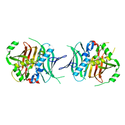
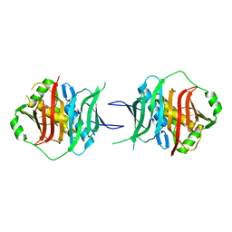
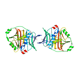
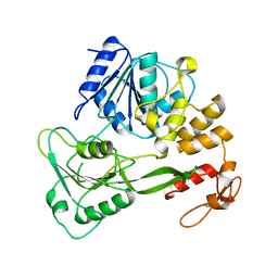
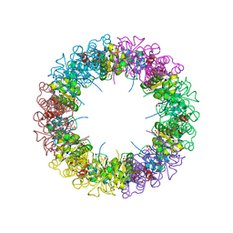
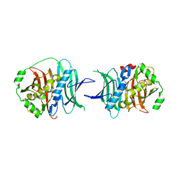
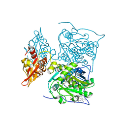
Dehydratase domain from CurH module of Curacin polyketide synthase
Descriptor: CurH
Authors:Akey, D.L, Smith, J.L.
Deposit date:2009-10-28
Release date:2010-01-19
Last modified:2017-11-01
Method:X-RAY DIFFRACTION (2.77 Å)
Cite:Crystal Structures of Dehydratase Domains from the Curacin Polyketide Biosynthetic Pathway.
Structure, 18, 2010
3BOF
DownloadVisualize
BU of 3bof by Molmil
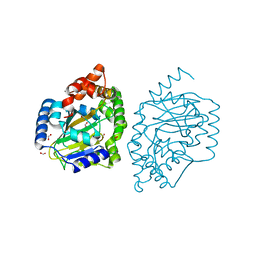
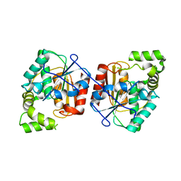
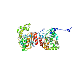
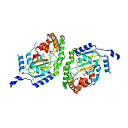
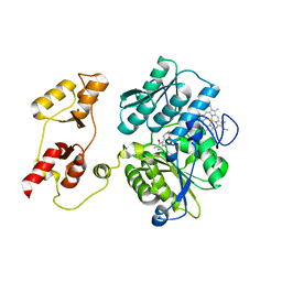
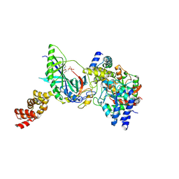
Cobalamin-dependent methionine synthase (1-566) from Thermotoga maritima complexed with Zn2+ and Homocysteine
Descriptor: 2-AMINO-4-MERCAPTO-BUTYRIC ACID, 5-methyltetrahydrofolate S-homocysteine methyltransferase, POTASSIUM ION, ...
Authors:Koutmos, M, Smith, J.L, Ludwig, M.L.
Deposit date:2007-12-17
Release date:2008-03-11
Last modified:2024-04-03
Method:X-RAY DIFFRACTION (1.7 Å)
Cite:Metal active site elasticity linked to activation of homocysteine in methionine synthases.
Proc.Natl.Acad.Sci.Usa, 105, 2008
2REF
DownloadVisualize
BU of 2ref by Molmil
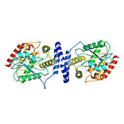
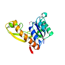
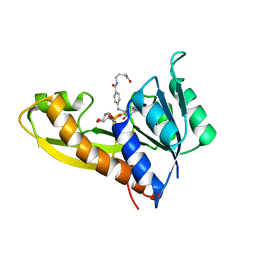
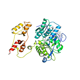
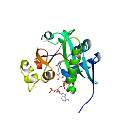
Crystal structure of the loading GNATL domain of CurA from Lyngbya majuscula soaked with malonyl-CoA
Descriptor: ACETYL COENZYME *A, CurA
Authors:Geders, T.W, Smith, J.L.
Deposit date:2007-09-26
Release date:2007-11-20
Last modified:2024-04-03
Method:X-RAY DIFFRACTION (2.75 Å)
Cite:GNAT-like strategy for polyketide chain initiation.
Science, 318, 2007
2Q34
DownloadVisualize
BU of 2q34 by Molmil
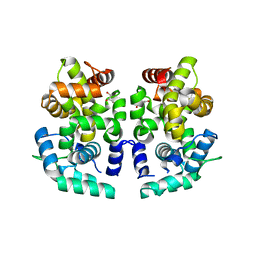
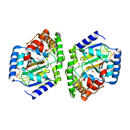
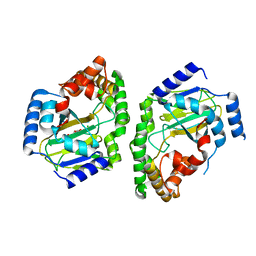
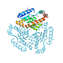
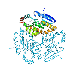
Crystal Structure of the ECH2 decarboxylase domain of CurF from Lyngbya majuscula, rhombohedral crystal form
Descriptor: CurF
Authors:Geders, T.W, Mowers, J.C, Smith, J.L.
Deposit date:2007-05-29
Release date:2007-10-09
Last modified:2023-11-15
Method:X-RAY DIFFRACTION (1.85 Å)
Cite:Crystal structure of the ECH2 catalytic domain of CurF from Lyngbya majuscula. Insights into a decarboxylase involved in polyketide chain beta-branching.
J.Biol.Chem., 282, 2007
2REE
DownloadVisualize
BU of 2ree by Molmil
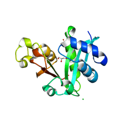
Crystal structure of the loading GNATL domain of CurA from Lyngbya majuscula
Descriptor: CHLORIDE ION, CurA, GLYCEROL, ...
Authors:Geders, T.W, Smith, J.L.
Deposit date:2007-09-26
Release date:2007-11-20
Last modified:2024-02-21
Method:X-RAY DIFFRACTION (1.95 Å)
Cite:GNAT-like strategy for polyketide chain initiation.
Science, 318, 2007
2Q35
DownloadVisualize
BU of 2q35 by Molmil
Crystal Structure of the Y82F variant of ECH2 decarboxylase domain of CurF from Lyngbya majuscula
Descriptor: CHLORIDE ION, CurF, GLYCEROL
Authors:Geders, T.W, Mowers, J.C, Smith, J.L.
Deposit date:2007-05-29
Release date:2007-10-09
Last modified:2023-08-30
Method:X-RAY DIFFRACTION (1.65 Å)
Cite:Crystal structure of the ECH2 catalytic domain of CurF from Lyngbya majuscula. Insights into a decarboxylase involved in polyketide chain beta-branching.
J.Biol.Chem., 282, 2007
2Q2X
DownloadVisualize
BU of 2q2x by Molmil
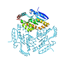
Crystal Structure of the ECH2 decarboxylase domain of CurF from Lyngbya majuscula
Descriptor: CurF, GLYCEROL
Authors:Geders, T.W, Mowers, J.C, Smith, J.L.
Deposit date:2007-05-29
Release date:2007-10-09
Last modified:2017-10-18
Method:X-RAY DIFFRACTION (2 Å)
Cite:Crystal structure of the ECH2 catalytic domain of CurF from Lyngbya majuscula. Insights into a decarboxylase involved in polyketide chain beta-branching.
J.Biol.Chem., 282, 2007
2XD4
DownloadVisualize
BU of 2xd4 by Molmil
Nucleotide-bound Structures of Bacillus subtilis Glycinamide Ribonucleotide Synthetase
Descriptor: ADENOSINE-5'-DIPHOSPHATE, MAGNESIUM ION, PHOSPHORIBOSYLAMINE--GLYCINE LIGASE
Authors:Bertrand, J.A, Chen, S, Zalkin, H, Smith, J.L.
Deposit date:2010-04-29
Release date:2011-03-23
Last modified:2023-12-20
Method:X-RAY DIFFRACTION (2.65 Å)
Cite:Nucleotide-Bound Structures of Bacillus Subtilis Glycinamide Ribonucleotide Synthetase
To be Published
2XCL
DownloadVisualize
BU of 2xcl by Molmil
Nucleotide-bound Structures of Bacillus subtilis Glycinamide Ribonucleotide Synthetase
Descriptor: MAGNESIUM ION, PHOSPHOAMINOPHOSPHONIC ACID-ADENYLATE ESTER, PHOSPHORIBOSYLAMINE--GLYCINE LIGASE
Authors:Bertrand, J.A, Chen, S, Zalkin, H, Smith, J.L.
Deposit date:2010-04-23
Release date:2011-03-23
Last modified:2011-07-13
Method:X-RAY DIFFRACTION (2.1 Å)
Cite:Nucleotide-Bound Structures of Bacillus Subtilis Glycinamide Ribonucleotide Synthetase
To be Published
7QOF
DownloadVisualize
BU of 7qof by Molmil
Icosahedral capsid of the phicrAss001 virion
Descriptor: Auxiliary capsid protein gp36, Head fiber dimer protein gp29, Head fiber trimer protein gp21, ...
Authors:Bayfield, O.W, Shkoporov, A.N, Yutin, N, Khokhlova, E.V, Smith, J.L.R, Hawkins, D.E.D.P, Koonin, E.V, Hill, C, Antson, A.A.
Deposit date:2021-12-24
Release date:2023-03-29
Last modified:2023-05-24
Method:ELECTRON MICROSCOPY (3.01 Å)
Cite:Structural atlas of a human gut crassvirus.
Nature, 617, 2023
7QOH
DownloadVisualize
BU of 7qoh by Molmil
Unique vertex of the phicrAss001 virion with C5 symmetry imposed
Descriptor: Auxiliary capsid protein gp36, Head fiber trimer protein gp21, MAGNESIUM ION, ...
Authors:Bayfield, O.W, Shkoporov, A.N, Yutin, N, Khokhlova, E.V, Smith, J.L.R, Hawkins, D.E.D.P, Koonin, E.V, Hill, C, Antson, A.A.
Deposit date:2021-12-24
Release date:2023-03-29
Last modified:2023-05-24
Method:ELECTRON MICROSCOPY (3.32 Å)
Cite:Structural atlas of a human gut crassvirus.
Nature, 617, 2023
7QOK
DownloadVisualize
BU of 7qok by Molmil
Tail muzzle assembly of the phicrAss001 virion with C6 symmetry imposed
Descriptor: MAGNESIUM ION, Muzzle bound helix, Muzzle protein gp44, ...
Authors:Bayfield, O.W, Shkoporov, A.N, Yutin, N, Khokhlova, E.V, Smith, J.L.R, Hawkins, D.E.D.P, Koonin, E.V, Hill, C, Antson, A.A.
Deposit date:2021-12-24
Release date:2023-03-29
Last modified:2023-05-24
Method:ELECTRON MICROSCOPY (3.38 Å)
Cite:Structural atlas of a human gut crassvirus.
Nature, 617, 2023
7QOG
DownloadVisualize
BU of 7qog by Molmil
Portal protein assembly of the phicrAss001 virion with C12 symmetry imposed
Descriptor: Cargo protein 1 gp45, Portal protein gp20, Ring protein 1 gp43, ...
Authors:Bayfield, O.W, Shkoporov, A.N, Yutin, N, Khokhlova, E.V, Smith, J.L.R, Hawkins, D.E.D.P, Koonin, E.V, Hill, C, Antson, A.A.
Deposit date:2021-12-24
Release date:2023-03-29
Last modified:2023-05-24
Method:ELECTRON MICROSCOPY (3.09 Å)
Cite:Structural atlas of a human gut crassvirus.
Nature, 617, 2023
7QOJ
DownloadVisualize
BU of 7qoj by Molmil
Tail barrel assembly of the phicrAss001 virion with C12 symmetry imposed
Descriptor: Cargo protein 1 gp45, MAGNESIUM ION, Portal protein gp20, ...
Authors:Bayfield, O.W, Shkoporov, A.N, Yutin, N, Khokhlova, E.V, Smith, J.L.R, Hawkins, D.E.D.P, Koonin, E.V, Hill, C, Antson, A.A.
Deposit date:2021-12-24
Release date:2023-03-29
Last modified:2023-05-24
Method:ELECTRON MICROSCOPY (3.21 Å)
Cite:Structural atlas of a human gut crassvirus.
Nature, 617, 2023
7QOL
DownloadVisualize
BU of 7qol by Molmil
Tail assembly of the phicrAss001 virion with C6 symmetry imposed
Descriptor: Cargo protein 1 gp45, MAGNESIUM ION, Muzzle bound helix, ...
Authors:Bayfield, O.W, Shkoporov, A.N, Yutin, N, Khokhlova, E.V, Smith, J.L.R, Hawkins, D.E.D.P, Koonin, E.V, Hill, C, Antson, A.A.
Deposit date:2021-12-24
Release date:2023-03-29
Last modified:2023-05-24
Method:ELECTRON MICROSCOPY (3.33 Å)
Cite:Structural atlas of a human gut crassvirus.
Nature, 617, 2023
7QOI
DownloadVisualize
BU of 7qoi by Molmil
Unique vertex of the phicrAss001 virion
Descriptor: Auxiliary capsid protein gp36, Cargo protein 1 gp45, Head fiber trimer protein gp21, ...
Authors:Bayfield, O.W, Shkoporov, A.N, Yutin, N, Khokhlova, E.V, Smith, J.L.R, Hawkins, D.E.D.P, Koonin, E.V, Hill, C, Antson, A.A.
Deposit date:2021-12-24
Release date:2023-03-29
Last modified:2023-05-24
Method:ELECTRON MICROSCOPY (3.62 Å)
Cite:Structural atlas of a human gut crassvirus.
Nature, 617, 2023
5THZ
DownloadVisualize
BU of 5thz by Molmil
Crystal structure of CurJ carbon methyltransferase
Descriptor: CITRATE ANION, CurJ, GLYCEROL, ...
Authors:Skiba, M.A, Smith, J.L.
Deposit date:2016-09-30
Release date:2016-10-19
Last modified:2023-10-04
Method:X-RAY DIFFRACTION (2.1 Å)
Cite:Domain Organization and Active Site Architecture of a Polyketide Synthase C-methyltransferase.
ACS Chem. Biol., 11, 2016
5THY
DownloadVisualize
BU of 5thy by Molmil
Crystal structure of SeMet-Substituted CurJ carbon methyltransferase
Descriptor: CurJ, OXIDIZED GLUTATHIONE DISULFIDE, S-ADENOSYL-L-HOMOCYSTEINE
Authors:Skiba, M.A, Smith, J.L.
Deposit date:2016-09-30
Release date:2016-10-19
Last modified:2019-12-04
Method:X-RAY DIFFRACTION (2.087 Å)
Cite:Domain Organization and Active Site Architecture of a Polyketide Synthase C-methyltransferase.
ACS Chem. Biol., 11, 2016
5TZ7
DownloadVisualize
BU of 5tz7 by Molmil
Crystal Structure of CurK Dehydratase D1169N Inactive Mutant
Descriptor: CITRATE ANION, CurK
Authors:Dodge, G.J, Smith, J.L.
Deposit date:2016-11-21
Release date:2017-01-11
Last modified:2023-10-04
Method:X-RAY DIFFRACTION (1.648 Å)
Cite:Vinylogous Dehydration by a Polyketide Dehydratase Domain in Curacin Biosynthesis.
J. Am. Chem. Soc., 138, 2016
5TZ6
DownloadVisualize
BU of 5tz6 by Molmil
Crystal Structure of CurJ Dehydratase H978F Inactive Mutant In Complex with Compound 21
Descriptor: (2E,5R)-5-hydroxy-2-methylhept-2-enoic acid, CurJ
Authors:Dodge, G.J, Smith, J.L.
Deposit date:2016-11-21
Release date:2017-01-11
Last modified:2023-10-04
Method:X-RAY DIFFRACTION (2.4 Å)
Cite:Vinylogous Dehydration by a Polyketide Dehydratase Domain in Curacin Biosynthesis.
J. Am. Chem. Soc., 138, 2016
6MCI
DownloadVisualize
BU of 6mci by Molmil
Solution structure of 7SK stem-loop 1
Descriptor: 7SK RNA
Authors:Pham, V.V, D'Souza, V.M.
Deposit date:2018-08-31
Release date:2018-10-31
Last modified:2024-05-01
Method:SOLUTION NMR
Cite:HIV-1 Tat interactions with cellular 7SK and viral TAR RNAs identifies dual structural mimicry.
Nat Commun, 9, 2018
6MCE
DownloadVisualize
BU of 6mce by Molmil
Solution structure of HIV-1 TAR with Tat RNA Binding Domain
Descriptor: Protein Tat, TAR RNA
Authors:Pham, V.V, D'Souza, V.M.
Deposit date:2018-08-31
Release date:2018-10-31
Last modified:2024-05-01
Method:SOLUTION NMR
Cite:HIV-1 Tat interactions with cellular 7SK and viral TAR RNAs identifies dual structural mimicry.
Nat Commun, 9, 2018
6MCF
DownloadVisualize
BU of 6mcf by Molmil
Solution structure of 7SK stem-loop 1 with HIV-1 Tat RNA Binding Domain
Descriptor: 7SK Stem-loop 1 RNA, Protein Tat
Authors:Pham, V.V, D'Souza, V.M.
Deposit date:2018-08-31
Release date:2018-10-31
Last modified:2024-05-01
Method:SOLUTION NMR
Cite:HIV-1 Tat interactions with cellular 7SK and viral TAR RNAs identifies dual structural mimicry.
Nat Commun, 9, 2018
1K2X
DownloadVisualize
BU of 1k2x by Molmil
Crystal structure of putative asparaginase encoded by Escherichia coli ybiK gene
Descriptor: CHLORIDE ION, Putative L-asparaginase, SODIUM ION
Authors:Borek, D, Jaskolski, M.
Deposit date:2001-09-30
Release date:2003-09-09
Last modified:2023-08-16
Method:X-RAY DIFFRACTION (1.65 Å)
Cite:Crystal packing of plant-type L-asparaginase from Escherichia coli.
Acta Crystallogr.,Sect.D, 64, 2008
8BGC
DownloadVisualize
BU of 8bgc by Molmil
Crystal structure of human Casein Kinase II subunit alpha (CK2a1) in complex with compound 2 (AA-CS-9-003)
Descriptor: 5-[(phenylmethyl)amino]pyrimido[4,5-c]quinoline-8-carboxylic acid, Casein kinase II subunit alpha, SULFATE ION
Authors:Kraemer, A, Axtman, A.D, Knapp, S, Structural Genomics Consortium (SGC)
Deposit date:2022-10-27
Release date:2022-11-23
Last modified:2024-01-31
Method:X-RAY DIFFRACTION (2.8 Å)
Cite:Discovery of a Potent and Selective Naphthyridine-Based Chemical Probe for Casein Kinase 2.
Acs Med.Chem.Lett., 14, 2023

221716

PDB entries from 2024-06-26

PDB statisticsPDBj update infoContact PDBjnumon