Loading
PDBj
MenuPDBj@FacebookPDBj@X(formerly Twitter)PDBj@BlueSkyPDBj@YouTubewwPDB FoundationwwPDBDonate
RCSB PDBPDBeBMRBAdv. SearchSearch help
Search by PDB author
2H9E
DownloadVisualize
BU of 2h9e by Molmil
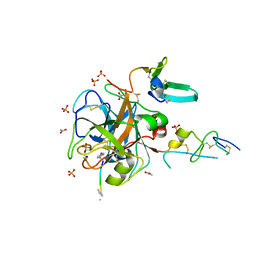
Crystal Structure of FXa/selectide/NAPC2 ternary complex
Descriptor: ACETATE ION, Anti-coagulant protein C2, Coagulation factor X heavy chain, ...
Authors:Murakami, M.T, Geiger, G, Tulinsky, A, Arni, R.K.
Deposit date:2006-06-09
Release date:2007-02-13
Last modified:2025-03-26
Method:X-RAY DIFFRACTION (2.2 Å)
Cite:Intermolecular Interactions and Characterization of the Novel Factor Xa Exosite Involved in Macromolecular Recognition and Inhibition: Crystal Structure of Human Gla-domainless Factor Xa Complexed with the Anticoagulant Protein NAPc2 from the Hematophagous Nematode Ancylostoma caninum.
J.Mol.Biol., 366, 2007
6WP2
DownloadVisualize
BU of 6wp2 by Molmil
The Crystal Structure of Apo Zinc-Bound Domain Swapped-Trimer Q108K:K40D:T53A:R58L:Q38F:Q4F:F57H Variant of HCRBPII
Descriptor: ACETATE ION, GLYCEROL, Retinol-binding protein 2, ...
Authors:Ghanbarpour, A, Geiger, J.
Deposit date:2020-04-26
Release date:2020-08-19
Last modified:2023-10-18
Method:X-RAY DIFFRACTION (2.48 Å)
Cite:Human Cellular Retinol Binding Protein II Forms a Domain-Swapped Trimer Representing a Novel Fold and a New Template for Protein Engineering.
Chembiochem, 21, 2020
6WNF
DownloadVisualize
BU of 6wnf by Molmil
The Crystal Structure of Apo Domain-Swapped Dimer Q108K:K40D:T53A:R58L:Q38F:Q4F:F57H Variant of HCRBPII
Descriptor: ACETATE ION, GLYCEROL, Retinol-binding protein 2
Authors:Ghanbarpour, A, Geiger, J.
Deposit date:2020-04-22
Release date:2020-08-19
Last modified:2023-10-18
Method:X-RAY DIFFRACTION (1.67 Å)
Cite:Human Cellular Retinol Binding Protein II Forms a Domain-Swapped Trimer Representing a Novel Fold and a New Template for Protein Engineering.
Chembiochem, 21, 2020
6WP1
DownloadVisualize
BU of 6wp1 by Molmil
The Crystal Structure of Apo Domain-Swapped Trimer Q108K:K40L:T51K Variant of HCRBPII
Descriptor: ACETATE ION, GLYCEROL, Retinol-binding protein 2
Authors:Ghanbarpour, A, Geiger, J.
Deposit date:2020-04-26
Release date:2020-08-19
Last modified:2023-10-18
Method:X-RAY DIFFRACTION (2.99 Å)
Cite:Human Cellular Retinol Binding Protein II Forms a Domain-Swapped Trimer Representing a Novel Fold and a New Template for Protein Engineering.
Chembiochem, 21, 2020
6WNJ
DownloadVisualize
BU of 6wnj by Molmil
The Crystal Structure of Apo Domain-Swapped Trimer Q108K:T51D:A28C:I32C of HCRBPII
Descriptor: GLYCEROL, Retinol-binding protein 2, SULFATE ION, ...
Authors:Ghanbarpour, A, Geiger, J.
Deposit date:2020-04-22
Release date:2020-08-19
Last modified:2024-11-13
Method:X-RAY DIFFRACTION (2.1 Å)
Cite:Human Cellular Retinol Binding Protein II Forms a Domain-Swapped Trimer Representing a Novel Fold and a New Template for Protein Engineering.
Chembiochem, 21, 2020
7MFX
DownloadVisualize
BU of 7mfx by Molmil
The Crystal Structure of Q108K:K40H:T53A:R58L:Q38F:Q4F Mutant of HCRBPII Bound with FR1 Chromophore Showing Excited State Intermolecular Proton Transfer
Descriptor: (4aP)-N,N-diethyl-9,9-dimethyl-7-[(1E)-prop-1-en-1-yl]-9H-fluoren-2-amine, GLYCEROL, Retinol-binding protein 2
Authors:Ghanbarpour, A, Geiger, J.
Deposit date:2021-04-11
Release date:2022-01-12
Last modified:2024-11-06
Method:X-RAY DIFFRACTION (1.59 Å)
Cite:Design of Large Stokes Shift Fluorescent Proteins Based on Excited State Proton Transfer of an Engineered Photobase.
J.Am.Chem.Soc., 143, 2021
6E51
DownloadVisualize
BU of 6e51 by Molmil
Crystal structure of the apo domain-swapped dimer Q108K:K40L:T51W mutant of human cellular retinol binding protein II
Descriptor: Retinol-binding protein 2
Authors:Ghanbarpour, A, Geiger, J.
Deposit date:2018-07-19
Release date:2019-10-16
Last modified:2023-10-11
Method:X-RAY DIFFRACTION (2.256 Å)
Cite:Engineering the hCRBPII Domain-Swapped Dimer into a New Class of Protein Switches.
J.Am.Chem.Soc., 141, 2019
6E50
DownloadVisualize
BU of 6e50 by Molmil
Crystal structure of the apo domain-swapped dimer Q108K:K40L:T51F mutant of human Cellular Retinol Binding Protein II
Descriptor: ACETATE ION, Retinol-binding protein 2
Authors:Ghanbarpour, A, Geiger, J.
Deposit date:2018-07-18
Release date:2019-10-16
Last modified:2023-10-11
Method:X-RAY DIFFRACTION (1.965 Å)
Cite:Engineering the hCRBPII Domain-Swapped Dimer into a New Class of Protein Switches.
J.Am.Chem.Soc., 141, 2019
6E5E
DownloadVisualize
BU of 6e5e by Molmil
Crystal structure of the apo domain-swapped dimer Q108K:T51D mutant of human cellular retinol binding protein II
Descriptor: ACETATE ION, Retinol-binding protein 2
Authors:Ghanbarpour, A, Geiger, J.
Deposit date:2018-07-20
Release date:2019-10-16
Last modified:2023-10-11
Method:X-RAY DIFFRACTION (1.696 Å)
Cite:Engineering the hCRBPII Domain-Swapped Dimer into a New Class of Protein Switches.
J.Am.Chem.Soc., 141, 2019
6E5Q
DownloadVisualize
BU of 6e5q by Molmil
Crystal structure of the apo domain-swapped dimer Q108K:T51D:A28H mutant of human Cellular Retinol Binding Protein II
Descriptor: ACETATE ION, Retinol-binding protein 2
Authors:Ghanbarpour, A, Geiger, J.
Deposit date:2018-07-21
Release date:2019-10-16
Last modified:2023-10-11
Method:X-RAY DIFFRACTION (1.99 Å)
Cite:Engineering the hCRBPII Domain-Swapped Dimer into a New Class of Protein Switches.
J.Am.Chem.Soc., 141, 2019
6E5R
DownloadVisualize
BU of 6e5r by Molmil
Crystal structure of the apo domain-swapped dimer Q108K:T51D:A28C mutant of human Cellular Retinol Binding Protein II
Descriptor: ACETATE ION, GLYCEROL, Retinol-binding protein 2
Authors:Ghanbarpour, A, Geiger, J.
Deposit date:2018-07-22
Release date:2019-08-07
Last modified:2024-11-20
Method:X-RAY DIFFRACTION (2.592 Å)
Cite:Engineering the hCRBPII Domain-Swapped Dimer into a New Class of Protein Switches.
J.Am.Chem.Soc., 141, 2019
6E7M
DownloadVisualize
BU of 6e7m by Molmil
Crystal structure of the holo retinal-bound domain-swapped dimer Q108K:T51D:A28C mutant of human Cellular Retinol Binding Protein II
Descriptor: ACETATE ION, GLYCEROL, RETINAL, ...
Authors:Ghanbarpour, A, Geiger, J.
Deposit date:2018-07-26
Release date:2019-10-16
Last modified:2024-11-13
Method:X-RAY DIFFRACTION (2.7 Å)
Cite:Engineering the hCRBPII Domain-Swapped Dimer into a New Class of Protein Switches.
J.Am.Chem.Soc., 141, 2019
6E5S
DownloadVisualize
BU of 6e5s by Molmil
Crystal structure of holo retinal-bound domain-swapped dimer Q108K:T51D mutant of human Cellular Retinol Binding Protein II
Descriptor: RETINAL, Retinol-binding protein 2
Authors:Nosrati, M, Ghanbarpour, A, Geiger, J.
Deposit date:2018-07-22
Release date:2019-07-24
Last modified:2024-11-13
Method:X-RAY DIFFRACTION (2.061 Å)
Cite:Engineering the hCRBPII Domain-Swapped Dimer into a New Class of Protein Switches.
J.Am.Chem.Soc., 141, 2019
7MFY
DownloadVisualize
BU of 7mfy by Molmil
The Crystal Structure of Q108K:K40L:T51V:T53S:R58W:Y19W:A33W:L117E Mutant of HCRBPII Bound with LizFluor
Descriptor: 4-{5-[(2E)-but-2-en-2-yl]thiophen-2-yl}-N,N-dimethylaniline, ACETATE ION, GLYCEROL, ...
Authors:Ghanbarpour, A, Geiger, J.
Deposit date:2021-04-12
Release date:2022-01-12
Last modified:2024-11-20
Method:X-RAY DIFFRACTION (1.26 Å)
Cite:Design of Large Stokes Shift Fluorescent Proteins Based on Excited State Proton Transfer of an Engineered Photobase.
J.Am.Chem.Soc., 143, 2021
7MFZ
DownloadVisualize
BU of 7mfz by Molmil
The Crystal Structure of Domain-Swapped Trimer Q108K:K40D:T53A:R58L:Q38F:Q4F Mutant of HCRBPII Bound with LizFluor3 Chromophore Showing Excited State Intermolecular Proton Transfer
Descriptor: 1-(4-{5-[(2E)-but-2-en-2-yl]thiophen-2-yl}phenyl)azetidine, ACETATE ION, GLYCEROL, ...
Authors:Ghanbarpour, A, Geiger, J.
Deposit date:2021-04-12
Release date:2022-01-12
Last modified:2024-10-09
Method:X-RAY DIFFRACTION (2.49 Å)
Cite:Design of Large Stokes Shift Fluorescent Proteins Based on Excited State Proton Transfer of an Engineered Photobase.
J.Am.Chem.Soc., 143, 2021
7LSQ
DownloadVisualize
BU of 7lsq by Molmil
The Crystal Structure of Q108K:K40E:T53A:R58W:Q38F:Q4F:Y19W Mutant of HCRBPII Bound with LizFluor Chromophore Showing Excited State Intermolecular Proton Transfer
Descriptor: 4-{5-[(2E)-but-2-en-2-yl]thiophen-2-yl}-N,N-dimethylaniline, ACETATE ION, Retinol-binding protein 2
Authors:Ghanbarpour, A, Geiger, J.
Deposit date:2021-02-18
Release date:2022-01-12
Last modified:2024-10-23
Method:X-RAY DIFFRACTION (1.67130542 Å)
Cite:Design of Large Stokes Shift Fluorescent Proteins Based on Excited State Proton Transfer of an Engineered Photobase.
J.Am.Chem.Soc., 143, 2021
3UNV
DownloadVisualize
BU of 3unv by Molmil
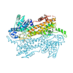
Pantoea agglomerans Phenylalanine Aminomutase
Descriptor: (3S)-3-amino-3-phenylpropanoic acid, 1,2-ETHANEDIOL, AdmH, ...
Authors:Geiger, J, Strom, S.
Deposit date:2011-11-16
Release date:2012-02-22
Last modified:2023-11-15
Method:X-RAY DIFFRACTION (1.54 Å)
Cite:Insights into the mechanistic pathway of the Pantoea agglomerans phenylalanine aminomutase.
Angew.Chem.Int.Ed.Engl., 51, 2012
6MCU
DownloadVisualize
BU of 6mcu by Molmil
Crystal structure of the holo retinal-bound domain-swapped dimer Q108K:T51D:A28H mutant of human Cellular Retinol Binding Protein II
Descriptor: GLYCEROL, RETINAL, Retinol-binding protein 2
Authors:Ghanbarpour, A, Geiger, J.
Deposit date:2018-09-02
Release date:2019-10-16
Last modified:2024-10-23
Method:X-RAY DIFFRACTION (2.572 Å)
Cite:Engineering the hCRBPII Domain-Swapped Dimer into a New Class of Protein Switches.
J.Am.Chem.Soc., 141, 2019
6MCV
DownloadVisualize
BU of 6mcv by Molmil
Crystal Structure of Holo Retinal-Bound Domain-Swapped Dimer of Wild Type Human Cellular Retinol Binding Protein II
Descriptor: RETINAL, Retinol-binding protein 2
Authors:Ghanbarpour, A, Geiger, J.
Deposit date:2018-09-02
Release date:2019-10-16
Last modified:2023-10-11
Method:X-RAY DIFFRACTION (3.3 Å)
Cite:Engineering the hCRBPII Domain-Swapped Dimer into a New Class of Protein Switches.
J.Am.Chem.Soc., 141, 2019
6MQJ
DownloadVisualize
BU of 6mqj by Molmil
Crystal Structure of the All-Trans Retinal bound R111K:Y134F:T54V:R132Q:P39Y:R59Y:L121Q mutant of Human Cellular Retinoic Acid Binding Protein II Irradiated with 532 nm Laser for 10 minutes at 2.1 Angstrom Resolution
Descriptor: Cellular retinoic acid-binding protein 2, RETINAL
Authors:Ghanbarpour, A, Geiger, J.
Deposit date:2018-10-09
Release date:2019-01-09
Last modified:2024-10-30
Method:X-RAY DIFFRACTION (2.125 Å)
Cite:Mimicking Microbial Rhodopsin Isomerization in a Single Crystal.
J. Am. Chem. Soc., 141, 2019
6MOQ
DownloadVisualize
BU of 6moq by Molmil
Crystal Structure of the All-trans Retinal-Bound R111K:Y134F:T54V:R132Q:P39Y:R59Y:L121W Mutant of Human Cellular Retinoic Acid Binding Protein II in the Dark at 2.2 Angstrom Resolution
Descriptor: Cellular retinoic acid-binding protein 2, RETINAL
Authors:Ghanbarpour, A, Geiger, J.
Deposit date:2018-10-04
Release date:2019-01-09
Last modified:2024-11-20
Method:X-RAY DIFFRACTION (2.198 Å)
Cite:Mimicking Microbial Rhodopsin Isomerization in a Single Crystal.
J. Am. Chem. Soc., 141, 2019
6MOP
DownloadVisualize
BU of 6mop by Molmil
Crystal Structure of the All-trans Retinal-Bound R111K:Y134F:T54V:R132Q:P39Y:R59Y:L121E Mutant of Human Cellular Retinoic Acid Binding Protein II in the Dark at 1.9 Angstrom Resolution
Descriptor: Cellular retinoic acid-binding protein 2, RETINAL
Authors:Ghanbarpour, A, Geiger, J.
Deposit date:2018-10-04
Release date:2019-01-09
Last modified:2024-10-16
Method:X-RAY DIFFRACTION (1.9 Å)
Cite:Mimicking Microbial Rhodopsin Isomerization in a Single Crystal.
J. Am. Chem. Soc., 141, 2019
6MPK
DownloadVisualize
BU of 6mpk by Molmil
Crystal Structure of the 13-cis Retinal-Bound R111K:Y134F:T54V:R132Q:P39Y:R59Y:L121E:A32Y Mutant of Human Cellular Retinoic Acid Binding Protein II in the Dark at 1.58 Angstrom Resolution
Descriptor: Cellular retinoic acid-binding protein 2, GLYCEROL, RETINAL
Authors:Ghanbarpour, A, Geiger, J.
Deposit date:2018-10-07
Release date:2019-01-09
Last modified:2024-10-23
Method:X-RAY DIFFRACTION (1.58 Å)
Cite:Mimicking Microbial Rhodopsin Isomerization in a Single Crystal.
J. Am. Chem. Soc., 141, 2019
6MQY
DownloadVisualize
BU of 6mqy by Molmil
Crystal Structure of the 13-cis Product of All-trans Retinal-Bound R111K:Y134F:T54V:R132Q:P39Y:R59Y:L121E Mutant of Human Cellular Retinoic Acid Binding Protein II Irradiated with 400 nm Laser (30 seconds) at 1.9 Angstrom
Descriptor: Cellular retinoic acid-binding protein 2, GLYCEROL, RETINAL
Authors:Ghanbarpour, A, Geiger, J.
Deposit date:2018-10-11
Release date:2019-01-09
Last modified:2024-10-23
Method:X-RAY DIFFRACTION (1.9 Å)
Cite:Mimicking Microbial Rhodopsin Isomerization in a Single Crystal.
J. Am. Chem. Soc., 141, 2019
6MLB
DownloadVisualize
BU of 6mlb by Molmil
Crystal structure of the holo retinal-bound domain-swapped dimer Q108K:K40L:T51F mutant of human cellular retinol binding protein II
Descriptor: ACETATE ION, GLYCEROL, RETINAL, ...
Authors:Ghanbarpour, A, Geiger, J.
Deposit date:2018-09-27
Release date:2019-10-16
Last modified:2024-11-13
Method:X-RAY DIFFRACTION (2.15 Å)
Cite:Engineering the hCRBPII Domain-Swapped Dimer into a New Class of Protein Switches.
J.Am.Chem.Soc., 141, 2019

238582

PDB entries from 2025-07-09

PDB statisticsPDBj update infoContact PDBjnumon