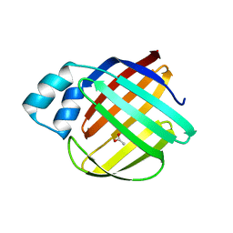
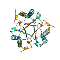
Loading
PDBj
MenuPDBj@FacebookPDBj@TwitterPDBj@YouTubewwPDB FoundationwwPDB
RCSB PDBPDBeBMRBAdv. SearchSearch help
PDB: 187 results

3UNV
DownloadVisualize
BU of 3unv by Molmil
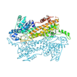
Pantoea agglomerans Phenylalanine Aminomutase
Descriptor: (3S)-3-amino-3-phenylpropanoic acid, 1,2-ETHANEDIOL, AdmH, ...
Authors:Geiger, J, Strom, S.
Deposit date:2011-11-16
Release date:2012-02-22
Last modified:2023-11-15
Method:X-RAY DIFFRACTION (1.54 Å)
Cite:Insights into the mechanistic pathway of the Pantoea agglomerans phenylalanine aminomutase.
Angew.Chem.Int.Ed.Engl., 51, 2012
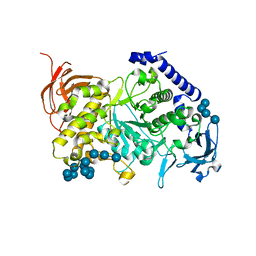
4EAT
DownloadVisualize
BU of 4eat by Molmil
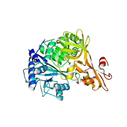
Crystal structure of a benzoate coenzyme A ligase
Descriptor: 1,2-ETHANEDIOL, BENZOIC ACID, Benzoate-coenzyme A ligase, ...
Authors:Geiger, J, Strom, S.
Deposit date:2012-03-22
Release date:2013-03-27
Last modified:2024-02-28
Method:X-RAY DIFFRACTION (1.798 Å)
Cite:Kinetically and Crystallographically Guided Mutations of a Benzoate CoA Ligase (BadA) Elucidate Mechanism and Expand Substrate Permissivity.
Biochemistry, 54, 2015
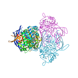
3GUH
DownloadVisualize
BU of 3guh by Molmil
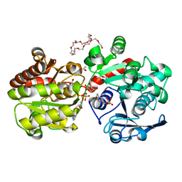
Crystal Structure of Wild-type E.coli GS in complex with ADP and DGM
Descriptor: (2R)-2-hydroxy-3-[4-(2-hydroxyethyl)piperazin-1-yl]propane-1-sulfonic acid, 1,5-anhydro-D-glucitol, 3,6,9,12,15,18,21,24,27,30,33,36,39-TRIDECAOXAHENTETRACONTANE-1,41-DIOL, ...
Authors:Sheng, F, Geiger, J.
Deposit date:2009-03-30
Release date:2009-04-28
Last modified:2023-09-06
Method:X-RAY DIFFRACTION (2.79 Å)
Cite:The Crystal Structures of the Open and Catalytically Competent Closed Conformation of Escherichia coli Glycogen Synthase.
J.Biol.Chem., 284, 2009
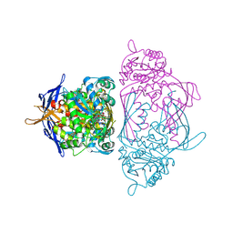
8SDB
DownloadVisualize
BU of 8sdb by Molmil
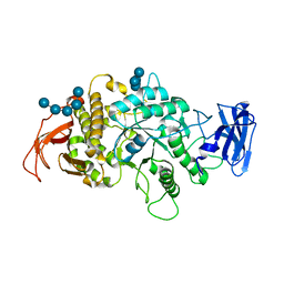
Crystal Structure of E.Coli Branching Enzyme in complex with malto-octose
Descriptor: 1,4-alpha-glucan branching enzyme GlgB, alpha-D-glucopyranose, alpha-D-glucopyranose-(1-4)-alpha-D-glucopyranose, ...
Authors:Bingham, C.R, Nayebi, H, Fawaz, R, Geiger, J.H.
Deposit date:2023-04-06
Release date:2023-07-12
Last modified:2023-08-16
Method:X-RAY DIFFRACTION (3 Å)
Cite:The Structure of Maltooctaose-Bound Escherichia coli Branching Enzyme Suggests a Mechanism for Donor Chain Specificity.
Molecules, 28, 2023
8VZZ
DownloadVisualize
BU of 8vzz by Molmil
Q108K:K40L:T51V:T53S:Y19W:R58W mutant of hCRBPII bound to synthetic fluorophore TD-1V
Descriptor: (2E)-3-{5-[4-(dimethylamino)phenyl]thiophen-2-yl}but-2-enal, ACETATE ION, Retinol-binding protein 2
Authors:Nosssoni, Z, Bingham, C.R, Geiger, J.H, Borhan, B.
Deposit date:2024-02-13
Release date:2024-04-03
Method:X-RAY DIFFRACTION (1.22 Å)
Cite:Regulation of Emission via a Protein-Bound Fluorophore
To Be Published
8VZX
DownloadVisualize
BU of 8vzx by Molmil
Q108K:K40L:T53A:R58F mutant of hCRBPII bound to synthetic fluorophore TD-1V
Descriptor: (2E)-3-{5-[4-(dimethylamino)phenyl]thiophen-2-yl}but-2-enal, ACETATE ION, Retinol-binding protein 2
Authors:Nossoni, Z, Bingham, C.R, Geiger, J.H.
Deposit date:2024-02-13
Release date:2024-04-03
Method:X-RAY DIFFRACTION (1.47 Å)
Cite:Regulation of Emission via a Protein-Bound Fluorophore
To Be Published
6C7Z
DownloadVisualize
BU of 6c7z by Molmil
Crystal structure of the Q108K:K40L:T51V:R58F mutant of human Cellular Retinol Binding Protein II in complex with synthetic Ligand Julolidine
Descriptor: (2E,4E)-3-methyl-5-(2,3,6,7-tetrahydro-1H,5H-pyrido[3,2,1-ij]quinolin-9-yl)penta-2,4-dienal, ACETATE ION, Retinol-binding protein 2
Authors:Nosrati, M, Geiger, J.H.
Deposit date:2018-01-23
Release date:2018-04-25
Last modified:2023-10-04
Method:X-RAY DIFFRACTION (1.42 Å)
Cite:A Genetically Encoded Ratiometric pH Probe: Wavelength Regulation-Inspired Design of pH Indicators.
Chembiochem, 19, 2018
7MFX
DownloadVisualize
BU of 7mfx by Molmil
The Crystal Structure of Q108K:K40H:T53A:R58L:Q38F:Q4F Mutant of HCRBPII Bound with FR1 Chromophore Showing Excited State Intermolecular Proton Transfer
Descriptor: (4aP)-N,N-diethyl-9,9-dimethyl-7-[(1E)-prop-1-en-1-yl]-9H-fluoren-2-amine, GLYCEROL, Retinol-binding protein 2
Authors:Ghanbarpour, A, Geiger, J.
Deposit date:2021-04-11
Release date:2022-01-12
Last modified:2023-10-18
Method:X-RAY DIFFRACTION (1.59 Å)
Cite:Design of Large Stokes Shift Fluorescent Proteins Based on Excited State Proton Transfer of an Engineered Photobase.
J.Am.Chem.Soc., 143, 2021
5U6G
DownloadVisualize
BU of 5u6g by Molmil
Crystal Structure of the holo Domain-Swapped Dimer mutant Q108K:K40D Human Cellular Retinol Binding Protein II bound with all trans retinal
Descriptor: RETINAL, Retinol-binding protein 2
Authors:Assar, Z, Geiger, J.H.
Deposit date:2016-12-08
Release date:2017-10-18
Last modified:2023-10-04
Method:X-RAY DIFFRACTION (2.6 Å)
Cite:Crystal Structure of the holo Domain-Swapped Dimer mutant Q108K:K40D Human Cellular Retinol Binding Protein II bound with all trans retinal
To Be Published
8W00
DownloadVisualize
BU of 8w00 by Molmil
Q108K:K40L:T51V:T53S:Y19W:R58W:L117E mutant of hCRBPII bound to synthetic fluorophore TD-1V
Descriptor: (2E)-3-{5-[4-(dimethylamino)phenyl]thiophen-2-yl}but-2-enal, ACETATE ION, Retinol-binding protein 2
Authors:Nossoni, Z, Bingham, C.R, Geiger, J.H.
Deposit date:2024-02-13
Release date:2024-04-03
Method:X-RAY DIFFRACTION (1.23 Å)
Cite:Regulation of Emission via a Protein-Bound Fluorophore
To Be Published
8VZY
DownloadVisualize
BU of 8vzy by Molmil
Q108K:K40L:T51V:T53S:R58Y mutant of hCRBPII bound to synthetic fluorophore TD-1V
Descriptor: (2E)-3-{5-[4-(dimethylamino)phenyl]thiophen-2-yl}but-2-enal, ACETATE ION, Retinol-binding protein 2
Authors:Nossoni, Z, Bingham, C.R, Geiger, J.H, Borhan, B.
Deposit date:2024-02-13
Release date:2024-04-03
Method:X-RAY DIFFRACTION (1.34 Å)
Cite:Regulation of Emission via a Protein-Bound Fluorophore
To Be Published
8W02
DownloadVisualize
BU of 8w02 by Molmil
Q108K:K40L:T51V:T53S:R58W mutant of hCRBPII bound to synthetic fluorophore TD-1V
Descriptor: (2E)-3-{5-[4-(dimethylamino)phenyl]thiophen-2-yl}but-2-enal, ACETATE ION, Retinol-binding protein 2
Authors:Ghanbarpour, A, Geiger, J.H, Borhan, B.
Deposit date:2024-02-13
Release date:2024-04-03
Method:X-RAY DIFFRACTION (1.499 Å)
Cite:Regulation of Emission via a Protein-Bound Fluorophore
To Be Published
7ML5
DownloadVisualize
BU of 7ml5 by Molmil
Structure of the Starch Branching Enzyme I (BEI) complexed with maltododecaose from Oryza sativa L
Descriptor: Isoform 2 of 1,4-alpha-glucan-branching enzyme, chloroplastic/amyloplastic, alpha-D-glucopyranose-(1-4)-alpha-D-glucopyranose-(1-4)-alpha-D-glucopyranose-(1-4)-alpha-D-glucopyranose, ...
Authors:Nayebi Gavgani, H, Fawaz, R, Geiger, J.H.
Deposit date:2021-04-27
Release date:2021-11-17
Last modified:2023-10-18
Method:X-RAY DIFFRACTION (2.35 Å)
Cite:A structural explanation for the mechanism and specificity of plant branching enzymes I and IIb.
J.Biol.Chem., 298, 2021
1JKI
DownloadVisualize
BU of 1jki by Molmil
myo-Inositol-1-phosphate Synthase Complexed with an Inhibitor, 2-deoxy-glucitol-6-phosphate
Descriptor: 1,4-DIHYDRONICOTINAMIDE ADENINE DINUCLEOTIDE, 2-DEOXY-GLUCITOL-6-PHOSPHATE, AMMONIUM ION, ...
Authors:Stein, A.J, Geiger, J.H.
Deposit date:2001-07-12
Release date:2002-04-10
Last modified:2023-08-16
Method:X-RAY DIFFRACTION (2.2 Å)
Cite:The crystal structure and mechanism of 1-L-myo-inositol- 1-phosphate synthase
J.Biol.Chem., 277, 2002
6MQW
DownloadVisualize
BU of 6mqw by Molmil
Crystal Structure of All-trans Retinal-Bound R111K:Y134F:T54V:R132Q:P39Y:R59Y:L121E Human Cellular Retinoic Acid Binding Protein II Irradiated with 400 nm laser (30 seconds) and subsequently dark adapted (10 minutes) at 2.1 Angstrom Resolution
Descriptor: Cellular retinoic acid-binding protein 2, RETINAL
Authors:Ghanbarpour, A, Geiger, J.
Deposit date:2018-10-11
Release date:2019-01-09
Last modified:2023-10-11
Method:X-RAY DIFFRACTION (2.09 Å)
Cite:Mimicking Microbial Rhodopsin Isomerization in a Single Crystal.
J. Am. Chem. Soc., 141, 2019
6ON5
DownloadVisualize
BU of 6on5 by Molmil
Crystal Structure of the Zn-bound Domain-Swapped Dimer Q108K:T51D:A28C:L36C:F57H Mutant of Human Cellular Retinol Binding Protein II
Descriptor: Retinol-binding protein 2, ZINC ION
Authors:Ghanbarpour, A, Geiger, J.
Deposit date:2019-04-20
Release date:2019-10-16
Last modified:2023-10-11
Method:X-RAY DIFFRACTION (1.638 Å)
Cite:Engineering the hCRBPII Domain-Swapped Dimer into a New Class of Protein Switches.
J.Am.Chem.Soc., 141, 2019
6ON8
DownloadVisualize
BU of 6on8 by Molmil
Crystal Structure of the Reduced Form of Apo Domain-Swapped Dimer Q108K:T51D:A28C:L36C:F57H Mutant of Human Cellular Retinol Binding Protein II
Descriptor: Retinol-binding protein 2
Authors:Ghanbarpour, A, Geiger, J.
Deposit date:2019-04-20
Release date:2019-10-16
Last modified:2023-10-11
Method:X-RAY DIFFRACTION (2.395 Å)
Cite:Engineering the hCRBPII Domain-Swapped Dimer into a New Class of Protein Switches.
J.Am.Chem.Soc., 141, 2019
6ON7
DownloadVisualize
BU of 6on7 by Molmil
Crystal Structure of Apo Domain-Swapped Dimer Q108K:T51D:A28C:L36C Mutant of Human Cellular Retinol Binding Protein II
Descriptor: Retinol-binding protein 2
Authors:Ghanbarpour, A, Geiger, J.
Deposit date:2019-04-20
Release date:2019-10-16
Last modified:2023-10-11
Method:X-RAY DIFFRACTION (1.981 Å)
Cite:Engineering the hCRBPII Domain-Swapped Dimer into a New Class of Protein Switches.
J.Am.Chem.Soc., 141, 2019
1JKF
DownloadVisualize
BU of 1jkf by Molmil
Holo 1L-myo-inositol-1-phosphate Synthase
Descriptor: NICOTINAMIDE-ADENINE-DINUCLEOTIDE, myo-inositol-1-phosphate synthase
Authors:Stein, A.J, Geiger, J.H.
Deposit date:2001-07-12
Release date:2002-04-10
Last modified:2018-01-31
Method:X-RAY DIFFRACTION (2.4 Å)
Cite:The crystal structure and mechanism of 1-L-myo-inositol- 1-phosphate synthase
J.Biol.Chem., 277, 2002
6WP0
DownloadVisualize
BU of 6wp0 by Molmil
The Crystal Structure of Domain-Swapped Trimer Q108K:T51D variant of HCRBPII
Descriptor: ACETATE ION, GLYCEROL, Retinol-binding protein 2
Authors:Ghanbarpour, A, Geiger, J.
Deposit date:2020-04-26
Release date:2020-08-19
Last modified:2023-10-18
Method:X-RAY DIFFRACTION (2.78 Å)
Cite:Human Cellular Retinol Binding Protein II Forms a Domain-Swapped Trimer Representing a Novel Fold and a New Template for Protein Engineering.
Chembiochem, 21, 2020
7MS9
DownloadVisualize
BU of 7ms9 by Molmil
Crystal structure of E114D mutant of Cg10062 with a covalent intermediate of the hydration of acetylenecarboxylic acid
Descriptor: 3-HYDROXY-PROPANOIC ACID, 4-oxalocrotonate tautomerase, SULFATE ION
Authors:Nayebi, G.H, Geiger, J.H, Draths, K.
Deposit date:2021-05-10
Release date:2022-02-02
Last modified:2023-10-18
Method:X-RAY DIFFRACTION (2.2 Å)
Cite:Cg10062 Catalysis Forges a Link between Acetylenecarboxylic Acid and Bacterial Metabolism.
Biochemistry, 60, 2021
7LSQ
DownloadVisualize
BU of 7lsq by Molmil
The Crystal Structure of Q108K:K40E:T53A:R58W:Q38F:Q4F:Y19W Mutant of HCRBPII Bound with LizFluor Chromophore Showing Excited State Intermolecular Proton Transfer
Descriptor: 4-{5-[(2E)-but-2-en-2-yl]thiophen-2-yl}-N,N-dimethylaniline, ACETATE ION, Retinol-binding protein 2
Authors:Ghanbarpour, A, Geiger, J.
Deposit date:2021-02-18
Release date:2022-01-12
Last modified:2023-10-18
Method:X-RAY DIFFRACTION (1.67130542 Å)
Cite:Design of Large Stokes Shift Fluorescent Proteins Based on Excited State Proton Transfer of an Engineered Photobase.
J.Am.Chem.Soc., 143, 2021
7MFZ
DownloadVisualize
BU of 7mfz by Molmil
The Crystal Structure of Domain-Swapped Trimer Q108K:K40D:T53A:R58L:Q38F:Q4F Mutant of HCRBPII Bound with LizFluor3 Chromophore Showing Excited State Intermolecular Proton Transfer
Descriptor: 1-(4-{5-[(2E)-but-2-en-2-yl]thiophen-2-yl}phenyl)azetidine, ACETATE ION, GLYCEROL, ...
Authors:Ghanbarpour, A, Geiger, J.
Deposit date:2021-04-12
Release date:2022-01-12
Last modified:2023-10-18
Method:X-RAY DIFFRACTION (2.49 Å)
Cite:Design of Large Stokes Shift Fluorescent Proteins Based on Excited State Proton Transfer of an Engineered Photobase.
J.Am.Chem.Soc., 143, 2021
7UD1
DownloadVisualize
BU of 7ud1 by Molmil
The Crystal Structure of Apo Monomer F57:H:H:H:R58 HCRBPII with Histidine Insertion in the Hinge Loop Region at 1.3 Angstrom Resolution
Descriptor: Retinol-binding protein 2
Authors:Ghanbarpour, A, Geiger, J.
Deposit date:2022-03-17
Release date:2023-10-18
Method:X-RAY DIFFRACTION (1.32 Å)
Cite:The Crystal Structure of Apo Monomer F57:H:H:H:R58 HCRBPII with Histidine Insertion in the Hinge Loop Region at 1.3 Angstrom Resolution
To Be Published
7UCZ
DownloadVisualize
BU of 7ucz by Molmil
The Crystal Structure of Apo Monomer F57:H:H:H:H:H:H:R58 Mutant of HCRBPII with Histidine Insertion in the Hinge Loop Region at 1.1 Angstrom Resolution
Descriptor: Retinol-binding protein 2
Authors:Ghanbarpour, A, Geiger, J.
Deposit date:2022-03-17
Release date:2023-10-18
Method:X-RAY DIFFRACTION (1.08 Å)
Cite:The Crystal Structure of Domain-Sawpped Dimer Q108K:T51D:A28C:L36C:F57:H:R58 Mutant of hCRBPII with a Histidine Insertion in the Hinge Loop Region at 1.96 Angstrom Resolution
To Be Published

218853

PDB entries from 2024-04-24

PDB statisticsPDBj update infoContact PDBjnumon