Loading
PDBj
MenuPDBj@FacebookPDBj@TwitterPDBj@YouTubewwPDB FoundationwwPDB
RCSB PDBPDBeBMRBAdv. SearchSearch help
Search by PDB author
3DZ1
DownloadVisualize
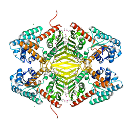
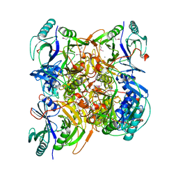
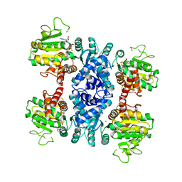
BU of 3dz1 by Molmil
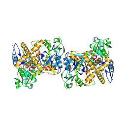
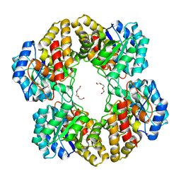
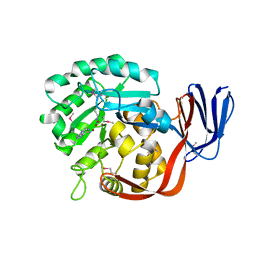
Crystal structure of Dihydrodipicolinate Synthase from Rhodopseudomonas palustris at 1.87A resolution
Descriptor: Dihydrodipicolinate synthase
Authors:Satyanarayana, L, Eswaramoorthy, S, Burley, S.K, Swaminathan, S, New York SGX Research Center for Structural Genomics (NYSGXRC)
Deposit date:2008-07-29
Release date:2008-08-12
Last modified:2024-10-30
Method:X-RAY DIFFRACTION (1.87 Å)
Cite:Crystal structure of Dihydrodipicolinate Synthase from Rhodopseudomonas palustris at 1.87A resolution
To be Published
3T2M
DownloadVisualize
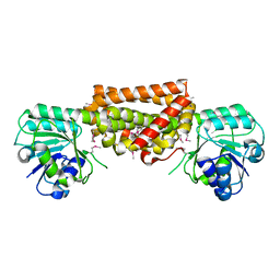
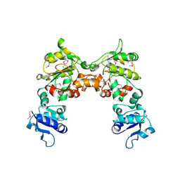
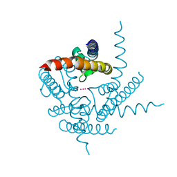
BU of 3t2m by Molmil
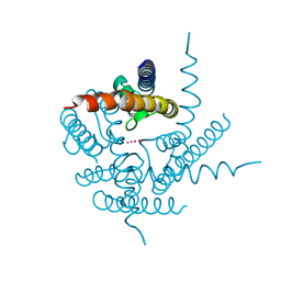
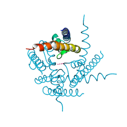
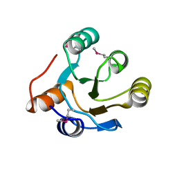
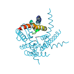
Crystal Structure of NaK Channel N68D Mutant
Descriptor: POTASSIUM ION, Potassium channel protein
Authors:Sauer, D.B, Zeng, W, Raghunathan, S, Jiang, Y.
Deposit date:2011-07-22
Release date:2011-10-05
Last modified:2024-02-28
Method:X-RAY DIFFRACTION (1.953 Å)
Cite:Protein interactions central to stabilizing the K+ channel selectivity filter in a four-sited configuration for selective K+ permeation.
Proc.Natl.Acad.Sci.USA, 108, 2011
3DTY
DownloadVisualize
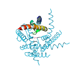
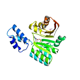
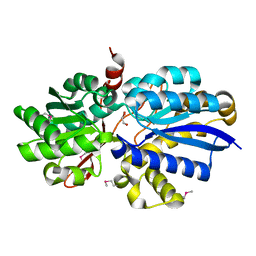
BU of 3dty by Molmil
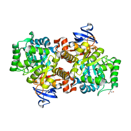
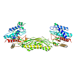
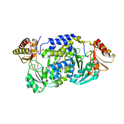
Crystal structure of an Oxidoreductase from Pseudomonas syringae
Descriptor: MAGNESIUM ION, Oxidoreductase, Gfo/Idh/MocA family
Authors:Eswaramoorthy, S, Mahmood, A, Burley, S.K, Swaminathan, S, New York SGX Research Center for Structural Genomics (NYSGXRC)
Deposit date:2008-07-16
Release date:2008-08-05
Last modified:2021-02-10
Method:X-RAY DIFFRACTION (2.04 Å)
Cite:Crystal structure of an Oxidoreductase from Pseudomonas syringae
To be Published
3DZB
DownloadVisualize
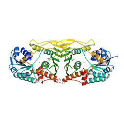
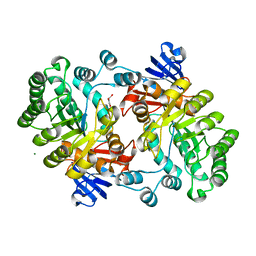
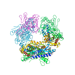
BU of 3dzb by Molmil
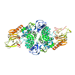
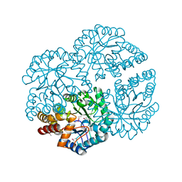
Crystal structure of Prephenate dehydrogenase from Streptococcus thermophilus
Descriptor: Prephenate dehydrogenase
Authors:Zhang, Z, Eswaramoorthy, S, Burley, S.K, Swaminathan, S, New York SGX Research Center for Structural Genomics (NYSGXRC)
Deposit date:2008-07-29
Release date:2008-08-12
Last modified:2024-10-16
Method:X-RAY DIFFRACTION (2.46 Å)
Cite:Crystal structure of Prephenate dehydrogenase from Streptococcus thermophilus
To be Published
3T1C
DownloadVisualize
BU of 3t1c by Molmil
Crystal Structure of NaK Channel D66Y Mutant
Descriptor: POTASSIUM ION, Potassium channel protein
Authors:Sauer, D.B, Zeng, W, Raghunathan, S, Jiang, Y.
Deposit date:2011-07-21
Release date:2011-10-05
Last modified:2024-02-28
Method:X-RAY DIFFRACTION (1.802 Å)
Cite:Protein interactions central to stabilizing the K+ channel selectivity filter in a four-sited configuration for selective K+ permeation.
Proc.Natl.Acad.Sci.USA, 108, 2011
3E82
DownloadVisualize
BU of 3e82 by Molmil
Crystal structure of a putative oxidoreductase from Klebsiella pneumoniae
Descriptor: CHLORIDE ION, Putative oxidoreductase
Authors:Eswaramoorthy, S, Mohammad, M.B, Thomas, C.A, Brown, A.C, Burley, S.K, Swaminathan, S, New York SGX Research Center for Structural Genomics (NYSGXRC)
Deposit date:2008-08-19
Release date:2008-08-26
Last modified:2024-10-09
Method:X-RAY DIFFRACTION (2.04 Å)
Cite:Crystal structure of a putative oxidoreductase from Klebsiella pneumoniae
To be Published
3B59
DownloadVisualize
BU of 3b59 by Molmil
Crystal structure of the Mn(II)-bound glyoxalase from Novosphingobium aromaticivorans
Descriptor: Glyoxalase/bleomycin resistance protein/dioxygenase, MANGANESE (II) ION
Authors:Madegowda, M, Eswaramoorthy, S, Burley, S.K, Swaminathan, S, New York SGX Research Center for Structural Genomics (NYSGXRC)
Deposit date:2007-10-25
Release date:2007-11-06
Last modified:2021-02-03
Method:X-RAY DIFFRACTION (2.53 Å)
Cite:Crystal structure of the Mn(II)-bound glyoxalase from Novosphingobium aromaticivorans.
To be Published
2G6T
DownloadVisualize
BU of 2g6t by Molmil
Crystal structure of an uncharacterized protein from Clostridium acetobutylicum
Descriptor: Uncharacterized protein, homolog HI1244 from Haemophilus influenzae
Authors:Rao, K.N, Swaminathan, S, Burley, S.K, New York SGX Research Center for Structural Genomics (NYSGXRC)
Deposit date:2006-02-25
Release date:2006-03-14
Last modified:2024-10-09
Method:X-RAY DIFFRACTION (3 Å)
Cite:Crystal structure of an uncharacterized protein from Clostridium acetobutylicum
To be Published
3B89
DownloadVisualize
BU of 3b89 by Molmil
Crystal structure of rRNA methylase from Escherichia coli
Descriptor: 16S rRNA methylase, GUANOSINE-5'-MONOPHOSPHATE
Authors:Eswaramoorthy, S, Burley, S.K, Swaminathan, S, New York SGX Research Center for Structural Genomics (NYSGXRC)
Deposit date:2007-10-31
Release date:2007-11-27
Last modified:2024-10-30
Method:X-RAY DIFFRACTION (2.6 Å)
Cite:Crystal structure of rRNA methylase from Escherichia coli.
To be Published
2GOK
DownloadVisualize
BU of 2gok by Molmil
Crystal structure of the imidazolonepropionase from Agrobacterium tumefaciens at 1.87 A resolution
Descriptor: CHLORIDE ION, FE (III) ION, GLYCEROL, ...
Authors:Tyagi, R, Kumaran, D, Swaminathan, S, Burley, S.K, New York SGX Research Center for Structural Genomics (NYSGXRC)
Deposit date:2006-04-13
Release date:2006-04-25
Last modified:2024-02-14
Method:X-RAY DIFFRACTION (1.87 Å)
Cite:X-ray structure of imidazolonepropionase from Agrobacterium tumefaciens at 1.87 A resolution.
Proteins, 69, 2007
2HAE
DownloadVisualize
BU of 2hae by Molmil
Crystal structure of a putative malic enzyme (malate oxidoreductase)
Descriptor: Malate oxidoreductase, NICOTINAMIDE-ADENINE-DINUCLEOTIDE, ZINC ION
Authors:Seetharaman, J, Swaminathan, S, Burley, S.K, New York SGX Research Center for Structural Genomics (NYSGXRC)
Deposit date:2006-06-12
Release date:2006-07-04
Last modified:2023-08-30
Method:X-RAY DIFFRACTION (3.13 Å)
Cite:Crystal structure of a putative malic enzyme (malate oxidoreductase)
To be Published
3EAF
DownloadVisualize
BU of 3eaf by Molmil
Crystal structure of ABC transporter, substrate binding protein Aeropyrum pernix
Descriptor: ABC transporter, substrate binding protein, GLYCEROL, ...
Authors:Zhang, Z, Eswaramoorthy, S, Burley, S.K, Swaminathan, S, New York SGX Research Center for Structural Genomics (NYSGXRC)
Deposit date:2008-08-25
Release date:2008-09-09
Last modified:2024-11-06
Method:X-RAY DIFFRACTION (2 Å)
Cite:Crystal structure of ABC transporter, substrate binding protein Aeropyrum pernix
To be Published
2GUW
DownloadVisualize
BU of 2guw by Molmil
Crystal structure of AMP Nucleosidase from Salmonella typhimurium LT2
Descriptor: AMP nucleosidase
Authors:Rao, K.N, Swaminathan, S, Burley, S.K, New York SGX Research Center for Structural Genomics (NYSGXRC)
Deposit date:2006-05-01
Release date:2006-06-06
Last modified:2024-02-14
Method:X-RAY DIFFRACTION (2.64 Å)
Cite:Crystal structure of AMP Nucleosidase from Salmonella typhimurium LT2
To be Published
3TET
DownloadVisualize
BU of 3tet by Molmil
Crystal Structure of NaK2K Channel Y66F Mutant
Descriptor: POTASSIUM ION, Potassium channel protein
Authors:Sauer, D.B, Zeng, W, Raghunathan, S, Jiang, Y.
Deposit date:2011-08-15
Release date:2011-10-19
Last modified:2024-02-28
Method:X-RAY DIFFRACTION (1.9 Å)
Cite:Protein interactions central to stabilizing the K+ channel selectivity filter in a four-sited configuration for selective K+ permeation.
Proc.Natl.Acad.Sci.USA, 108, 2011
2I6E
DownloadVisualize
BU of 2i6e by Molmil
Crystal structure of protein DR0370 from Deinococcus radiodurans, Pfam DUF178
Descriptor: Hypothetical protein, SULFATE ION
Authors:Tyagi, R, Kumaran, D, Burley, S.K, Swaminathan, S, New York SGX Research Center for Structural Genomics (NYSGXRC)
Deposit date:2006-08-28
Release date:2006-09-05
Last modified:2024-10-30
Method:X-RAY DIFFRACTION (2.5 Å)
Cite:X-ray structures of two proteins belonging to Pfam DUF178 revealed unexpected structural similarity to the DUF191 Pfam family.
Bmc Struct.Biol., 7, 2007
2I9U
DownloadVisualize
BU of 2i9u by Molmil
Crystal Structure of Guanine Deaminase from C. acetobutylicum with bound guanine in the active site
Descriptor: Cytosine/guanine deaminase related protein, FE (III) ION, GLYCEROL, ...
Authors:Kumaran, D, Burley, S.K, Swaminathan, S, New York SGX Research Center for Structural Genomics (NYSGXRC)
Deposit date:2006-09-06
Release date:2006-09-19
Last modified:2024-02-21
Method:X-RAY DIFFRACTION (2.05 Å)
Cite:Crystal Structure of Guanine Deaminase from C. acetobutylicum with bound guanine in the active site
To be Published
3T4D
DownloadVisualize
BU of 3t4d by Molmil
Crystal Structure of NaK2K Channel Y55F Mutant
Descriptor: POTASSIUM ION, Potassium channel protein
Authors:Sauer, D.B, Zeng, W, Raghunathan, S, Jiang, Y.
Deposit date:2011-07-25
Release date:2011-10-05
Last modified:2024-02-28
Method:X-RAY DIFFRACTION (1.7 Å)
Cite:Protein interactions central to stabilizing the K+ channel selectivity filter in a four-sited configuration for selective K+ permeation.
Proc.Natl.Acad.Sci.USA, 108, 2011
3T81
DownloadVisualize
BU of 3t81 by Molmil
Crystal Structure of diiron adenine deaminase
Descriptor: Adenine deaminase 2, FE (III) ION
Authors:Bagaria, A, Kumaran, D, Burley, S.K, Swaminathan, S, New York SGX Research Center for Structural Genomics (NYSGXRC)
Deposit date:2011-08-01
Release date:2011-11-02
Last modified:2023-12-06
Method:X-RAY DIFFRACTION (2.63 Å)
Cite:Crystal Structure of diiron adenine deaminase
TO BE PUBLISHED
3T4Z
DownloadVisualize
BU of 3t4z by Molmil
Crystal Structure of NaK2K Channel Y55W Mutant
Descriptor: POTASSIUM ION, Potassium channel protein
Authors:Sauer, D.B, Zeng, W, Raghunathan, S, Jiang, Y.
Deposit date:2011-07-26
Release date:2011-10-05
Last modified:2024-02-28
Method:X-RAY DIFFRACTION (1.901 Å)
Cite:Protein interactions central to stabilizing the K+ channel selectivity filter in a four-sited configuration for selective K+ permeation.
Proc.Natl.Acad.Sci.USA, 108, 2011
3EB2
DownloadVisualize
BU of 3eb2 by Molmil
Crystal structure of Dihydrodipicolinate Synthase from Rhodopseudomonas palustris at 2.0A resolution
Descriptor: Putative dihydrodipicolinate synthetase, TRIETHYLENE GLYCOL
Authors:Satyanarayana, L, Eswaramoorthy, S, Burley, S.K, Swaminathan, S, New York SGX Research Center for Structural Genomics (NYSGXRC)
Deposit date:2008-08-26
Release date:2008-09-02
Last modified:2024-02-21
Method:X-RAY DIFFRACTION (2.04 Å)
Cite:Crystal structure of Dihydrodipicolinate Synthase from Rhodopseudomonas palustris at 2.0A resolution
To be Published
2IMO
DownloadVisualize
BU of 2imo by Molmil
Crystal structure of allantoate amidohydrolase from Escherichia coli at pH 4.6
Descriptor: Allantoate amidohydrolase
Authors:Agarwal, R, Burley, S.K, Swaminathan, S, New York SGX Research Center for Structural Genomics (NYSGXRC)
Deposit date:2006-10-04
Release date:2006-10-17
Last modified:2024-10-09
Method:X-RAY DIFFRACTION (2.8 Å)
Cite:Structural Analysis of a Ternary Complex of Allantoate Amidohydrolase from Escherichia coli Reveals its Mechanics.
J.Mol.Biol., 368, 2007
2IMG
DownloadVisualize
BU of 2img by Molmil
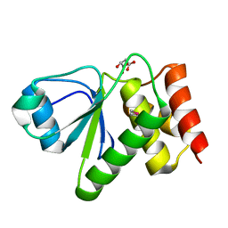
Crystal structure of dual specificity protein phosphatase 23 from Homo sapiens in complex with ligand malate ion
Descriptor: D-MALATE, Dual specificity protein phosphatase 23
Authors:Agarwal, R, Burley, S.K, Swaminathan, S, New York SGX Research Center for Structural Genomics (NYSGXRC)
Deposit date:2006-10-04
Release date:2006-10-17
Last modified:2024-10-30
Method:X-RAY DIFFRACTION (1.93 Å)
Cite:Structure of human dual specificity protein phosphatase 23, VHZ, enzyme-substrate/product complex.
J.Biol.Chem., 283, 2008
2ICS
DownloadVisualize
BU of 2ics by Molmil
Crystal structure of an adenine deaminase
Descriptor: ADENINE, Adenine Deaminase, ZINC ION
Authors:Sugadev, R, Kumaran, D, Burley, S.K, Swaminathan, S, New York SGX Research Center for Structural Genomics (NYSGXRC)
Deposit date:2006-09-13
Release date:2006-10-17
Last modified:2021-02-03
Method:X-RAY DIFFRACTION (2.3 Å)
Cite:Crystal structure of an adenine deaminase
TO BE PUBLISHED
2IOJ
DownloadVisualize
BU of 2ioj by Molmil
Crystal structure of protein AF1212 from Archaeoglobus fulgidus, Pfam DRTGG
Descriptor: Hypothetical protein AF_1212
Authors:Tyagi, R, Burley, S.K, Swaminathan, S, New York SGX Research Center for Structural Genomics (NYSGXRC)
Deposit date:2006-10-10
Release date:2006-10-31
Last modified:2024-10-30
Method:X-RAY DIFFRACTION (2.15 Å)
Cite:The crystal structure of a hypothetical protein (Y1212_ARCFU) from Archeoglobus fulgidus
To be Published
3FKD
DownloadVisualize
BU of 3fkd by Molmil
The crystal structure of L-threonine-O-3-phosphate decarboxylase from Porphyromonas gingivalis
Descriptor: L-threonine-O-3-phosphate decarboxylase
Authors:Zhang, Z, Eswaramoorthy, S, Burley, S.K, Swaminathan, S, New York SGX Research Center for Structural Genomics (NYSGXRC)
Deposit date:2008-12-16
Release date:2009-02-03
Last modified:2024-11-06
Method:X-RAY DIFFRACTION (2.5 Å)
Cite:The crystal structure of L-threonine-O-3-phosphate decarboxylase from Porphyromonas gingivalis
To be Published

227111

PDB entries from 2024-11-06

PDB statisticsPDBj update infoContact PDBjnumon