Loading
PDBj
MenuPDBj@FacebookPDBj@TwitterPDBj@YouTubewwPDB FoundationwwPDB
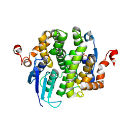
RCSB PDBPDBeBMRBAdv. SearchSearch help
Search by PDB author
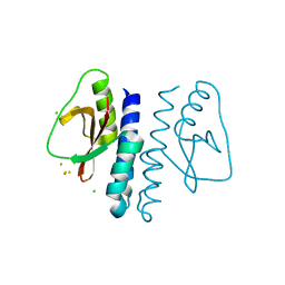
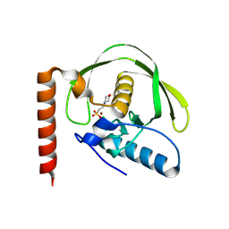
3LB5
DownloadVisualize
BU of 3lb5 by Molmil
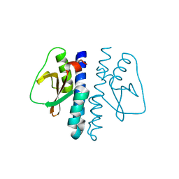
Crystal structure of Hit-like protein involved in cell-cycle regulation from Bartonella henselae with unknown ligand
Descriptor: Hit-like protein involved in cell-cycle regulation, UNKNOWN LIGAND
Authors:Seattle Structural Genomics Center for Infectious Disease (SSGCID)
Deposit date:2010-01-07
Release date:2010-01-19
Last modified:2023-09-06
Method:X-RAY DIFFRACTION (1.9 Å)
Cite:Crystal structure of Hit-like protein involved in cell-cycle regulation from Bartonella henselae with unknown ligand
To be Published
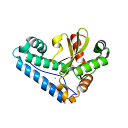
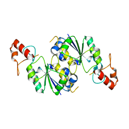
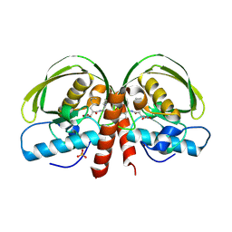
3LG6
DownloadVisualize
BU of 3lg6 by Molmil
Crystal structure of putative glutathione transferase from Coccidioides immitis
Descriptor: SULFATE ION, putative glutathione transferase
Authors:Seattle Structural Genomics Center for Infectious Disease (SSGCID)
Deposit date:2010-01-19
Release date:2010-01-26
Last modified:2023-09-06
Method:X-RAY DIFFRACTION (2.2 Å)
Cite:Structures of a putative zeta-class glutathione S-transferase from the pathogenic fungus Coccidioides immitis.
Acta Crystallogr.,Sect.F, 67, 2011
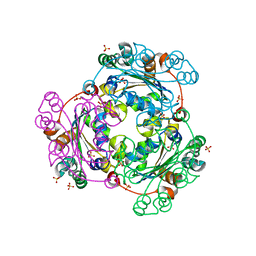
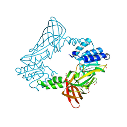
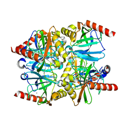
3LA9
DownloadVisualize
BU of 3la9 by Molmil
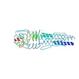
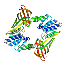
Crystal structure of the trimeric autotransporter adhesin head domain BpaA from Burkholderia pseudomallei, iodide phased
Descriptor: Haemagglutinin family protein, IODIDE ION
Authors:Seattle Structural Genomics Center for Infectious Disease (SSGCID)
Deposit date:2010-01-06
Release date:2010-05-12
Last modified:2024-02-21
Method:X-RAY DIFFRACTION (2.05 Å)
Cite:Structure of a Burkholderia pseudomallei trimeric autotransporter adhesin head.
Plos One, 5, 2010
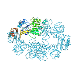
3LR3
DownloadVisualize
BU of 3lr3 by Molmil
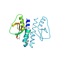
Periplasmic domain of the risS sensor protein from Burkholderia pesuromallei, low pH native structure
Descriptor: Sensor protein
Authors:Seattle Structural Genomics Center for Infectious Disease (SSGCID)
Deposit date:2010-02-10
Release date:2010-06-16
Last modified:2023-09-06
Method:X-RAY DIFFRACTION (2.1 Å)
Cite:Structural basis for pH sensing by the periplasmic domain of the risS histidine kinase from Burkholderia pseudomallei
To be Published
3LR0
DownloadVisualize
BU of 3lr0 by Molmil
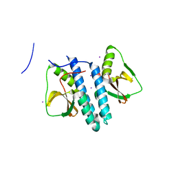
Periplasmic domain of the risS sensor protein from Burkholderia pseudomallei, iodide phased at low pH
Descriptor: IODIDE ION, Sensor protein
Authors:Seattle Structural Genomics Center for Infectious Disease (SSGCID)
Deposit date:2010-02-10
Release date:2010-06-16
Last modified:2024-02-21
Method:X-RAY DIFFRACTION (1.9 Å)
Cite:Structural basis for pH sensing by the periplasmic domain of the risS histidine kinase from Bukholderia pseudomallei
To be Published
3LR5
DownloadVisualize
BU of 3lr5 by Molmil
Periplasmic domain of the risS sensor protein from Burkholderia pseudomallei, iodide phased at neutral pH
Descriptor: IODIDE ION, Sensor protein
Authors:Seattle Structural Genomics Center for Infectious Disease (SSGCID)
Deposit date:2010-02-10
Release date:2010-06-16
Last modified:2024-02-21
Method:X-RAY DIFFRACTION (2.3 Å)
Cite:Structural basis for pH sensing by the periplsamic domain of the risS histidine kinase from Burkholderia pseudomallei
To be Published
3LR4
DownloadVisualize
BU of 3lr4 by Molmil
Periplasmic domain of the risS sensor protein from Burkholderia pseudomallei, barium phased at low pH
Descriptor: BARIUM ION, CHLORIDE ION, Sensor protein
Authors:Seattle Structural Genomics Center for Infectious Disease (SSGCID)
Deposit date:2010-02-10
Release date:2010-06-16
Last modified:2024-02-21
Method:X-RAY DIFFRACTION (1.9 Å)
Cite:Structural basis for pH sensing by the periplasmic domain of the risS histidine kinase from Burkholderia pseudomallei
To be Published
3JS4
DownloadVisualize
BU of 3js4 by Molmil
Crystal structure of iron superoxide dismutase from Anaplasma phagocytophilum
Descriptor: FE (III) ION, SODIUM ION, Superoxide dismutase
Authors:Seattle Structural Genomics Center for Infectious Disease (SSGCID)
Deposit date:2009-09-09
Release date:2009-09-22
Last modified:2023-09-06
Method:X-RAY DIFFRACTION (1.95 Å)
Cite:Crystal structure of Iron superoxide dismutase from Anaplasma phagocytophilum
To be Published
3JS9
DownloadVisualize
BU of 3js9 by Molmil
Crystal structure of nucleoside diphosphate kinase family protein from Babesia bovis
Descriptor: Nucleoside diphosphate kinase family protein, SULFATE ION
Authors:Seattle Structural Genomics Center for Infectious Disease (SSGCID)
Deposit date:2009-09-09
Release date:2009-09-22
Last modified:2023-09-06
Method:X-RAY DIFFRACTION (2.5 Å)
Cite:Crystal structure of nucleoside diphosphate kinase family protein from Babesia bovis
To be Published
3K5P
DownloadVisualize
BU of 3k5p by Molmil
Crystal structure of amino acid-binding ACT: D-isomer specific 2-hydroxyacid dehydrogenase catalytic domain from Brucella melitensis
Descriptor: 1,2-ETHANEDIOL, D-3-phosphoglycerate dehydrogenase, GLYCEROL
Authors:Seattle Structural Genomics Center for Infectious Disease (SSGCID)
Deposit date:2009-10-07
Release date:2009-11-03
Last modified:2023-09-06
Method:X-RAY DIFFRACTION (2.15 Å)
Cite:Crystal structure of amino acid-binding ACT: D-isomer specific 2-hydroxyacid dehydrogenase catalytic domain from Brucella melitensis
To be Published
3KNU
DownloadVisualize
BU of 3knu by Molmil
Crystal structure of tRNA (guanine-N1)-methyltransferase from Anaplasma phagocytophilum
Descriptor: tRNA (guanine-N(1)-)-methyltransferase
Authors:Seattle Structural Genomics Center for Infectious Disease (SSGCID)
Deposit date:2009-11-12
Release date:2009-11-24
Last modified:2023-09-06
Method:X-RAY DIFFRACTION (2.25 Å)
Cite:Crystal structure of tRNA (guanine-N1)-methyltransferase from Anaplasma phagocytophilum
To be Published
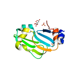
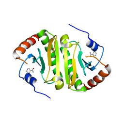
3MXU
DownloadVisualize
BU of 3mxu by Molmil
Crystal structure of glycine cleavage system protein H from Bartonella henselae
Descriptor: 1,2-ETHANEDIOL, CITRIC ACID, Glycine cleavage system H protein, ...
Authors:Seattle Structural Genomics Center for Infectious Disease (SSGCID)
Deposit date:2010-05-07
Release date:2010-05-26
Last modified:2023-09-06
Method:X-RAY DIFFRACTION (1.8 Å)
Cite:Crystal structure of glycine cleavage system protein H from Bartonella henselae
To be Published
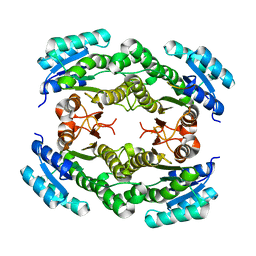
3N74
DownloadVisualize
BU of 3n74 by Molmil
Crystal structure of 3-ketoacyl-(acyl-carrier-protein) reductase from Brucella melitensis
Descriptor: 3-ketoacyl-(acyl-carrier-protein) reductase
Authors:Seattle Structural Genomics Center for Infectious Disease (SSGCID)
Deposit date:2010-05-26
Release date:2010-06-16
Last modified:2023-09-06
Method:X-RAY DIFFRACTION (2.2 Å)
Cite:Crystal structure of 3-ketoacyl-(acyl-carrier-protein) reductase from Brucella melitensis
TO BE PUBLISHED
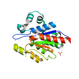
3N7T
DownloadVisualize
BU of 3n7t by Molmil
Crystal structure of a macrophage binding protein from Coccidioides immitis
Descriptor: 1,2-ETHANEDIOL, CHLORIDE ION, Macrophage binding protein, ...
Authors:Seattle Structural Genomics Center for Infectious Disease (SSGCID)
Deposit date:2010-05-27
Release date:2010-06-16
Last modified:2023-09-06
Method:X-RAY DIFFRACTION (2.1 Å)
Cite:Crystal structure of a macrophage binding protein from Coccidioides immitis
TO BE PUBLISHED
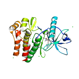
7TNH
DownloadVisualize
BU of 7tnh by Molmil
Crystal structure of CSF1R kinase domain in complex with DP-6233
Descriptor: 2,2-dimethyl-N-[(6-methyl-5-{[2-(1-methyl-1H-pyrazol-4-yl)pyridin-4-yl]oxy}pyridin-2-yl)carbamoyl]propanamide, CHLORIDE ION, Macrophage colony-stimulating factor 1 receptor,Fibroblast growth factor receptor 1 chimera, ...
Authors:Edwards, T.E, Arakaki, T.L, Chun, L, Flynn, D.L.
Deposit date:2022-01-21
Release date:2022-08-24
Last modified:2023-10-18
Method:X-RAY DIFFRACTION (2.7 Å)
Cite:Discovery of acyl ureas as highly selective small molecule CSF1R kinase inhibitors.
Bioorg.Med.Chem.Lett., 74, 2022
6CU3
DownloadVisualize
BU of 6cu3 by Molmil
Crystal structure of a protein arginine N-methyltransferase from Naegleria fowleri
Descriptor: 1,2-ETHANEDIOL, protein arginine N-methyltransferase
Authors:Seattle Structural Genomics Center for Infectious Disease (SSGCID)
Deposit date:2018-03-23
Release date:2018-04-04
Last modified:2024-10-23
Method:X-RAY DIFFRACTION (2.5 Å)
Cite:Crystal structure of a protein arginine N-methyltransferase from Naegleria fowleri
To Be Published
6CK7
DownloadVisualize
BU of 6ck7 by Molmil
Crystal structure of a peptide deformylase from Legionella pneumophila bound to actinonin
Descriptor: ACTINONIN, Peptide deformylase, SULFATE ION, ...
Authors:Seattle Structural Genomics Center for Infectious Disease (SSGCID)
Deposit date:2018-02-27
Release date:2018-03-07
Last modified:2023-10-04
Method:X-RAY DIFFRACTION (1.65 Å)
Cite:Crystal structure of a peptide deformylase from Legionella pneumophila bound to actinonin
To be Published
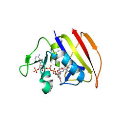
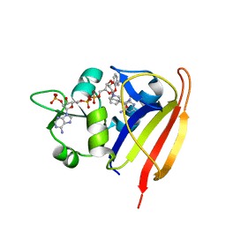
6CXM
DownloadVisualize
BU of 6cxm by Molmil
Crystal structure of a dihydrofolate reductase from Mycobacterium smegmatis in complex with NADP and P218
Descriptor: 3-(2-{3-[(2,4-diamino-6-ethylpyrimidin-5-yl)oxy]propoxy}phenyl)propanoic acid, Dihydrofolate reductase, NADP NICOTINAMIDE-ADENINE-DINUCLEOTIDE PHOSPHATE
Authors:Seattle Structural Genomics Center for Infectious Disease (SSGCID)
Deposit date:2018-04-03
Release date:2018-04-11
Last modified:2023-10-04
Method:X-RAY DIFFRACTION (2.65 Å)
Cite:Crystal structure of a dihydrofolate reductase from Mycobacterium smegmatis in complex with NADP and P218
To Be Published
6CU5
DownloadVisualize
BU of 6cu5 by Molmil
Crystal structure of a protein arginine N-methyltransferase from Naegleria fowleri bound to SAH
Descriptor: S-ADENOSYL-L-HOMOCYSTEINE, protein arginine N-methyltransferase
Authors:Seattle Structural Genomics Center for Infectious Disease (SSGCID)
Deposit date:2018-03-23
Release date:2018-04-04
Last modified:2023-10-04
Method:X-RAY DIFFRACTION (2.7 Å)
Cite:Crystal structure of a protein arginine N-methyltransferase from Naegleria fowleri bound to SAH
To Be Published
6D46
DownloadVisualize
BU of 6d46 by Molmil
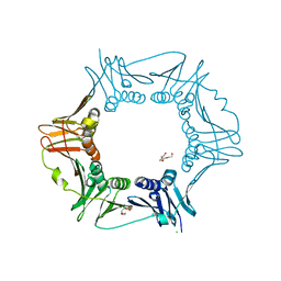
Crystal structure of a DNA polymerase III subunit beta DnaN sliding clamp from Rickettsia typhi str. Wilmington
Descriptor: Beta sliding clamp, CHLORIDE ION, TETRAETHYLENE GLYCOL
Authors:Seattle Structural Genomics Center for Infectious Disease (SSGCID)
Deposit date:2018-04-17
Release date:2018-05-02
Last modified:2023-10-04
Method:X-RAY DIFFRACTION (2 Å)
Cite:Crystal structure of a DNA polymerase III subunit beta DnaN sliding clamp from Rickettsia typhi str. Wilmington
To Be Published
6D47
DownloadVisualize
BU of 6d47 by Molmil
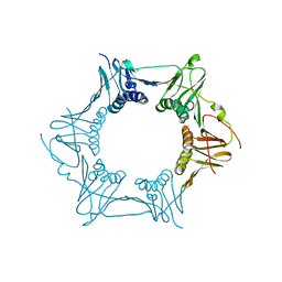
Crystal structure of a DNA polymerase III subunit beta DnaN sliding clamp from Mycobacterium marinum
Descriptor: Beta sliding clamp
Authors:Seattle Structural Genomics Center for Infectious Disease (SSGCID)
Deposit date:2018-04-17
Release date:2018-05-02
Last modified:2023-10-04
Method:X-RAY DIFFRACTION (2.45 Å)
Cite:Crystal structure of a DNA polymerase III subunit beta DnaN sliding clamp from Mycobacterium marinum
To Be Published
6DEG
DownloadVisualize
BU of 6deg by Molmil
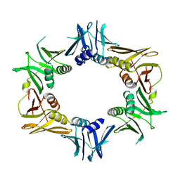
Crystal structure of a DNA polymerase III subunit beta DnaN sliding clamp from Bartonella birtlesii LL-WM9
Descriptor: Beta sliding clamp
Authors:Seattle Structural Genomics Center for Infectious Disease (SSGCID)
Deposit date:2018-05-11
Release date:2018-05-30
Last modified:2024-03-13
Method:X-RAY DIFFRACTION (2.45 Å)
Cite:Crystal structure of a DNA polymerase III subunit beta DnaN sliding clamp from Bartonella birtlesii LL-WM9
To Be Published
6CAZ
DownloadVisualize
BU of 6caz by Molmil
Crystal structure of a peptide deformylase from Legionella pneumophila
Descriptor: 2-(N-MORPHOLINO)-ETHANESULFONIC ACID, CHLORIDE ION, NICKEL (II) ION, ...
Authors:Seattle Structural Genomics Center for Infectious Disease (SSGCID)
Deposit date:2018-02-01
Release date:2018-02-14
Last modified:2023-10-04
Method:X-RAY DIFFRACTION (1.5 Å)
Cite:Crystal structure of a peptide deformylase from Legionella pneumophila
To Be Published
6E4E
DownloadVisualize
BU of 6e4e by Molmil
Crystal structure of dihydrofolate reductase from Staphylococcus aureus MW2 bound to NADP and p218
Descriptor: 3-(2-{3-[(2,4-diamino-6-ethylpyrimidin-5-yl)oxy]propoxy}phenyl)propanoic acid, Dihydrofolate reductase, NADP NICOTINAMIDE-ADENINE-DINUCLEOTIDE PHOSPHATE
Authors:Seattle Structural Genomics Center for Infectious Disease (SSGCID)
Deposit date:2018-07-17
Release date:2018-07-25
Last modified:2023-10-11
Method:X-RAY DIFFRACTION (1.9 Å)
Cite:Crystal structure of dihydrofolate reductase from Staphylococcus aureus MW2 bound to NADP and p218
To Be Published
5TEA
DownloadVisualize
BU of 5tea by Molmil
Crystal structure of an inorganic pyrophosphatase from Neisseria gonorrhoeae
Descriptor: CALCIUM ION, CHLORIDE ION, Inorganic pyrophosphatase
Authors:Seattle Structural Genomics Center for Infectious Disease (SSGCID)
Deposit date:2016-09-20
Release date:2016-10-05
Last modified:2023-10-04
Method:X-RAY DIFFRACTION (1.85 Å)
Cite:Crystal structure of an inorganic pyrophosphatase from
To Be Published

227111

PDB entries from 2024-11-06

PDB statisticsPDBj update infoContact PDBjnumon