Loading
PDBj
MenuPDBj@FacebookPDBj@X(formerly Twitter)PDBj@BlueSkyPDBj@YouTubewwPDB FoundationwwPDB
RCSB PDBPDBeBMRBAdv. SearchSearch help
Search by PDB author
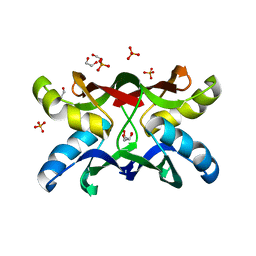
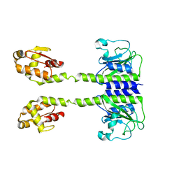
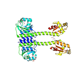
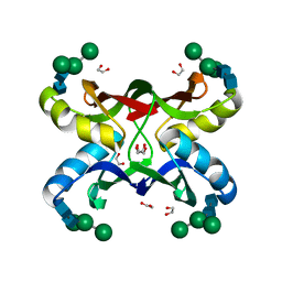
1ZHS
DownloadVisualize
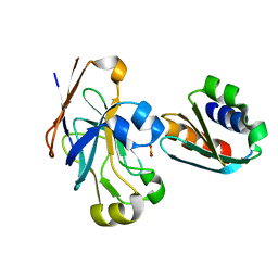
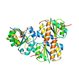
BU of 1zhs by Molmil
Crystal structure of MVL bound to Man3GlcNAc2
Descriptor: 1,2-ETHANEDIOL, PHOSPHATE ION, alpha-D-mannopyranose-(1-3)-[alpha-D-mannopyranose-(1-6)]beta-D-mannopyranose-(1-4)-2-acetamido-2-deoxy-beta-D-glucopyranose-(1-4)-2-acetamido-2-deoxy-beta-D-glucopyranose, ...
Authors:Williams, D.C, Lee, J.Y, Cai, M, Bewley, C.A, Clore, G.M.
Deposit date:2005-04-26
Release date:2005-06-07
Last modified:2024-02-14
Method:X-RAY DIFFRACTION (1.8 Å)
Cite:Crystal Structures of the HIV-1 Inhibitory Cyanobacterial Protein MVL Free and Bound to Man3GlcNAc2: STRUCTURAL BASIS FOR SPECIFICITY AND HIGH-AFFINITY BINDING TO THE CORE PENTASACCHARIDE FROM N-LINKED OLIGOMANNOSIDE.
J.Biol.Chem., 280, 2005
1ZHQ
DownloadVisualize
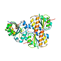
BU of 1zhq by Molmil
Crystal structure of apo MVL
Descriptor: 1,2-ETHANEDIOL, PHOSPHATE ION, mannan-binding lectin
Authors:Williams, D.C, Lee, J.Y, Cai, M, Bewley, C.A, Clore, G.M.
Deposit date:2005-04-26
Release date:2005-06-07
Last modified:2024-02-14
Method:X-RAY DIFFRACTION (1.9 Å)
Cite:Crystal Structures of the HIV-1 Inhibitory Cyanobacterial Protein MVL Free and Bound to Man3GlcNAc2: STRUCTURAL BASIS FOR SPECIFICITY AND HIGH-AFFINITY BINDING TO THE CORE PENTASACCHARIDE FROM N-LINKED OLIGOMANNOSIDE.
J.Biol.Chem., 280, 2005
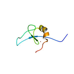
9DE2
DownloadVisualize
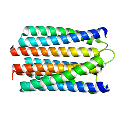
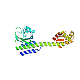
BU of 9de2 by Molmil
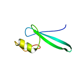
ETO2 MYND bound to MPPL peptide from GATAD2A
Descriptor: GATAD2A-MPPL motif and ETO2 NHR4 domain fusion protein, ZINC ION
Authors:Williams, D.C.
Deposit date:2024-08-28
Release date:2024-09-18
Method:SOLUTION NMR
Cite:The role of multivalency in the association of the eight twenty-one protein 2 (ETO2) with the nucleosome remodeling and deacetylase (NuRD) complex
To Be Published
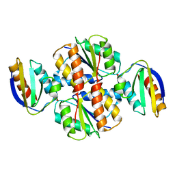
1VRC
DownloadVisualize
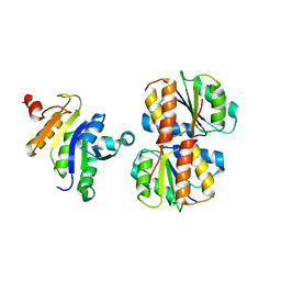
BU of 1vrc by Molmil
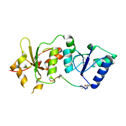
Complex of enzyme IIAmannose and the histidine-containing phosphocarrier protein HPr from escherichia coli nmr, restrained regularized mean structure
Descriptor: PHOSPHITE ION, PTS system, mannose-specific IIAB component, ...
Authors:Clore, G.M, Williams, D.C.
Deposit date:2005-02-21
Release date:2005-04-19
Last modified:2023-12-27
Method:SOLUTION NMR
Cite:Solution NMR structure of the 48-kDa IIAMannose-HPr complex of the Escherichia coli mannose phosphotransferase system.
J.Biol.Chem., 280, 2005
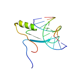
2MOE
DownloadVisualize
BU of 2moe by Molmil
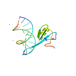
Solution structure of MBD4 methyl-cytosine binding domain bound to methylated DNA
Descriptor: DNA (5'-D(*GP*AP*GP*CP*(5CM)P*GP*AP*TP*CP*C)-3'), DNA (5'-D(*GP*GP*AP*TP*(5CM)P*GP*GP*CP*TP*C)-3'), Methyl-CpG-binding domain protein 4
Authors:Williams, D.C, Walavalkar, N.M.
Deposit date:2014-04-24
Release date:2014-09-10
Last modified:2024-05-15
Method:SOLUTION NMR
Cite:Solution structure and intramolecular exchange of methyl-cytosine binding domain protein 4 (MBD4) on DNA suggests a mechanism to scan for mCpG/TpG mismatches.
Nucleic Acids Res., 42, 2015
1O4X
DownloadVisualize
BU of 1o4x by Molmil
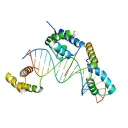
TERNARY COMPLEX OF THE DNA BINDING DOMAINS OF THE OCT1 AND SOX2 TRANSCRIPTION FACTORS WITH A 19MER OLIGONUCLEOTIDE FROM THE HOXB1 REGULATORY ELEMENT
Descriptor: 5'-D(*CP*AP*TP*TP*AP*GP*CP*AP*TP*GP*AP*CP*AP*AP*AP*GP*AP*CP*A)-3', 5'-D(*TP*GP*TP*CP*TP*TP*TP*GP*TP*CP*AP*TP*GP*CP*TP*AP*AP*TP*G)-3', Transcription factor SOX-2, ...
Authors:Clore, G.M, Williams, D.C.
Deposit date:2003-07-17
Release date:2004-01-27
Last modified:2023-12-27
Method:SOLUTION NMR
Cite:Molecular basis for synergistic transcriptional activation by Oct1 and Sox2 revealed from the solution structure of the 42-kDa Oct1.Sox2.Hoxb1-DNA ternary transcription factor complex.
J.Biol.Chem., 279, 2004
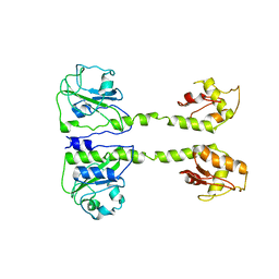
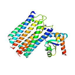
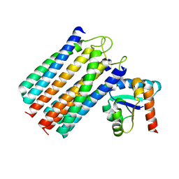
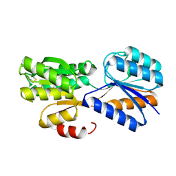
6CYM
DownloadVisualize
BU of 6cym by Molmil
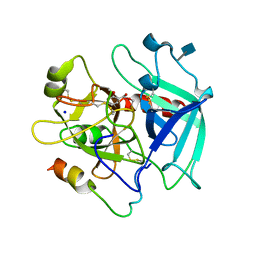
Reversible Covalent Direct Thrombin Inhibitors
Descriptor: 2-acetamido-2-deoxy-beta-D-glucopyranose, 2-methoxybenzoic acid, Prothrombin, ...
Authors:Sivaraja, M, Williams, D.C.
Deposit date:2018-04-06
Release date:2018-05-09
Last modified:2024-11-13
Method:X-RAY DIFFRACTION (2.9 Å)
Cite:Reversible covalent direct thrombin inhibitors.
PLoS ONE, 13, 2018
2L2L
DownloadVisualize
BU of 2l2l by Molmil
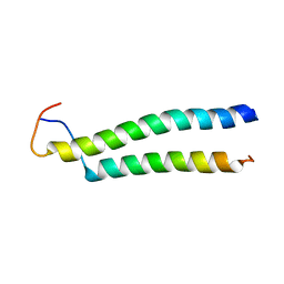
Solution structure of the coiled-coil complex between MBD2 and p66alpha
Descriptor: Methyl-CpG-binding domain protein 2, Transcriptional repressor p66-alpha
Authors:Williams Jr, D.C, Scarsdale Jr, N.
Deposit date:2010-08-20
Release date:2011-05-04
Last modified:2024-05-01
Method:SOLUTION NMR
Cite:p66Alpha-MBD2 coiled-coil interaction and recruitment of Mi-2 are critical for globin gene silencing by the MBD2-NuRD complex.
Proc.Natl.Acad.Sci.USA, 108, 2011
2JZH
DownloadVisualize
BU of 2jzh by Molmil
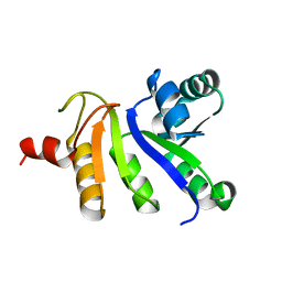
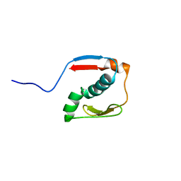
structure of IIB domain of the mannose transporter of E. coli
Descriptor: PTS system mannose-specific EIIAB component
Authors:Komlosh, M, Williams Jr, D.C.
Deposit date:2008-01-08
Release date:2008-02-19
Last modified:2024-05-29
Method:SOLUTION NMR
Cite:Solution NMR Structures of Productive and Non-productive Complexes between the A and B Domains of the Cytoplasmic Subunit of the Mannose Transporter of the Escherichia coli Phosphotransferase System.
J.Biol.Chem., 283, 2008
2MB7
DownloadVisualize
BU of 2mb7 by Molmil
Solution structure of MBD3 methylcytosine binding domain
Descriptor: Methyl-CpG-binding domain protein 3
Authors:Williams Jr, D.C, Scarsdale, J.N.
Deposit date:2013-07-26
Release date:2013-12-11
Last modified:2024-05-15
Method:SOLUTION NMR
Cite:Probing the Dynamic Distribution of Bound States for Methylcytosine-binding Domains on DNA.
J.Biol.Chem., 289, 2014
2KY8
DownloadVisualize
BU of 2ky8 by Molmil
Solution structure and dynamic analysis of chicken MBD2 methyl binding domain bound to a target methylated DNA sequence
Descriptor: DNA (5'-D(*GP*AP*GP*CP*(5CM)P*GP*AP*TP*(TED)P*CP*C)-3'), DNA (5'-D(*GP*GP*AP*AP*TP*(5CM)P*GP*GP*CP*(TED)P*C)-3'), MANGANESE (II) ION, ...
Authors:Williams Jr, D.C, Scarsdale Jr, J.N.
Deposit date:2010-05-18
Release date:2011-05-18
Last modified:2024-05-01
Method:SOLUTION NMR
Cite:Solution structure and dynamic analysis of chicken MBD2 methyl binding domain bound to a target-methylated DNA sequence.
Nucleic Acids Res., 39, 2011
4OFB
DownloadVisualize
BU of 4ofb by Molmil
Crystal structure of human BRCA1 BRCT in complex with nonphosphopeptide inhibitor
Descriptor: Breast cancer type 1 susceptibility protein, nonphosphopeptide inhibitor
Authors:Sun, L, Edwards, R.A, Glover, J.N.M.
Deposit date:2014-01-14
Release date:2015-04-15
Last modified:2017-11-22
Method:X-RAY DIFFRACTION (3.05 Å)
Cite:Peptide Library Approach to Uncover Phosphomimetic Inhibitors of the BRCA1 C-Terminal Domain.
Acs Chem.Biol., 10, 2015
2N2Y
DownloadVisualize
BU of 2n2y by Molmil
Solution structure of the meiosis-expressed gene 1 (Meig1)
Descriptor: Meiosis-expressed gene 1 protein
Authors:Williams Jr, D.C, Walavalkar, N.M, Buchwald, W.A.
Deposit date:2015-05-16
Release date:2016-01-20
Last modified:2024-05-15
Method:SOLUTION NMR
Cite:Dissecting the structural basis of MEIG1 interaction with PACRG.
Sci Rep, 6, 2016
1O2F
DownloadVisualize
BU of 1o2f by Molmil
COMPLEX OF ENZYME IIAGLC AND IIBGLC PHOSPHOCARRIER PROTEIN HPR FROM ESCHERICHIA COLI NMR, RESTRAINED REGULARIZED MEAN STRUCTURE
Descriptor: PHOSPHITE ION, PTS system, glucose-specific IIA component, ...
Authors:Clore, G.M, Cai, M, Williams, D.C.
Deposit date:2003-03-11
Release date:2003-05-13
Last modified:2023-12-27
Method:SOLUTION NMR
Cite:Solution Structure of the Phosphoryl Transfer Complex between the Signal-transducing Protein IIAGlucose and the Cytoplasmic Domain of the Glucose Transporter IICBGlucose of the Escherichia coli Glucose Phosphotransferase System.
J.Biol.Chem., 278, 2003
1VSQ
DownloadVisualize
BU of 1vsq by Molmil
Solution NMR structure of the productive complex between IIAMannose and IIBMannose of the mannose transporter of the E. coli phosphotransferase system
Descriptor: Mannose-specific phosphotransferase enzyme IIA component, Mannose-specific phosphotransferase enzyme IIB component
Authors:Clore, G.M, Hu, J, Hu, K.
Deposit date:2008-01-10
Release date:2008-02-19
Last modified:2023-12-27
Method:SOLUTION NMR
Cite:Solution NMR Structures of Productive and Non-productive Complexes between the A and B Domains of the Cytoplasmic Subunit of the Mannose Transporter of the Escherichia coli Phosphotransferase System.
J.Biol.Chem., 283, 2008
1WCR
DownloadVisualize
BU of 1wcr by Molmil
Trimeric Structure of the Enzyme IIA from Escherichia coli Phosphotransferase System Specific for N,N'-Diacetylchitobiose
Descriptor: PTS SYSTEM, N, N'-DIACETYLCHITOBIOSE-SPECIFIC IIA COMPONENT
Authors:Tang, C, Clore, G.M.
Deposit date:2004-11-19
Release date:2005-01-19
Last modified:2024-05-15
Method:SOLUTION NMR
Cite:Solution Structure of Enzyme Iiachitobiose from the N,N'-Diacetylchitobiose Branch of the Escherichia Coli Phosphotransferase System
J.Biol.Chem., 280, 2005
2JZO
DownloadVisualize
BU of 2jzo by Molmil
Solution NMR structure of the non-productive complex between IIAMannose and IIBMannose of the mannose transporter of the E. coli phosphotransferase system
Descriptor: PTS system mannose-specific EIIAB component
Authors:Clore, G, Hu, J, Hu, K.
Deposit date:2008-01-10
Release date:2008-02-19
Last modified:2024-05-29
Method:SOLUTION NMR
Cite:Solution NMR Structures of Productive and Non-productive Complexes between the A and B Domains of the Cytoplasmic Subunit of the Mannose Transporter of the Escherichia coli Phosphotransferase System.
J.Biol.Chem., 283, 2008
2JZN
DownloadVisualize
BU of 2jzn by Molmil
Solution NMR structure of the productive complex between IIAMannose and IIBMannose of the mannose transporter of the E. coli phosphotransferase system
Descriptor: Mannose-specific phosphotransferase enzyme IIA component, Mannose-specific phosphotransferase enzyme IIB component
Authors:Clore, G.M, Hu, J, Hu, K.
Deposit date:2008-01-10
Release date:2008-02-19
Last modified:2024-05-29
Method:SOLUTION NMR
Cite:Solution NMR Structures of Productive and Non-productive Complexes between the A and B Domains of the Cytoplasmic Subunit of the Mannose Transporter of the Escherichia coli Phosphotransferase System.
J.Biol.Chem., 283, 2008
5LOE
DownloadVisualize
BU of 5loe by Molmil
Structure of full length Cody from Bacillus subtilis in complex with Ile
Descriptor: GTP-sensing transcriptional pleiotropic repressor CodY, ISOLEUCINE
Authors:Wilkinson, A.J, Levdikov, V.M, Blagova, E.V.
Deposit date:2016-08-09
Release date:2017-01-11
Last modified:2024-01-10
Method:X-RAY DIFFRACTION (3 Å)
Cite:Structure of the Branched-chain Amino Acid and GTP-sensing Global Regulator, CodY, from Bacillus subtilis.
J. Biol. Chem., 292, 2017
5LOJ
DownloadVisualize
BU of 5loj by Molmil
Structure of full length unliganded CodY from Bacillus subtilis
Descriptor: GTP-sensing transcriptional pleiotropic repressor CodY
Authors:Wilkinson, A.J, Levdikov, V.M, Blagova, E.V.
Deposit date:2016-08-09
Release date:2017-01-11
Last modified:2024-01-10
Method:X-RAY DIFFRACTION (3.71 Å)
Cite:Structure of the Branched-chain Amino Acid and GTP-sensing Global Regulator, CodY, from Bacillus subtilis.
J. Biol. Chem., 292, 2017
5LNH
DownloadVisualize
BU of 5lnh by Molmil
Structure of full length Unliganded CodY from Bacillus subtilis
Descriptor: GTP-sensing transcriptional pleiotropic repressor CodY, SULFATE ION
Authors:Wilkinson, A.J, Levdikov, V.M, Blagova, E.V.
Deposit date:2016-08-04
Release date:2017-01-11
Last modified:2024-11-13
Method:X-RAY DIFFRACTION (3 Å)
Cite:Structure of the Branched-chain Amino Acid and GTP-sensing Global Regulator, CodY, from Bacillus subtilis.
J. Biol. Chem., 292, 2017
5LOO
DownloadVisualize
BU of 5loo by Molmil
Structure of full length unliganded CodY from Bacillus subtilis
Descriptor: GTP-sensing transcriptional pleiotropic repressor CodY
Authors:Wilkinson, A.J, Levdikov, V.M, Blagova, E.V.
Deposit date:2016-08-09
Release date:2017-01-11
Last modified:2024-01-10
Method:X-RAY DIFFRACTION (4.5 Å)
Cite:Structure of the Branched-chain Amino Acid and GTP-sensing Global Regulator, CodY, from Bacillus subtilis.
J. Biol. Chem., 292, 2017
2WY2
DownloadVisualize
BU of 2wy2 by Molmil
NMR structure of the IIAchitobiose-IIBchitobiose phosphoryl transition state complex of the N,N'-diacetylchitoboise brance of the E. coli phosphotransferase system.
Descriptor: N,N'-DIACETYLCHITOBIOSE-SPECIFIC PHOSPHOTRANSFERASE ENZYME IIA COMPONENT, N,N'-DIACETYLCHITOBIOSE-SPECIFIC PHOSPHOTRANSFERASE ENZYME IIB COMPONENT, PHOSPHITE ION
Authors:Sang, Y.S, Cai, M, Clore, G.M.
Deposit date:2009-11-11
Release date:2009-12-08
Last modified:2024-05-15
Method:SOLUTION NMR
Cite:Solution Structure of the Iiachitobose-Iibchitobiose Complex of the N,N'-Diacetylchitobiose Branch of the Escherichia Coli Phosphotransfer System
J.Biol.Chem., 285, 2010
2WWV
DownloadVisualize
BU of 2wwv by Molmil
NMR structure of the IIAchitobiose-IIBchitobiose complex of the N,N'- diacetylchitoboise brance of the E. coli phosphotransferase system.
Descriptor: N,N'-DIACETYLCHITOBIOSE-SPECIFIC PHOSPHOTRANSFERASE ENZYME IIA COMPONENT, N,N'-DIACETYLCHITOBIOSE-SPECIFIC PHOSPHOTRANSFERASE ENZYME IIB COMPONENT
Authors:Sang, Y.S, Cai, M, Clore, G.M.
Deposit date:2009-10-29
Release date:2009-12-08
Last modified:2024-05-15
Method:SOLUTION NMR
Cite:Solution Structure of the Iiachitobose-Iibchitobiose Complex of the N,N'-Diacetylchitobiose Branch of the Escherichia Coli Phosphotransfer System
J.Biol.Chem., 285, 2010
2VK2
DownloadVisualize
BU of 2vk2 by Molmil
Crystal structure of a galactofuranose binding protein
Descriptor: ABC TRANSPORTER PERIPLASMIC-BINDING PROTEIN YTFQ, beta-D-galactofuranose
Authors:Muller, A, Horler, R.S.P, Thomas, G.H, Wilson, K.S.
Deposit date:2007-12-16
Release date:2009-05-19
Last modified:2023-12-13
Method:X-RAY DIFFRACTION (1.2 Å)
Cite:Furanose-Specific Sugar Transport: Characterization of a Bacterial Galactofuranose-Binding Protein.
J.Biol.Chem., 284, 2009

227344

數據於2024-11-13公開中

PDB statisticsPDBj update infoContact PDBjnumon