Loading
PDBj
MenuPDBj@FacebookPDBj@TwitterPDBj@YouTubewwPDB FoundationwwPDB
RCSB PDBPDBeBMRBAdv. SearchSearch help
Search by PDB author
1WTU
DownloadVisualize
BU of 1wtu by Molmil
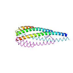
TRANSCRIPTION FACTOR 1, NMR, MINIMIZED AVERAGE STRUCTURE
Descriptor: TRANSCRIPTION FACTOR 1
Authors:Jia, X, Grove, A, Ivancic, M, Hsu, V.L, Geiduschek, E.P, Kearns, D.R.
Deposit date:1996-07-29
Release date:1997-02-12
Last modified:2024-05-22
Method:SOLUTION NMR
Cite:Structure of the Bacillus subtilis phage SPO1-encoded type II DNA-binding protein TF1 in solution.
J.Mol.Biol., 263, 1996
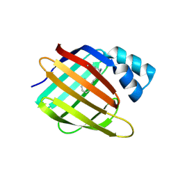
3CR6
DownloadVisualize
BU of 3cr6 by Molmil
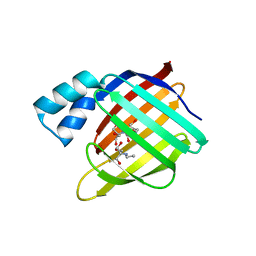
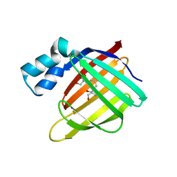
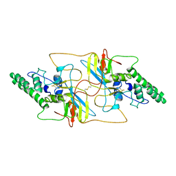
Crystal Structure of the R132K:R111L:A32E Mutant of Cellular Retinoic Acid Binding Protein Type II Complexed with C15-aldehyde (a retinal analog) at 1.22 Angstrom resolution.
Descriptor: 1,3,3-trimethyl-2-[(1E,3E)-3-methylpenta-1,3-dien-1-yl]cyclohexene, Cellular retinoic acid-binding protein 2
Authors:Jia, X, Geiger, J.H.
Deposit date:2008-04-04
Release date:2009-03-17
Last modified:2023-08-30
Method:X-RAY DIFFRACTION (1.22 Å)
Cite:Two distinctive orientations of binding determined by a single mutation in the CRABPII mutant-C15-aldehyde complexes
To be Published
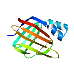
3FA6
DownloadVisualize
BU of 3fa6 by Molmil
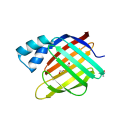
Crystal structure of the R132K:Y134F:R111L:L121D:T54V mutant of cellular retinoic acid-binding protein II complexed with C15-aldehyde (a retinal analog) at 1.54 angstrom resolution
Descriptor: 1,3,3-trimethyl-2-[(1E,3E)-3-methylpenta-1,3-dien-1-yl]cyclohexene, Cellular retinoic acid-binding protein 2
Authors:Jia, X, Geiger, J.H.
Deposit date:2008-11-15
Release date:2009-10-27
Last modified:2024-10-30
Method:X-RAY DIFFRACTION (1.54 Å)
Cite:Two distinctive orientations of binding determined by a single mutation in the CRABPII mutant-C15-aldehyde complexes
To be Published
3F9D
DownloadVisualize
BU of 3f9d by Molmil
Crystal structure of the R132K:R111L:T54E mutant of cellular retinoic acid-binding protein II complexed with C15-aldehyde (a retinal analog) at 2.00 angstrom resolution
Descriptor: 1,3,3-trimethyl-2-[(1E,3E)-3-methylpenta-1,3-dien-1-yl]cyclohexene, Cellular retinoic acid-binding protein 2
Authors:Jia, X, Geiger, J.H.
Deposit date:2008-11-13
Release date:2009-10-27
Last modified:2023-09-06
Method:X-RAY DIFFRACTION (2 Å)
Cite:Two distinctive orientations of binding determined by a single mutation in the CRABPII mutant-C15-aldehyde complexes
To be Published
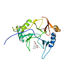
8EPY
DownloadVisualize
BU of 8epy by Molmil
The solution structure of abxF in complex with its product (-)-ABX, an enzyme catalyzing the formation of the chiral spiroketal of an anthrabenzoxocinone antibiotic, (-)-ABX
Descriptor: (6R,16R)-3,11,13,15-tetrahydroxy-1,6,9,9-tetramethyl-6,7,9,16-tetrahydro-14H-6,16-epoxyanthra[2,3-e]benzo[b]oxocin-14-one, Glyoxalase
Authors:Jia, X, Yan, X, Qu, X, Mobli, M.
Deposit date:2022-10-06
Release date:2024-04-10
Last modified:2024-05-15
Method:SOLUTION NMR
Cite:The solution structure of abxF, an enzyme catalyzing the formation of chiral spiroketal of an antibiotics, (-)-ABX.
To Be Published
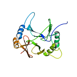
8EO9
DownloadVisualize
BU of 8eo9 by Molmil
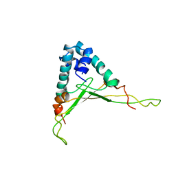
The solution structure of abxF, an enzyme catalyzing the formation of chiral spiroketal of an antibiotics, (-)-ABX
Descriptor: Glyoxalase
Authors:Jia, X, Yan, X, Mobli, M, Qu, X.
Deposit date:2022-10-02
Release date:2024-04-03
Last modified:2024-05-15
Method:SOLUTION NMR
Cite:The solution structure of abxF, an enzyme catalyzing the formation of chiral spiroketal of an antibiotics, (-)-ABX.
To Be Published
3FEL
DownloadVisualize
BU of 3fel by Molmil
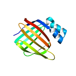
Crystal structure of the R132K:R111L:T54E mutant of cellular retinoic acid-binding protein II at 1.85 angstrom resolution
Descriptor: 2-[3-(2-HYDROXY-1,1-DIHYDROXYMETHYL-ETHYLAMINO)-PROPYLAMINO]-2-HYDROXYMETHYL-PROPANE-1,3-DIOL, Cellular retinoic acid-binding protein 2
Authors:Jia, X, Geiger, J.H.
Deposit date:2008-11-30
Release date:2009-11-10
Last modified:2023-09-06
Method:X-RAY DIFFRACTION (1.85 Å)
Cite:Crystal structures of apo cellular retinoic acid-binding protein II mutants: Structural integrity investigated through multiple site mutations
To be Published
3FA9
DownloadVisualize
BU of 3fa9 by Molmil
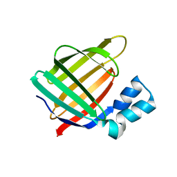
Crystal structure of the apo R132K:Y134F:R111L:L121D mutant of cellular retinoic acid-binding protein II at 1.94 angstrom resolution
Descriptor: ACETATE ION, Cellular retinoic acid-binding protein 2
Authors:Jia, X, Geiger, J.H.
Deposit date:2008-11-16
Release date:2009-10-27
Last modified:2023-12-27
Method:X-RAY DIFFRACTION (1.94 Å)
Cite:Crystal structures of apo cellular retinoic acid-binding protein II mutants: Structural integrity investigated through multiple site mutations
To be Published
3FA8
DownloadVisualize
BU of 3fa8 by Molmil
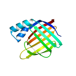
Crystal structure of the apo R132K:Y134F:R111L:L121E mutant of cellular retinoic acid-binding protein II at 1.78 angstrom resolution
Descriptor: Cellular retinoic acid-binding protein 2
Authors:Jia, X, Geiger, J.H.
Deposit date:2008-11-16
Release date:2009-10-27
Last modified:2023-09-06
Method:X-RAY DIFFRACTION (1.78 Å)
Cite:Crystal structures of apo cellular retinoic acid-binding protein II mutants: Structural integrity investigated through multiple site mutations
To be Published
3FEN
DownloadVisualize
BU of 3fen by Molmil
Crystal structure of the R132K:R111L:A32E mutant of cellular retinoic acid-binding protein II at 1.56 angstrom resolution
Descriptor: CHLORIDE ION, Cellular retinoic acid-binding protein 2
Authors:Jia, X, Geiger, J.H.
Deposit date:2008-11-30
Release date:2009-11-10
Last modified:2023-09-06
Method:X-RAY DIFFRACTION (1.56 Å)
Cite:Crystal structures of apo cellular retinoic acid-binding protein II mutants: Structural integrity investigated through multiple site mutations
To be Published
3FA7
DownloadVisualize
BU of 3fa7 by Molmil
Crystal structure of the apo R132K:R111L:L121E:R59E mutant of cellular retinoic acid-binding protein II at 1.90 angstrom resolution
Descriptor: 2-[3-(2-HYDROXY-1,1-DIHYDROXYMETHYL-ETHYLAMINO)-PROPYLAMINO]-2-HYDROXYMETHYL-PROPANE-1,3-DIOL, Cellular retinoic acid-binding protein 2
Authors:Jia, X, Geiger, J.H.
Deposit date:2008-11-15
Release date:2009-10-27
Last modified:2023-12-27
Method:X-RAY DIFFRACTION (1.9 Å)
Cite:Crystal structures of apo cellular retinoic acid-binding protein II mutants: Structural integrity investigated through multiple site mutations
To be Published
3FEK
DownloadVisualize
BU of 3fek by Molmil
Crystal structure of the R132K:Y134F:R111L:L121D:T54V mutant of cellular retinoic acid-binding protein II at 1.51 angstrom resolution
Descriptor: ACETATE ION, Cellular retinoic acid-binding protein 2, DI(HYDROXYETHYL)ETHER
Authors:Jia, X, Geiger, J.H.
Deposit date:2008-11-30
Release date:2009-11-10
Last modified:2023-09-06
Method:X-RAY DIFFRACTION (1.51 Å)
Cite:Crystal structures of apo cellular retinoic acid-binding protein II mutants: Structural integrity investigated through multiple site mutations
To be Published
7YR7
DownloadVisualize
BU of 7yr7 by Molmil
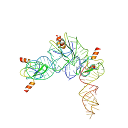
Cryo-EM structure of Pseudomonas aeruginosa RsmZ RNA in complex with three RsmA protein dimers
Descriptor: RsmZ RNA (118-MER), Translational regulator CsrA
Authors:Jia, X, Pan, Z, Yuan, Y, Luo, B, Luo, Y, Mukherjee, S, Jia, G, Liu, L, Ling, X, Yang, X, Wu, Y, Liu, T, Miao, Z, Wei, X, Bujnicki, J.M, Zhao, K, Su, Z.
Deposit date:2022-08-09
Release date:2023-05-17
Last modified:2024-07-03
Method:ELECTRON MICROSCOPY (3.8 Å)
Cite:Structural basis of sRNA RsmZ regulation of Pseudomonas aeruginosa virulence.
Cell Res., 33, 2023
7YR6
DownloadVisualize
BU of 7yr6 by Molmil
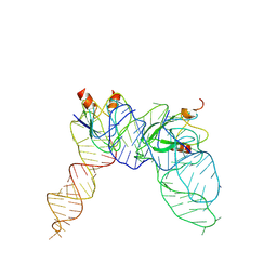
Cryo-EM structure of Pseudomonas aeruginosa RsmZ RNA in complex with two RsmA protein dimers
Descriptor: RsmZ RNA, Translational regulator CsrA
Authors:Jia, X, Pan, Z, Yuan, Y, Luo, B, Luo, Y, Mukherjee, S, Jia, G, Ling, X, Yang, X, Wu, Y, Liu, T, Wei, X, Bujnick, J.M, Zhao, K, Su, Z.
Deposit date:2022-08-09
Release date:2023-05-17
Last modified:2024-07-03
Method:ELECTRON MICROSCOPY (4.8 Å)
Cite:Structural basis of sRNA RsmZ regulation of Pseudomonas aeruginosa virulence.
Cell Res., 33, 2023
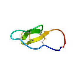
2MH1
DownloadVisualize
BU of 2mh1 by Molmil
Enzymatic cyclisation of kalata B1 using sortase A
Descriptor: Kalata-B1
Authors:Jia, X, Schroeder, C.I.
Deposit date:2013-11-12
Release date:2014-01-22
Last modified:2024-10-16
Method:SOLUTION NMR
Cite:Semienzymatic Cyclization of Disulfide-rich Peptides Using Sortase A.
J.Biol.Chem., 289, 2014
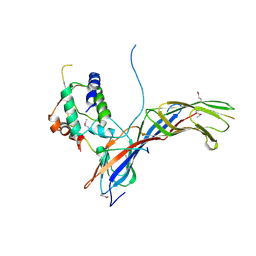
4EMZ
DownloadVisualize
BU of 4emz by Molmil
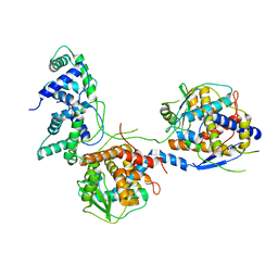
HIV-1 Nef in complex with MHC-I cytoplasmic domain and Mu1 adaptin subunit of AP1 adaptor (second domain)
Descriptor: AP-1 complex subunit mu-1, MHC-I, Protein Nef
Authors:Jia, X, Xiong, Y.
Deposit date:2012-04-12
Release date:2012-06-20
Last modified:2012-07-25
Method:X-RAY DIFFRACTION (2.9 Å)
Cite:Structural basis of evasion of cellular adaptive immunity by HIV-1 Nef.
Nat.Struct.Mol.Biol., 19, 2012
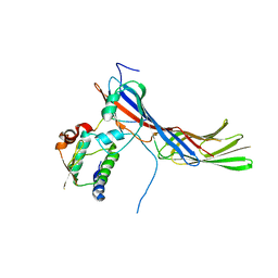
4EN2
DownloadVisualize
BU of 4en2 by Molmil
HIV-1 Nef in complex with MHC-I cytoplasmic domain and Mu1 adaptin subunit of AP1 adaptor (second domain)
Descriptor: AP-1 complex subunit mu-1, MHC-I, Protein Nef
Authors:Jia, X, Xiong, Y.
Deposit date:2012-04-12
Release date:2012-06-20
Last modified:2024-02-28
Method:X-RAY DIFFRACTION (2.58 Å)
Cite:Structural basis of evasion of cellular adaptive immunity by HIV-1 Nef.
Nat.Struct.Mol.Biol., 19, 2012
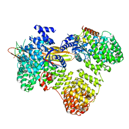
4P6Z
DownloadVisualize
BU of 4p6z by Molmil
Crystal structure of the human BST2 cytoplasmic domain and the HIV-1 Vpu cytoplasmic domain bound to the clathrin adaptor protein complex 1 (AP1) core
Descriptor: AP-1 complex subunit beta-1, AP-1 complex subunit gamma-1, AP-1 complex subunit mu-1, ...
Authors:Jia, X, Xiong, Y.
Deposit date:2014-03-25
Release date:2014-05-21
Last modified:2023-12-27
Method:X-RAY DIFFRACTION (3 Å)
Cite:Structural basis of HIV-1 Vpu-mediated BST2 antagonism via hijacking of the clathrin adaptor protein complex 1.
Elife, 3, 2014
5YEP
DownloadVisualize
BU of 5yep by Molmil
Crystal structure of SO_3166-SO_3165 from Shewanella oneidensis
Descriptor: Toxin-antitoxin system antidote Mnt family, Toxin-antitoxin system toxin HepN family
Authors:Jia, X, Gao, Z.Q, Zhang, H, Dong, Y.H.
Deposit date:2017-09-19
Release date:2018-03-28
Last modified:2019-04-10
Method:X-RAY DIFFRACTION (3 Å)
Cite:Structure-function analyses reveal the molecular architecture and neutralization mechanism of a bacterial HEPN-MNT toxin-antitoxin system.
J. Biol. Chem., 293, 2018
8WHK
DownloadVisualize
BU of 8whk by Molmil
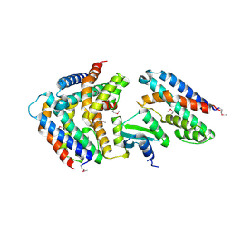
Crystal structure of CLASP2 in complex with LL5beta
Descriptor: CHLORIDE ION, CLIP-associating protein 2, Pleckstrin homology-like domain family B member 2
Authors:Jia, X, Wei, Z.
Deposit date:2023-09-23
Release date:2024-08-28
Method:X-RAY DIFFRACTION (2.401 Å)
Cite:CLASP-mediated competitive binding in protein condensates directs microtubule growth.
Nat Commun, 15, 2024
8WHJ
DownloadVisualize
BU of 8whj by Molmil
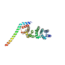
Crystal structure of CLASP2 TOG4 fused with LL5beta
Descriptor: CHLORIDE ION, CLIP-associating protein 2,Pleckstrin homology-like domain family B member 2, DI(HYDROXYETHYL)ETHER
Authors:Jia, X, Wei, Z.
Deposit date:2023-09-23
Release date:2024-08-28
Method:X-RAY DIFFRACTION (1.4 Å)
Cite:CLASP-mediated competitive binding in protein condensates directs microtubule growth.
Nat Commun, 15, 2024
8WHL
DownloadVisualize
BU of 8whl by Molmil
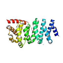
Crystal structure of CLASP2 in complex with CENP-E
Descriptor: ACETATE ION, CLIP-associating protein 2, Centromere-associated protein E, ...
Authors:Jia, X, Wei, Z.
Deposit date:2023-09-23
Release date:2024-08-28
Method:X-RAY DIFFRACTION (3.2 Å)
Cite:CLASP-mediated competitive binding in protein condensates directs microtubule growth.
Nat Commun, 15, 2024
8WHM
DownloadVisualize
BU of 8whm by Molmil
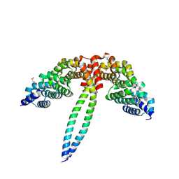
Crystal structure of the ELKS2/LL5beta complex
Descriptor: ERC protein 2, Pleckstrin homology-like domain family B member 2
Authors:Jia, X, Wei, Z.
Deposit date:2023-09-23
Release date:2024-08-28
Method:X-RAY DIFFRACTION (2.3 Å)
Cite:CLASP-mediated competitive binding in protein condensates directs microtubule growth.
Nat Commun, 15, 2024
8I8A
DownloadVisualize
BU of 8i8a by Molmil
Cryo-EM structure of the major capsid protein VP39 of Autographa californica multiple nucleopolyhedrovirus (AcMNPV)
Descriptor: Major viral capsid protein
Authors:Jia, X, Zhang, Q.
Deposit date:2023-02-03
Release date:2023-12-13
Last modified:2024-10-16
Method:ELECTRON MICROSCOPY (3.21 Å)
Cite:Architecture of the baculovirus nucleocapsid revealed by cryo-EM.
Nat Commun, 14, 2023
8I8C
DownloadVisualize
BU of 8i8c by Molmil
Plug structure of the Autographa californica multiple nucleopolyhedrovirus (AcMNPV)
Descriptor: Occlusion-derived virus envelope/capsid protein, P40
Authors:Jia, X, Gao, Y, Zhang, Q.
Deposit date:2023-02-03
Release date:2023-12-13
Method:ELECTRON MICROSCOPY (4.93 Å)
Cite:Architecture of the baculovirus nucleocapsid revealed by cryo-EM.
Nat Commun, 14, 2023

226707

數據於2024-10-30公開中

PDB statisticsPDBj update infoContact PDBjnumon