Loading
PDBj
MenuPDBj@FacebookPDBj@TwitterPDBj@YouTubewwPDB FoundationwwPDB
RCSB PDBPDBeBMRBAdv. SearchSearch help
Search by PDB author
7LSQ
DownloadVisualize
BU of 7lsq by Molmil
The Crystal Structure of Q108K:K40E:T53A:R58W:Q38F:Q4F:Y19W Mutant of HCRBPII Bound with LizFluor Chromophore Showing Excited State Intermolecular Proton Transfer
Descriptor: 4-{5-[(2E)-but-2-en-2-yl]thiophen-2-yl}-N,N-dimethylaniline, ACETATE ION, Retinol-binding protein 2
Authors:Ghanbarpour, A, Geiger, J.
Deposit date:2021-02-18
Release date:2022-01-12
Last modified:2023-10-18
Method:X-RAY DIFFRACTION (1.67130542 Å)
Cite:Design of Large Stokes Shift Fluorescent Proteins Based on Excited State Proton Transfer of an Engineered Photobase.
J.Am.Chem.Soc., 143, 2021
4YGG
DownloadVisualize
BU of 4ygg by Molmil
CRYSTAL STRUCTURE OF R111K:Y134F:T54V:R132Q:P39Y:R59Y MUTANT OF HUMAN CELLULAR RETINOIC ACID BINDING PROTEIN II WITH RETINAL AT 1.9 ANGSTROM RESOLUTION - VISIBLE LIGHT IRRADIATED CRYSTAL - 2ND CYCLE
Descriptor: Cellular retinoic acid-binding protein 2, RETINAL
Authors:Nosrati, M, Geiger, J.H.
Deposit date:2015-02-26
Release date:2016-03-02
Last modified:2023-09-27
Method:X-RAY DIFFRACTION (1.9 Å)
Cite:A Photoisomerizing Rhodopsin Mimic Observed at Atomic Resolution.
J.Am.Chem.Soc., 138, 2016
4QZU
DownloadVisualize
BU of 4qzu by Molmil
Crystal Structure of wild type Human Cellular Retinol Binding Protein II (hCRBPII) bound to retinol at 11 KeV beam energy
Descriptor: ACETATE ION, GLYCEROL, RETINOL, ...
Authors:Assar, Z, Geiger, J.H.
Deposit date:2014-07-28
Release date:2014-12-10
Last modified:2023-09-20
Method:X-RAY DIFFRACTION (1.5 Å)
Cite:Structures of holo wild-type human cellular retinol-binding protein II (hCRBPII) bound to retinol and retinal.
Acta Crystallogr.,Sect.D, 70, 2014
7MFZ
DownloadVisualize
BU of 7mfz by Molmil
The Crystal Structure of Domain-Swapped Trimer Q108K:K40D:T53A:R58L:Q38F:Q4F Mutant of HCRBPII Bound with LizFluor3 Chromophore Showing Excited State Intermolecular Proton Transfer
Descriptor: 1-(4-{5-[(2E)-but-2-en-2-yl]thiophen-2-yl}phenyl)azetidine, ACETATE ION, GLYCEROL, ...
Authors:Ghanbarpour, A, Geiger, J.
Deposit date:2021-04-12
Release date:2022-01-12
Last modified:2023-10-18
Method:X-RAY DIFFRACTION (2.49 Å)
Cite:Design of Large Stokes Shift Fluorescent Proteins Based on Excited State Proton Transfer of an Engineered Photobase.
J.Am.Chem.Soc., 143, 2021
7MFX
DownloadVisualize
BU of 7mfx by Molmil
The Crystal Structure of Q108K:K40H:T53A:R58L:Q38F:Q4F Mutant of HCRBPII Bound with FR1 Chromophore Showing Excited State Intermolecular Proton Transfer
Descriptor: (4aP)-N,N-diethyl-9,9-dimethyl-7-[(1E)-prop-1-en-1-yl]-9H-fluoren-2-amine, GLYCEROL, Retinol-binding protein 2
Authors:Ghanbarpour, A, Geiger, J.
Deposit date:2021-04-11
Release date:2022-01-12
Last modified:2023-10-18
Method:X-RAY DIFFRACTION (1.59 Å)
Cite:Design of Large Stokes Shift Fluorescent Proteins Based on Excited State Proton Transfer of an Engineered Photobase.
J.Am.Chem.Soc., 143, 2021
7MFY
DownloadVisualize
BU of 7mfy by Molmil
The Crystal Structure of Q108K:K40L:T51V:T53S:R58W:Y19W:A33W:L117E Mutant of HCRBPII Bound with LizFluor
Descriptor: 4-{5-[(2E)-but-2-en-2-yl]thiophen-2-yl}-N,N-dimethylaniline, ACETATE ION, GLYCEROL, ...
Authors:Ghanbarpour, A, Geiger, J.
Deposit date:2021-04-12
Release date:2022-01-12
Last modified:2023-10-18
Method:X-RAY DIFFRACTION (1.26 Å)
Cite:Design of Large Stokes Shift Fluorescent Proteins Based on Excited State Proton Transfer of an Engineered Photobase.
J.Am.Chem.Soc., 143, 2021
4QZT
DownloadVisualize
BU of 4qzt by Molmil
Crystal Structure of wild type Human Cellular Retinol Binding Protein II (hCRBPII) bound to retinol at 7 KeV beam energy
Descriptor: ACETATE ION, RETINOL, Retinol-binding protein 2
Authors:Assar, Z, Geiger, J.H.
Deposit date:2014-07-28
Release date:2014-12-10
Last modified:2023-09-20
Method:X-RAY DIFFRACTION (1.9 Å)
Cite:Structures of holo wild-type human cellular retinol-binding protein II (hCRBPII) bound to retinol and retinal.
Acta Crystallogr.,Sect.D, 70, 2014
4YH0
DownloadVisualize
BU of 4yh0 by Molmil
Crystal structure of the R111K:Y134F:T54V:R132Q:P39Y:R59Y mutant of human Cellular Retinoic Acid Binding Protein II in complex with Retinal at 2.14 angstrom resolution - UV irradiated crystal - 3rd cycle
Descriptor: Cellular retinoic acid-binding protein 2, RETINAL
Authors:Nosrati, M, Geiger, J.H.
Deposit date:2015-02-26
Release date:2016-03-02
Last modified:2023-09-27
Method:X-RAY DIFFRACTION (2.144 Å)
Cite:A Photoisomerizing Rhodopsin Mimic Observed at Atomic Resolution.
J.Am.Chem.Soc., 138, 2016
6HRP
DownloadVisualize
BU of 6hrp by Molmil
CRYSTAL STRUCTURE OF BTK KINASE DOMAIN COMPLEXED WITH 6-(dimethylamino)-2-[2-(hydroxymethyl)-3-[1-methyl-5-[[5-(morpholine-4-carbonyl)-2-pyridyl]amino]-6-oxo-3-pyridyl]phenyl]-3,4-dihydroisoquinolin-1-one
Descriptor: 6-~{tert}-butyl-8-fluoranyl-2-[2-(hydroxymethyl)-3-[1-methyl-5-[(5-morpholin-4-ylcarbonylpyridin-2-yl)amino]-6-oxidanylidene-pyridazin-3-yl]phenyl]phthalazin-1-one, Tyrosine-protein kinase BTK
Authors:Janson, C, Kuglstatter, A.
Deposit date:2018-09-28
Release date:2019-03-20
Last modified:2024-05-15
Method:X-RAY DIFFRACTION (1.12 Å)
Cite:A potent seven-membered cyclic BTK (Bruton's tyrosine Kinase) chiral inhibitor conceived by structure-based drug design to lock its bioactive conformation.
Bioorg.Med.Chem.Lett., 29, 2019
6HRT
DownloadVisualize
BU of 6hrt by Molmil
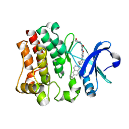
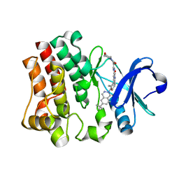
CRYSTAL STRUCTURE OF BTK KINASE DOMAIN COMPLEXED WITH 12-(6-tert-butyl-8-fluoro-1-oxo-phthalazin-2-yl)-9-hydroxy-6-methyl-4-[[5-(morpholine-4-carbonyl)-2-pyridyl]amino]-6-azatricyclo[9.4.0.02,7]pentadeca-1(15),2(7),3,11,13-pentaen-5-one
Descriptor: (9~{S})-12-(6-~{tert}-butyl-8-fluoranyl-1-oxidanylidene-phthalazin-2-yl)-6-methyl-4-[(5-morpholin-4-ylcarbonylpyridin-2-yl)amino]-9-oxidanyl-6-azatricyclo[9.4.0.0^{2,7}]pentadeca-1(15),2(7),3,11,13-pentaen-5-one, Tyrosine-protein kinase BTK
Authors:Kuglstatter, A, Janson, C.
Deposit date:2018-09-28
Release date:2019-03-20
Last modified:2024-05-15
Method:X-RAY DIFFRACTION (1.36 Å)
Cite:A potent seven-membered cyclic BTK (Bruton's tyrosine Kinase) chiral inhibitor conceived by structure-based drug design to lock its bioactive conformation.
Bioorg.Med.Chem.Lett., 29, 2019
4YBU
DownloadVisualize
BU of 4ybu by Molmil
Crystal structure of the R111K:Y134F:T54V:R132Q:P39Q:R59Y mutant of human Cellular Retinoic Acid Binding ProteinII in complex with Retinal after 24 h incubation and 1 hour UV irradiation at 1.92 angstrom - 1st cycle
Descriptor: Cellular retinoic acid-binding protein 2, RETINAL
Authors:Nosrati, M, Geiger, J.H.
Deposit date:2015-02-19
Release date:2016-02-24
Last modified:2023-09-27
Method:X-RAY DIFFRACTION (1.924 Å)
Cite:A Photoisomerizing Rhodopsin Mimic Observed at Atomic Resolution.
J.Am.Chem.Soc., 138, 2016
4YCH
DownloadVisualize
BU of 4ych by Molmil
Crystal Structure of R111K:Y134F:T54V:R132Q:P39Q:R59Y mutant of human Cellular Retinoic Acid Binding ProteinII with Retinal at 1.96 Angstrom - UV irradiated Crystal for 1 hour - 2nd cycle
Descriptor: Cellular retinoic acid-binding protein 2, RETINAL
Authors:Nosrati, M, Geiger, J.H.
Deposit date:2015-02-20
Release date:2016-02-24
Last modified:2023-09-27
Method:X-RAY DIFFRACTION (1.96 Å)
Cite:A Photoisomerizing Rhodopsin Mimic Observed at Atomic Resolution.
J.Am.Chem.Soc., 138, 2016
4RUU
DownloadVisualize
BU of 4ruu by Molmil
Crystal structure of the Q108K:K40L mutant of human Cellular Retinol Binding ProteinII in complex with All-trans-Retinal after 24 hour incubation at 1.4 Angstrom Resolution
Descriptor: ACETATE ION, RETINAL, Retinol-binding protein 2
Authors:Nosrati, M, Geiger, J.H.
Deposit date:2014-11-21
Release date:2014-12-10
Last modified:2023-09-20
Method:X-RAY DIFFRACTION (1.4 Å)
Cite:Tuning the electronic absorption of protein-embedded all-trans-retinal.
Science, 338, 2012
4YGH
DownloadVisualize
BU of 4ygh by Molmil
Crystal Structure of R111K:Y134F:T54V:R132Q:P39Y:R59Y mutant of human Cellular Retinoic Acid Binding Protein II with Retinal at 2.1 Angstrom resolution - UV irradiated crystal - 2nd cycle
Descriptor: Cellular retinoic acid-binding protein 2, RETINAL
Authors:Nosrati, M, Geiger, J.H.
Deposit date:2015-02-26
Release date:2016-03-02
Last modified:2023-09-27
Method:X-RAY DIFFRACTION (2.1 Å)
Cite:A Photoisomerizing Rhodopsin Mimic Observed at Atomic Resolution.
J.Am.Chem.Soc., 138, 2016
4YKM
DownloadVisualize
BU of 4ykm by Molmil
Crystal structure of the R111K:Y134F:T54V:R132Q:P39Q:R59Y:A32W:F3Q mutant of human Cellular Retinoic Acid Binding Protein II with Retinal at 1.58 angstrom resolution
Descriptor: Cellular retinoic acid-binding protein 2, RETINAL
Authors:Nosrati, M, Geiger, J.H.
Deposit date:2015-03-04
Release date:2016-03-09
Last modified:2023-09-27
Method:X-RAY DIFFRACTION (1.58 Å)
Cite:A Photoisomerizing Rhodopsin Mimic Observed at Atomic Resolution.
J.Am.Chem.Soc., 138, 2016
4YKO
DownloadVisualize
BU of 4yko by Molmil
Crystal structure of the R111K:Y134F:T54V:R132Q:P39Q:R59Y:A32Y:F3Q mutant of human Cellular Retinoic Acid Binding Protein II with Retinal at 1.58 angstrom resolution
Descriptor: Cellular retinoic acid-binding protein 2, RETINAL
Authors:Nosrati, M, Geiger, J.
Deposit date:2015-03-04
Release date:2016-03-09
Last modified:2023-09-27
Method:X-RAY DIFFRACTION (1.57 Å)
Cite:A Photoisomerizing Rhodopsin Mimic Observed at Atomic Resolution.
J.Am.Chem.Soc., 138, 2016
4YBP
DownloadVisualize
BU of 4ybp by Molmil
Crystal structure of the R111K:Y134F:T54V:R132Q:P39Q:R59Y mutant of human cellular retinoic acid binding proteinii with retinal after 24 hour incubation at 1.83 angstrom resolution - thermodynamic product - 1st cycle
Descriptor: Cellular retinoic acid-binding protein 2, RETINAL
Authors:Nosrati, M, Geiger, J.H.
Deposit date:2015-02-19
Release date:2016-02-24
Last modified:2023-09-27
Method:X-RAY DIFFRACTION (1.831 Å)
Cite:A Photoisomerizing Rhodopsin Mimic Observed at Atomic Resolution.
J.Am.Chem.Soc., 138, 2016
4YGZ
DownloadVisualize
BU of 4ygz by Molmil
CRYSTAL STRUCTURE OF THE R111K:Y134F:T54V:R132Q:P39Y:R59Y MUTANT OF HUMAN CELLULAR RETINOIC ACID BINDING PROTEIN II WITH RETINAL AT 2.06 ANGSTROM RESOLUTION - VISIBLE LIGHT IRRADIATED CRYSTAL -3RD CYCLE
Descriptor: Cellular retinoic acid-binding protein 2, RETINAL
Authors:Nosrati, M, Geiger, J.H.
Deposit date:2015-02-26
Release date:2016-03-02
Last modified:2023-09-27
Method:X-RAY DIFFRACTION (2.06 Å)
Cite:A Photoisomerizing Rhodopsin Mimic Observed at Atomic Resolution.
J.Am.Chem.Soc., 138, 2016
6C7Z
DownloadVisualize
BU of 6c7z by Molmil
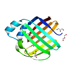
Crystal structure of the Q108K:K40L:T51V:R58F mutant of human Cellular Retinol Binding Protein II in complex with synthetic Ligand Julolidine
Descriptor: (2E,4E)-3-methyl-5-(2,3,6,7-tetrahydro-1H,5H-pyrido[3,2,1-ij]quinolin-9-yl)penta-2,4-dienal, ACETATE ION, Retinol-binding protein 2
Authors:Nosrati, M, Geiger, J.H.
Deposit date:2018-01-23
Release date:2018-04-25
Last modified:2023-10-04
Method:X-RAY DIFFRACTION (1.42 Å)
Cite:A Genetically Encoded Ratiometric pH Probe: Wavelength Regulation-Inspired Design of pH Indicators.
Chembiochem, 19, 2018
3CWK
DownloadVisualize
BU of 3cwk by Molmil
Crystal Structure of the R132K:Y134F:R111L:T54V:L121E Mutant of Cellular Retinoic Acid Binding Protein Type II in Complex with All-trans-Retinoic Acid at 1.57 Angstroms Resolution
Descriptor: Cellular retinoic acid-binding protein 2, RETINOIC ACID, SULFATE ION
Authors:Vaezeslami, S, Geiger, J.H.
Deposit date:2008-04-22
Release date:2008-09-09
Last modified:2024-02-21
Method:X-RAY DIFFRACTION (1.6 Å)
Cite:Structural analysis of site-directed mutants of cellular retinoic acid-binding protein II addresses the relationship between structural integrity and ligand binding.
Acta Crystallogr.,Sect.D, 64, 2008
4M83
DownloadVisualize
BU of 4m83 by Molmil
Ensemble refinement of protein crystal structure (2IYF) of macrolide glycosyltransferases OleD complexed with UDP and Erythromycin A
Descriptor: ERYTHROMYCIN A, MAGNESIUM ION, Oleandomycin glycosyltransferase, ...
Authors:Wang, F, Helmich, K.E, Xu, W, Singh, S, Olmos Jr, J.L, Martinez iii, E, Bingman, C.A, Thorson, J.S, Phillips Jr, G.N, Enzyme Discovery for Natural Product Biosynthesis (NatPro)
Deposit date:2013-08-12
Release date:2013-09-11
Last modified:2024-02-28
Method:X-RAY DIFFRACTION (1.698 Å)
Cite:Crystal structure of macrolide glycosyltransferases OleD
To be Published
8B7H
DownloadVisualize
BU of 8b7h by Molmil
Crystal structure of human Gremlin-1 in complex with Fab
Descriptor: Fab antibody fragment (heavy chain), Fab antibody fragment (light chain), Gremlin-1
Authors:Schulze, M.-S, Martinez-Fleites, C.
Deposit date:2022-09-30
Release date:2023-12-27
Method:X-RAY DIFFRACTION (1.95 Å)
Cite:Discovery of ginisortamab, a potent and novel anti-gremlin-1 antibody in clinical development for the treatment of cancer.
Mabs, 15, 2023
2FRS
DownloadVisualize
BU of 2frs by Molmil
Crystal structure of the f15w mutant of apo-cellular retinoic acid binding protein type ii at 1.51 angstroms resolution
Descriptor: Cellular retinoic acid-binding protein 2, SODIUM ION
Authors:Vaezeslami, S, Geiger, J.H.
Deposit date:2006-01-20
Release date:2006-09-19
Last modified:2024-02-14
Method:X-RAY DIFFRACTION (1.51 Å)
Cite:The structure of Apo-wild-type cellular retinoic acid binding protein II at 1.4 A and its relationship to ligand binding and nuclear translocation.
J.Mol.Biol., 363, 2006
2FS6
DownloadVisualize
BU of 2fs6 by Molmil
Crystal Structure of Apo-Cellular Retinoic Acid Binding Protein Type II At 1.35 Angstroms Resolution
Descriptor: ACETATE ION, CHLORIDE ION, Cellular retinoic acid-binding protein 2, ...
Authors:Vaezeslami, S, Geiger, J.H.
Deposit date:2006-01-21
Release date:2006-09-19
Last modified:2024-02-14
Method:X-RAY DIFFRACTION (1.35 Å)
Cite:The structure of Apo-wild-type cellular retinoic acid binding protein II at 1.4 A and its relationship to ligand binding and nuclear translocation.
J.Mol.Biol., 363, 2006
4I9S
DownloadVisualize
BU of 4i9s by Molmil
Crystal Structure of the R111K:R132L:Y134F:T54V:R59W Mutant of the Cellular Retinoic Acid Binding Protein Type II in Complex with All-Trans Retinal at 2.58 Angstrom Resolution
Descriptor: Cellular retinoic acid-binding protein 2, RETINAL
Authors:Nosrati, M, Geiger, J.H.
Deposit date:2012-12-05
Release date:2013-10-09
Last modified:2023-09-20
Method:X-RAY DIFFRACTION (2.58 Å)
Cite:Rational Design of a Colorimetric pH Sensor from a Soluble Retinoic Acid Chaperone.
J.Am.Chem.Soc., 135, 2013

225399

數據於2024-09-25公開中

PDB statisticsPDBj update infoContact PDBjnumon