Loading
PDBj
MenuPDBj@FacebookPDBj@TwitterPDBj@YouTubewwPDB FoundationwwPDB
RCSB PDBPDBeBMRBAdv. SearchSearch help
Search by PDB author
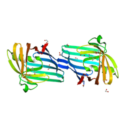
4EPS
DownloadVisualize
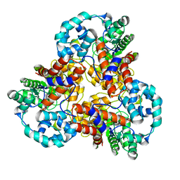
BU of 4eps by Molmil
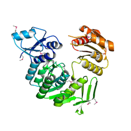
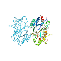
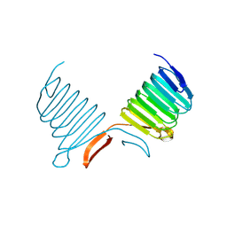
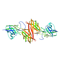
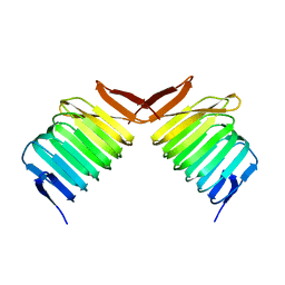
Crystal structure of a fimbrial protein (BACOVA_04982) from Bacteroides ovatus ATCC 8483 at 1.85 A resolution
Descriptor: CHLORIDE ION, GLYCEROL, SULFATE ION, ...
Authors:Joint Center for Structural Genomics (JCSG)
Deposit date:2012-04-17
Release date:2012-06-13
Last modified:2023-02-01
Method:X-RAY DIFFRACTION (1.85 Å)
Cite:A Distinct Type of Pilus from the Human Microbiome.
Cell, 165, 2016
2H1T
DownloadVisualize
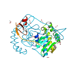
BU of 2h1t by Molmil
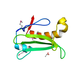
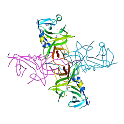
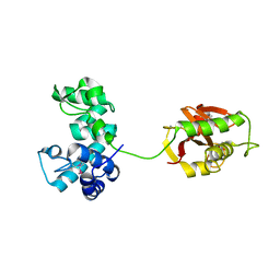
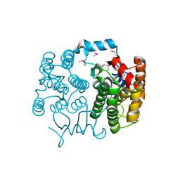
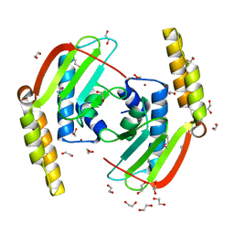
Crystal structure of a duf1089 family protein (pa1994) from pseudomonas aeruginosa at 1.80 A resolution
Descriptor: (4S)-2-METHYL-2,4-PENTANEDIOL, 1,2-ETHANEDIOL, Hypothetical protein
Authors:Joint Center for Structural Genomics (JCSG)
Deposit date:2006-05-16
Release date:2006-05-30
Last modified:2023-01-25
Method:X-RAY DIFFRACTION (1.8 Å)
Cite:The structure of the first representative of Pfam family PF06475 reveals a new fold with possible involvement in glycolipid metabolism.
Acta Crystallogr.,Sect.F, 66, 2010
1J5S
DownloadVisualize
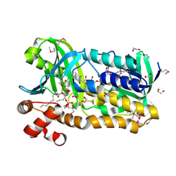
BU of 1j5s by Molmil
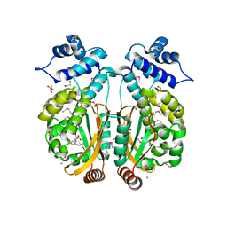
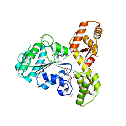
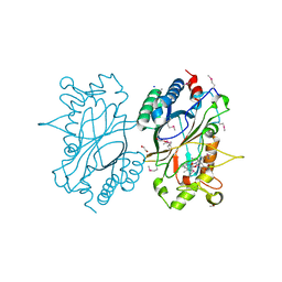
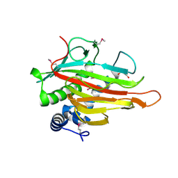
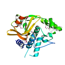
Crystal structure of uronate isomerase (TM0064) from Thermotoga maritima at 2.85 A resolution
Descriptor: URONATE ISOMERASE
Authors:Joint Center for Structural Genomics (JCSG)
Deposit date:2002-07-02
Release date:2002-07-17
Last modified:2024-05-22
Method:X-RAY DIFFRACTION (2.85 Å)
Cite:Crystal structure of uronate isomerase (TM0064) from Thermotoga maritima at 2.85 A resolution.
Proteins, 53, 2003
2GVI
DownloadVisualize
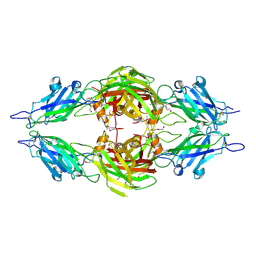
BU of 2gvi by Molmil
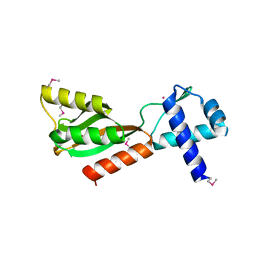
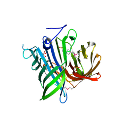
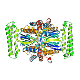
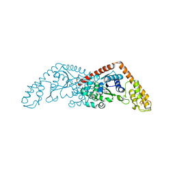
Crystal structure of a putative formylmethanofuran dehydrogenase subunit e (ta1109) from thermoplasma acidophilum at 1.87 A resolution
Descriptor: 1,2-ETHANEDIOL, ACETIC ACID, UNKNOWN LIGAND, ...
Authors:Joint Center for Structural Genomics (JCSG)
Deposit date:2006-05-02
Release date:2006-06-13
Last modified:2023-01-25
Method:X-RAY DIFFRACTION (1.87 Å)
Cite:Structures of three members of Pfam PF02663 (FmdE) implicated in microbial methanogenesis reveal a conserved alpha+beta core domain and an auxiliary C-terminal treble-clef zinc finger.
Acta Crystallogr.,Sect.F, 66, 2010
3OZ2
DownloadVisualize
BU of 3oz2 by Molmil
Crystal structure of a geranylgeranyl bacteriochlorophyll reductase-like (Ta0516) from Thermoplasma acidophilum at 1.60 A resolution
Descriptor: (2R)-3-{[(R)-{[(2S)-2,3-dihydroxypropyl]oxy}(hydroxy)phosphoryl]oxy}-2-[(6Z)-tridec-6-enoyloxy]propyl (9Z)-octadec-9-enoate, 1,2-ETHANEDIOL, Digeranylgeranylglycerophospholipid reductase, ...
Authors:Joint Center for Structural Genomics (JCSG)
Deposit date:2010-09-24
Release date:2010-10-27
Last modified:2024-10-09
Method:X-RAY DIFFRACTION (1.6 Å)
Cite:Insights into substrate specificity of geranylgeranyl reductases revealed by the structure of digeranylgeranylglycerophospholipid reductase, an essential enzyme in the biosynthesis of archaeal membrane lipids.
J.Mol.Biol., 404, 2010
3PAY
DownloadVisualize
BU of 3pay by Molmil
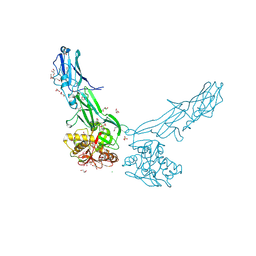
Crystal structure of a putative adhesin (BACOVA_04077) from Bacteroides ovatus at 2.50 A resolution
Descriptor: DI(HYDROXYETHYL)ETHER, putative adhesin
Authors:Joint Center for Structural Genomics (JCSG)
Deposit date:2010-10-19
Release date:2010-11-10
Last modified:2020-04-22
Method:X-RAY DIFFRACTION (2.5 Å)
Cite:A Distinct Type of Pilus from the Human Microbiome.
Cell, 165, 2016
1J6U
DownloadVisualize
BU of 1j6u by Molmil
Crystal structure of UDP-N-acetylmuramate-alanine ligase MurC (TM0231) from Thermotoga maritima at 2.3 A resolution
Descriptor: UDP-N-acetylmuramate-alanine ligase MurC
Authors:Joint Center for Structural Genomics (JCSG)
Deposit date:2002-08-29
Release date:2002-11-06
Last modified:2023-01-25
Method:X-RAY DIFFRACTION (2.3 Å)
Cite:Crystal structure of an Udp-n-acetylmuramate-alanine ligase MurC (TM0231) from Thermotoga maritima at 2.3 A resolution.
Proteins, 55, 2004
2IAY
DownloadVisualize
BU of 2iay by Molmil
Crystal structure of a duf1831 family protein (lp2179) from lactobacillus plantarum at 1.20 A resolution
Descriptor: CHLORIDE ION, GLYCEROL, Hypothetical protein
Authors:Joint Center for Structural Genomics (JCSG)
Deposit date:2006-09-08
Release date:2006-10-10
Last modified:2023-01-25
Method:X-RAY DIFFRACTION (1.2 Å)
Cite:Structure of LP2179, the first representative of Pfam family PF08866, suggests a new fold with a role in amino-acid metabolism.
Acta Crystallogr.,Sect.F, 66, 2010
3PXP
DownloadVisualize
BU of 3pxp by Molmil
Crystal structure of a PAS and DNA binding domain containing protein (Caur_2278) from CHLOROFLEXUS AURANTIACUS J-10-FL at 2.30 A resolution
Descriptor: 1,2-ETHANEDIOL, CHLORIDE ION, Helix-turn-helix domain protein, ...
Authors:Joint Center for Structural Genomics (JCSG)
Deposit date:2010-12-10
Release date:2011-01-19
Last modified:2024-10-09
Method:X-RAY DIFFRACTION (2.3 Å)
Cite:Structure of an MmyB-Like Regulator from C. aurantiacus, Member of a New Transcription Factor Family Linked to Antibiotic Metabolism in Actinomycetes.
Plos One, 7, 2012
1J5Y
DownloadVisualize
BU of 1j5y by Molmil
Crystal structure of transcriptional regulator (TM1602) from Thermotoga maritima at 2.3 A resolution
Descriptor: NICKEL (II) ION, POTASSIUM ION, TRANSCRIPTIONAL REGULATOR, ...
Authors:Joint Center for Structural Genomics (JCSG)
Deposit date:2002-07-05
Release date:2002-07-31
Last modified:2024-10-16
Method:X-RAY DIFFRACTION (2.3 Å)
Cite:Crystal structure of a transcription regulator (TM1602) from Thermotoga maritima at 2.3 A resolution.
Proteins, 67, 2007
2HAG
DownloadVisualize
BU of 2hag by Molmil
Crystal structure of a putative dyp-type peroxidase protein (so_0740) from shewanella oneidensis at 2.75 A resolution
Descriptor: Melanin biosynthesis protein TyrA, putative
Authors:Joint Center for Structural Genomics (JCSG)
Deposit date:2006-06-12
Release date:2006-08-08
Last modified:2023-01-25
Method:X-RAY DIFFRACTION (2.75 Å)
Cite:Crystal structures of two novel dye-decolorizing peroxidases reveal a beta-barrel fold with a conserved heme-binding motif.
Proteins, 69, 2007
4DGU
DownloadVisualize
BU of 4dgu by Molmil
Crystal structure of a putative cell adhesion protein (BT0320) from Bacteroides thetaiotaomicron VPI-5482 at 2.37 A resolution
Descriptor: CHLORIDE ION, ZINC ION, putative cell adhesion protein
Authors:Joint Center for Structural Genomics (JCSG)
Deposit date:2012-01-26
Release date:2012-02-15
Last modified:2023-02-01
Method:X-RAY DIFFRACTION (2.37 Å)
Cite:A Distinct Type of Pilus from the Human Microbiome.
Cell, 165, 2016
2FNA
DownloadVisualize
BU of 2fna by Molmil
Crystal structure of an archaeal aaa+ atpase (sso1545) from sulfolobus solfataricus p2 at 2.00 A resolution
Descriptor: 1,2-ETHANEDIOL, ADENOSINE-5'-DIPHOSPHATE, Conserved hypothetical protein, ...
Authors:Joint Center for Structural Genomics (JCSG)
Deposit date:2006-01-10
Release date:2006-02-07
Last modified:2024-10-09
Method:X-RAY DIFFRACTION (2 Å)
Cite:Crystal structure of a novel archaeal AAA+ ATPase SSO1545 from Sulfolobus solfataricus.
Proteins, 74, 2009
2ICH
DownloadVisualize
BU of 2ich by Molmil
CRYSTAL STRUCTURE OF A PUTATIVE ATTH (NE1406) FROM NITROSOMONAS EUROPAEA AT 2.00 A RESOLUTION
Descriptor: 2-[N-CYCLOHEXYLAMINO]ETHANE SULFONIC ACID, GLYCEROL, Putative AttH, ...
Authors:Joint Center for Structural Genomics (JCSG)
Deposit date:2006-09-12
Release date:2006-10-03
Last modified:2024-10-30
Method:X-RAY DIFFRACTION (2 Å)
Cite:Structure of the first representative of Pfam family PF09410 (DUF2006) reveals a structural signature of the calycin superfamily that suggests a role in lipid metabolism.
Acta Crystallogr.,Sect.F, 66, 2010
1K4Z
DownloadVisualize
BU of 1k4z by Molmil
C-terminal Domain of Cyclase Associated Protein
Descriptor: Adenylyl Cyclase-Associated Protein
Authors:Rozwarski, D.A, Fedorov, A.A, Dodatko, T, Almo, S.C, Burley, S.K, New York SGX Research Center for Structural Genomics (NYSGXRC)
Deposit date:2001-10-09
Release date:2002-03-13
Last modified:2024-10-30
Method:X-RAY DIFFRACTION (2.3 Å)
Cite:Crystal structure of the actin binding domain of the cyclase-associated protein
Biochemistry, 43, 2004
4FDY
DownloadVisualize
BU of 4fdy by Molmil
Crystal structure of a similar to lipoprotein, NLP/P60 family (SAV0400) from Staphylococcus aureus subsp. aureus Mu50 at 2.23 A resolution
Descriptor: Similar to lipoprotein, NLP/P60 family
Authors:Joint Center for Structural Genomics (JCSG)
Deposit date:2012-05-29
Release date:2012-07-25
Last modified:2023-02-01
Method:X-RAY DIFFRACTION (2.23 Å)
Cite:Structures of a Bifunctional Cell Wall Hydrolase CwlT Containing a Novel Bacterial Lysozyme and an NlpC/P60 dl-Endopeptidase.
J.Mol.Biol., 426, 2014
2IIZ
DownloadVisualize
BU of 2iiz by Molmil
Crystal structure of putative melanin biosynthesis protein TyrA with bound heme (NP_716371.1) from Shewanella Oneidensis at 2.30 A resolution
Descriptor: 1,2-ETHANEDIOL, ISOPROPYL ALCOHOL, Melanin biosynthesis protein TyrA, ...
Authors:Joint Center for Structural Genomics (JCSG)
Deposit date:2006-09-28
Release date:2006-11-14
Last modified:2023-11-15
Method:X-RAY DIFFRACTION (2.3 Å)
Cite:Identification and structural characterization of heme binding in a novel dye-decolorizing peroxidase, TyrA.
Proteins, 69, 2007
1KQ4
DownloadVisualize
BU of 1kq4 by Molmil
CRYSTAL STRUCTURE OF A THY1-COMPLEMENTING PROTEIN (TM0449) FROM THERMOTOGA MARITIMA AT 2.25 A RESOLUTION
Descriptor: FLAVIN-ADENINE DINUCLEOTIDE, HYPOTHETICAL PROTEIN TM0449
Authors:Wilson, I.A, Miller, M.D, Joint Center for Structural Genomics (JCSG)
Deposit date:2002-01-03
Release date:2002-02-27
Last modified:2017-10-11
Method:X-RAY DIFFRACTION (2.25 Å)
Cite:Structural genomics of the Thermotoga maritima proteome implemented in a high-throughput structure determination pipeline
Proc.Natl.Acad.Sci.USA, 99, 2002
4H40
DownloadVisualize
BU of 4h40 by Molmil
Crystal structure of a putative cell adhesion protein (BF2867) from Bacteroides fragilis NCTC 9343 at 2.57 A resolution
Descriptor: CHLORIDE ION, putative cell adhesion protein
Authors:Joint Center for Structural Genomics (JCSG)
Deposit date:2012-09-14
Release date:2012-10-03
Last modified:2023-02-01
Method:X-RAY DIFFRACTION (2.57 Å)
Cite:A Distinct Type of Pilus from the Human Microbiome.
Cell, 165, 2016
2FNO
DownloadVisualize
BU of 2fno by Molmil
Crystal structure of a glutathione s-transferase (atu5508) from agrobacterium tumefaciens str. c58 at 2.00 A resolution
Descriptor: AGR_pAT_752p, THIOCYANATE ION
Authors:Joint Center for Structural Genomics (JCSG)
Deposit date:2006-01-11
Release date:2006-02-14
Last modified:2023-01-25
Method:X-RAY DIFFRACTION (2 Å)
Cite:Comparative structural analysis of a novel glutathioneS-transferase (ATU5508) from Agrobacterium tumefaciens at 2.0 A resolution.
Proteins, 65, 2006
3PL0
DownloadVisualize
BU of 3pl0 by Molmil
Crystal structure of a bsmA homolog (Mpe_A2762) from Methylobium petroleophilum PM1 at 1.91 A resolution
Descriptor: CHLORIDE ION, GLYCEROL, Uncharacterized protein
Authors:Joint Center for Structural Genomics (JCSG)
Deposit date:2010-11-12
Release date:2010-12-08
Last modified:2023-02-01
Method:X-RAY DIFFRACTION (1.91 Å)
Cite:Crystal structure of a member of a novel family of dioxygenases (PF10014) reveals a conserved cupin fold and active site.
Proteins, 82, 2014
2G36
DownloadVisualize
BU of 2g36 by Molmil
Crystal structure of Tryptophanyl-tRNA synthetase (EC 6.1.1.2) (Tryptophan-tRNA ligase)(TrpRS) (tm0492) from THERMOTOGA MARITIMA at 2.50 A resolution
Descriptor: IRON/SULFUR CLUSTER, TRYPTOPHAN, Tryptophanyl-tRNA synthetase
Authors:Joint Center for Structural Genomics (JCSG)
Deposit date:2006-02-17
Release date:2006-03-28
Last modified:2024-04-03
Method:X-RAY DIFFRACTION (2.5 Å)
Cite:Structure of a tryptophanyl-tRNA synthetase containing an iron-sulfur cluster.
Acta Crystallogr.,Sect.F, 66, 2010
2GLZ
DownloadVisualize
BU of 2glz by Molmil
Crystal structure of a formylmethanofuran dehydrogenase subunit e-like protein (dhaf_2992) from desulfitobacterium hafniense dcb-2 at 1.45 A resolution
Descriptor: 1,2-ETHANEDIOL, NICKEL (II) ION, ZINC ION, ...
Authors:Joint Center for Structural Genomics (JCSG)
Deposit date:2006-04-05
Release date:2006-04-18
Last modified:2023-01-25
Method:X-RAY DIFFRACTION (1.45 Å)
Cite:Structures of three members of Pfam PF02663 (FmdE) implicated in microbial methanogenesis reveal a conserved alpha+beta core domain and an auxiliary C-terminal treble-clef zinc finger.
Acta Crystallogr.,Sect.F, 66, 2010
4H4J
DownloadVisualize
BU of 4h4j by Molmil
Crystal structure of a N-acetylmuramoyl-L-alanine amidase (BACUNI_02947) from Bacteroides uniformis ATCC 8492 at 1.15 A resolution
Descriptor: hypothetical protein
Authors:Joint Center for Structural Genomics (JCSG)
Deposit date:2012-09-17
Release date:2012-10-17
Last modified:2024-10-30
Method:X-RAY DIFFRACTION (1.15 Å)
Cite:Structure-guided functional characterization of DUF1460 reveals a highly specific NlpC/P60 amidase family.
Structure, 22, 2014
1K8F
DownloadVisualize
BU of 1k8f by Molmil
CRYSTAL STRUCTURE OF THE HUMAN C-TERMINAL CAP1-ADENYLYL CYCLASE ASSOCIATED PROTEIN
Descriptor: ADENYLYL CYCLASE-ASSOCIATED PROTEIN
Authors:Patskovsky, Y.V, Chance, M, Almo, S.C, Burley, S.K, New York SGX Research Center for Structural Genomics (NYSGXRC)
Deposit date:2001-10-24
Release date:2003-07-01
Last modified:2023-08-16
Method:X-RAY DIFFRACTION (2.8 Å)
Cite:Crystal structure of the actin binding domain of the cyclase-associated protein.
Biochemistry, 43, 2004

226707

數據於2024-10-30公開中

PDB statisticsPDBj update infoContact PDBjnumon