Loading
PDBj
MenuPDBj@FacebookPDBj@TwitterPDBj@YouTubewwPDB FoundationwwPDB
RCSB PDBPDBeBMRBAdv. SearchSearch help
Search by PDB author
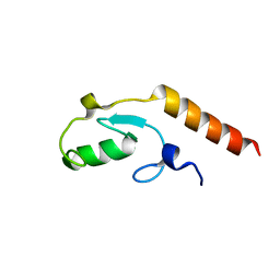
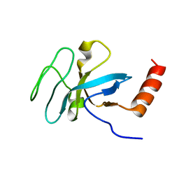
2KRE
DownloadVisualize
BU of 2kre by Molmil
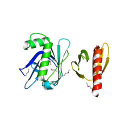
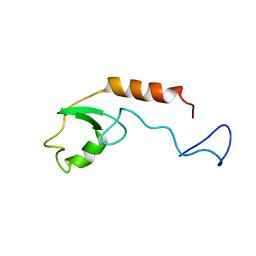
Solution structure of E4B/UFD2A U-Box domain
Descriptor: Ubiquitin conjugation factor E4 B
Authors:Nomine, Y, Wasielewski, E, Botuyan, M, Mer, G.
Deposit date:2009-12-16
Release date:2009-12-29
Last modified:2024-05-01
Method:SOLUTION NMR
Cite:Molecular Basis for the Association of Human E4B U Box Ubiquitin Ligase with E2-Conjugating Enzymes UbcH5c and Ubc4.
Structure, 18, 2010
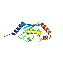
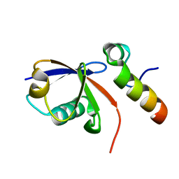
3SZM
DownloadVisualize
BU of 3szm by Molmil
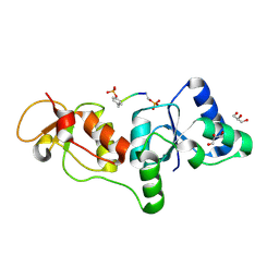
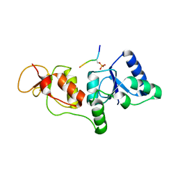
STRUCTURE OF HUMAN MICROCEPHALIN (MCPH1) TANDEM BRCT DOMAINS IN COMPLEX WITH A GAMMA-H2AX PHOSPHOPEPTIDE
Descriptor: Histone H2A.x, Microcephalin
Authors:Singh, N, Thompson, J.R, Mer, G.
Deposit date:2011-07-19
Release date:2011-11-30
Last modified:2012-09-19
Method:X-RAY DIFFRACTION (2.63 Å)
Cite:Dual recognition of phosphoserine and phosphotyrosine in histone variant H2A.X by DNA damage response protein MCPH1.
Proc.Natl.Acad.Sci.USA, 109, 2012
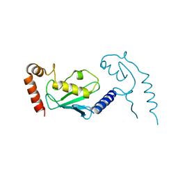
3TVV
DownloadVisualize
BU of 3tvv by Molmil
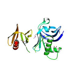
Structure of the tandem PH domains of Rtt106 (residues 68-315)
Descriptor: Histone chaperone RTT106
Authors:Su, D, Thompson, J.R, Mer, G.
Deposit date:2011-09-20
Release date:2012-02-01
Last modified:2023-12-06
Method:X-RAY DIFFRACTION (2.589 Å)
Cite:Structural basis for recognition of H3K56-acetylated histone H3-H4 by the chaperone Rtt106.
Nature, 483, 2012
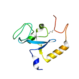
3TW1
DownloadVisualize
BU of 3tw1 by Molmil
Structure of Rtt106-AHN
Descriptor: GLYCEROL, Histone chaperone RTT106, N-[2-(1H-IMIDAZOL-4-YL)ETHYL]ACETAMIDE
Authors:Su, D, Thompson, J.R, Mer, G.
Deposit date:2011-09-21
Release date:2012-02-01
Last modified:2024-10-16
Method:X-RAY DIFFRACTION (1.772 Å)
Cite:Structural basis for recognition of H3K56-acetylated histone H3-H4 by the chaperone Rtt106.
Nature, 483, 2012
3U3Z
DownloadVisualize
BU of 3u3z by Molmil
Structure of human microcephalin (MCPH1) tandem BRCT domains in complex with an H2A.X peptide phosphorylated at Ser139 and Tyr142
Descriptor: GLYCEROL, Histone H2A.X peptide, Microcephalin
Authors:Singh, N, Thompson, J.R, Heroux, A, Mer, G.
Deposit date:2011-10-06
Release date:2012-07-25
Last modified:2024-10-30
Method:X-RAY DIFFRACTION (1.5 Å)
Cite:Dual recognition of phosphoserine and phosphotyrosine in histone variant H2A.X by DNA damage response protein MCPH1.
Proc.Natl.Acad.Sci.USA, 109, 2012
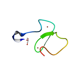
1A3Z
DownloadVisualize
BU of 1a3z by Molmil
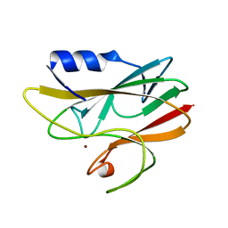
REDUCED RUSTICYANIN AT 1.9 ANGSTROMS
Descriptor: COPPER (I) ION, RUSTICYANIN
Authors:Zhao, D, Shoham, M.
Deposit date:1998-01-27
Release date:1998-07-29
Last modified:2024-05-22
Method:X-RAY DIFFRACTION (1.9 Å)
Cite:Rusticyanin: Extremes in acid stability and redox potential explained by the crystal structure.
Biophys.J., 74, 1998
3L1X
DownloadVisualize
BU of 3l1x by Molmil
Crystal Structure of U-box Domain of Human E4B Ubiquitin Ligase
Descriptor: Ubiquitin conjugation factor E4 B
Authors:Benirschke, R, Thompson, J.R, Mer, G.
Deposit date:2009-12-14
Release date:2010-05-05
Last modified:2023-09-06
Method:X-RAY DIFFRACTION (2.6 Å)
Cite:Molecular Basis for the Association of Human E4B U Box Ubiquitin Ligase with E2-Conjugating Enzymes UbcH5c and Ubc4.
Structure, 18, 2010
3L1Y
DownloadVisualize
BU of 3l1y by Molmil
Crystal structure of human UBC4 E2 conjugating enzyme
Descriptor: Ubiquitin-conjugating enzyme E2 D2
Authors:Benirschke, R, Thompson, J.R, Mer, G.
Deposit date:2009-12-14
Release date:2010-05-05
Last modified:2023-09-06
Method:X-RAY DIFFRACTION (1.6 Å)
Cite:Molecular Basis for the Association of Human E4B U Box Ubiquitin Ligase with E2-Conjugating Enzymes UbcH5c and Ubc4.
Structure, 18, 2010
3L1Z
DownloadVisualize
BU of 3l1z by Molmil
Crystal structure of the U-BOX domain of human E4B ubiquitin ligase in complex with UBCH5C E2 ubiquitin conjugating enzyme
Descriptor: Ubiquitin conjugation factor E4 B, Ubiquitin-conjugating enzyme E2 D3
Authors:Benirschke, R, Thompson, J.R, Mer, G.
Deposit date:2009-12-14
Release date:2010-05-05
Last modified:2023-09-06
Method:X-RAY DIFFRACTION (3.17 Å)
Cite:Molecular Basis for the Association of Human E4B U Box Ubiquitin Ligase with E2-Conjugating Enzymes UbcH5c and Ubc4.
Structure, 18, 2010
2K3Y
DownloadVisualize
BU of 2k3y by Molmil
Solution structure of EAF3 chromo barrel domain bound to histone h3 with a dimethyllysine analog H3K36ME2
Descriptor: Chromatin modification-related protein EAF3
Authors:Mer, G, Xu, C.
Deposit date:2008-05-19
Release date:2008-09-16
Last modified:2023-11-15
Method:SOLUTION NMR
Cite:Structural Basis for the Recognition of Methylated Histone H3K36 by the Eaf3 Subunit of Histone Deacetylase Complex Rpd3S.
Structure, 16, 2008
2K3X
DownloadVisualize
BU of 2k3x by Molmil
Solution structure of EAF3 chromo barrel domain
Descriptor: Chromatin modification-related protein EAF3
Authors:Mer, G, Xu, C.
Deposit date:2008-05-19
Release date:2008-09-16
Last modified:2024-05-29
Method:SOLUTION NMR
Cite:Structural Basis for the Recognition of Methylated Histone H3K36 by the Eaf3 Subunit of Histone Deacetylase Complex Rpd3S.
Structure, 16, 2008
2KTF
DownloadVisualize
BU of 2ktf by Molmil
Solution NMR structure of human polymerase iota UBM2 in complex with ubiquitin
Descriptor: DNA polymerase iota, Ubiquitin
Authors:Cui, G, Benirschke, R, Mer, G.
Deposit date:2010-02-01
Release date:2010-11-03
Last modified:2024-05-01
Method:SOLUTION NMR
Cite:Structural Basis of Ubiquitin Recognition by Translesion Synthesis DNA Polymerase iota.
Biochemistry, 49, 2010
5TAB
DownloadVisualize
BU of 5tab by Molmil
Crystal Structure of the PHD Finger of PHF20
Descriptor: GLYCEROL, PHD finger protein 20, ZINC ION
Authors:Klein, B.J, Kutateladze, T.G.
Deposit date:2016-09-09
Release date:2016-10-26
Last modified:2024-03-06
Method:X-RAY DIFFRACTION (1.25 Å)
Cite:PHF20 Readers Link Methylation of Histone H3K4 and p53 with H4K16 Acetylation.
Cell Rep, 17, 2016
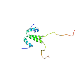
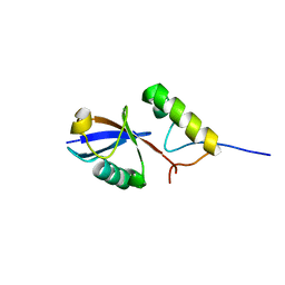
2LH0
DownloadVisualize
BU of 2lh0 by Molmil
NMR structure of the histone-interacting N-terminal homodimeric region of Rtt106
Descriptor: Histone chaperone RTT106
Authors:Hu, Q, Cui, G, Mer, G.
Deposit date:2011-08-04
Release date:2012-02-01
Last modified:2024-05-01
Method:SOLUTION NMR
Cite:Structural basis for recognition of H3K56-acetylated histone H3-H4 by the chaperone Rtt106.
Nature, 483, 2012
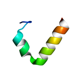
2L0G
DownloadVisualize
BU of 2l0g by Molmil
Solution NMR structure of ubiquitin-binding motif (UBM2) of human polymerase iota
Descriptor: DNA polymerase iota
Authors:Cui, G, Benirschke, R, Mer, G.
Deposit date:2010-07-01
Release date:2010-11-03
Last modified:2024-05-01
Method:SOLUTION NMR
Cite:Structural Basis of Ubiquitin Recognition by Translesion Synthesis DNA Polymerase iota.
Biochemistry, 49, 2010
2L0F
DownloadVisualize
BU of 2l0f by Molmil
Solution NMR structure of human polymerase iota UBM2 (P692A mutant) in complex with ubiquitin
Descriptor: DNA polymerase iota, Ubiquitin
Authors:Cui, G, Benirschke, R, Mer, G.
Deposit date:2010-07-01
Release date:2010-11-03
Last modified:2024-05-01
Method:SOLUTION NMR
Cite:Structural Basis of Ubiquitin Recognition by Translesion Synthesis DNA Polymerase iota.
Biochemistry, 49, 2010
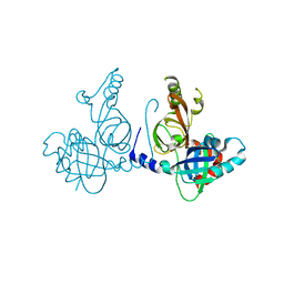
8S9K
DownloadVisualize
BU of 8s9k by Molmil
Structure of dimeric FAM111A SPD S541A Mutant
Descriptor: GLYCEROL, Serine protease FAM111A
Authors:Palani, S, Alvey, J.A, Cong, A.T.Q, Schellenberg, M.J, Machida, Y.
Deposit date:2023-03-29
Release date:2024-03-20
Method:X-RAY DIFFRACTION (2.72 Å)
Cite:Dimerization-dependent serine protease activity of FAM111A prevents replication fork stalling at topoisomerase 1 cleavage complexes.
Nat Commun, 15, 2024
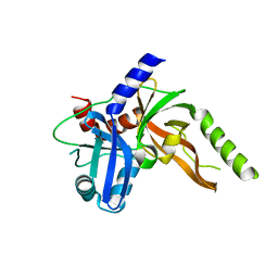
8S9L
DownloadVisualize
BU of 8s9l by Molmil
Structure of monomeric FAM111A SPD V347D Mutant
Descriptor: SULFATE ION, Serine protease FAM111A
Authors:Palani, S, Alvey, J.A, Cong, A.T.Q, Schellenberg, M.J, Machida, Y.
Deposit date:2023-03-29
Release date:2024-03-20
Method:X-RAY DIFFRACTION (1.85 Å)
Cite:Dimerization-dependent serine protease activity of FAM111A prevents replication fork stalling at topoisomerase 1 cleavage complexes.
Nat Commun, 15, 2024
<1234


226707

數據於2024-10-30公開中

PDB statisticsPDBj update infoContact PDBjnumon