Loading
PDBj
MenuPDBj@FacebookPDBj@TwitterPDBj@YouTubewwPDB FoundationwwPDB
RCSB PDBPDBeBMRBAdv. SearchSearch help
Search by PDB author
7EPT
DownloadVisualize
BU of 7ept by Molmil
Structural basis for the tethered peptide activation of adhesion GPCRs
Descriptor: Adhesion G-protein coupled receptor D1, Guanine nucleotide-binding protein G(I)/G(S)/G(O) subunit gamma-2, Guanine nucleotide-binding protein G(I)/G(S)/G(T) subunit beta-1, ...
Authors:Ping, Y.-Q, Xiao, P, Yang, F, Zhao, R.-J, Guo, S.-C, Yan, X, Wu, X, Liebscher, I, Xu, H.E, Sun, J.-P.
Deposit date:2021-04-27
Release date:2022-05-11
Method:ELECTRON MICROSCOPY (3 Å)
Cite:Structural basis for the tethered peptide activation of adhesion GPCRs.
Nature, 604, 2022
5YLX
DownloadVisualize
BU of 5ylx by Molmil
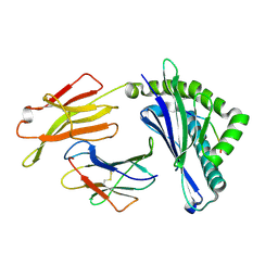
Integrated illustration of a valid epitope based on the SLA class I structure and tetramer technique could carry forward the development of molecular vaccine in swine species
Descriptor: Beta-2-microglobulin, MHC class I antigen, PRRSV-NSP9-TMP9 peptide
Authors:Pan, X.C, Wei, X.H, Zhang, N, Xia, C.
Deposit date:2017-10-20
Release date:2018-10-24
Last modified:2020-05-06
Method:X-RAY DIFFRACTION (2.2 Å)
Cite:Illumination of PRRSV Cytotoxic T Lymphocyte Epitopes by the Three-Dimensional Structure and Peptidome of Swine Lymphocyte Antigen Class I (SLA-I).
Front Immunol, 10, 2019
8GKA
DownloadVisualize
BU of 8gka by Molmil
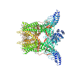
Human TRPV3 tetramer structure, closed conformation
Descriptor: (2S)-3-(hexadecanoyloxy)-2-[(9Z)-octadec-9-enoyloxy]propyl 2-(trimethylammonio)ethyl phosphate, SODIUM ION, Transient receptor potential cation channel subfamily V member 3
Authors:Lansky, S, Betancourt, J.M, Scheuring, S.
Deposit date:2023-03-17
Release date:2023-09-06
Last modified:2023-09-20
Method:ELECTRON MICROSCOPY (2.55 Å)
Cite:A pentameric TRPV3 channel with a dilated pore.
Nature, 621, 2023
8GKG
DownloadVisualize
BU of 8gkg by Molmil
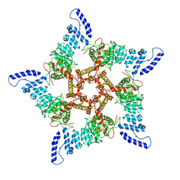
Human TRPV3 pentamer structure
Descriptor: Transient receptor potential cation channel subfamily V member 3
Authors:Lansky, S, Betancourt, J.M, Scheuring, S.
Deposit date:2023-03-18
Release date:2023-09-06
Last modified:2023-09-20
Method:ELECTRON MICROSCOPY (4.38 Å)
Cite:A pentameric TRPV3 channel with a dilated pore.
Nature, 621, 2023
7VRD
DownloadVisualize
BU of 7vrd by Molmil
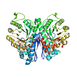
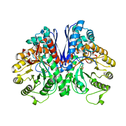
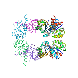
Crystal structure of Enolase1 from Candida albicans complexed with 2'-phosphoglyceric acid sodium
Descriptor: 2-PHOSPHOGLYCERIC ACID, Enolase 1, MAGNESIUM ION
Authors:Zhang, M, Zhang, X.
Deposit date:2021-10-22
Release date:2022-07-06
Last modified:2023-11-29
Method:X-RAY DIFFRACTION (1.7 Å)
Cite:Baicalein Acts against Candida albicans by Targeting Eno1 and Inhibiting Glycolysis.
Microbiol Spectr, 10, 2022
7V67
DownloadVisualize
BU of 7v67 by Molmil
Crystal Structure of Enolase1 from Candida albicans
Descriptor: Enolase 1, SULFATE ION
Authors:Zhang, M, Zhang, X.
Deposit date:2021-08-19
Release date:2022-07-06
Last modified:2023-11-29
Method:X-RAY DIFFRACTION (2 Å)
Cite:Baicalein Acts against Candida albicans by Targeting Eno1 and Inhibiting Glycolysis.
Microbiol Spectr, 10, 2022
7C2E
DownloadVisualize
BU of 7c2e by Molmil
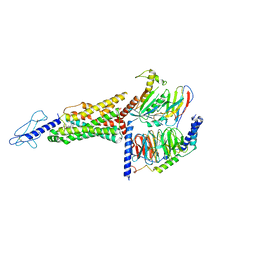
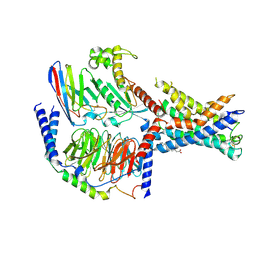
GLP-1R-Gs complex structure with a small molecule full agonist
Descriptor: 2-[[4-[6-[(4-cyano-2-fluoranyl-phenyl)methoxy]pyridin-2-yl]-3,6-dihydro-2~{H}-pyridin-1-yl]methyl]-3-[[(2~{S})-oxetan-2-yl]methyl]imidazo[4,5-b]pyridine-5-carboxylic acid, Glucagon-like peptide 1 receptor, Guanine nucleotide-binding protein G(I)/G(S)/G(O) subunit gamma-2, ...
Authors:Ma, H, Yuan, D.P, Huang, W, Wenge, Z, Xu, E.
Deposit date:2020-05-07
Release date:2020-08-26
Last modified:2020-12-16
Method:ELECTRON MICROSCOPY (4.2 Å)
Cite:Structural insights into the activation of GLP-1R by a small molecule agonist.
Cell Res., 30, 2020
2HMW
DownloadVisualize
BU of 2hmw by Molmil
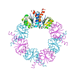
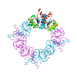
Square-shaped octameric ring structure of an RCK domain with ATP bound
Descriptor: ADENOSINE-5'-TRIPHOSPHATE, YuaA protein
Authors:Albright, R.A, Morais-Cabral, J.H.
Deposit date:2006-07-11
Release date:2006-09-26
Last modified:2023-08-30
Method:X-RAY DIFFRACTION (3 Å)
Cite:The RCK Domain of the KtrAB K(+) Transporter: Multiple Conformations of an Octameric Ring.
Cell(Cambridge,Mass.), 126, 2006
2HMT
DownloadVisualize
BU of 2hmt by Molmil
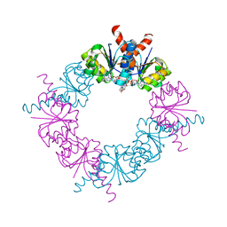
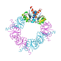
Diamond-shaped octameric ring structure of an RCK domain with NADH bound
Descriptor: 1,4-DIHYDRONICOTINAMIDE ADENINE DINUCLEOTIDE, YuaA protein
Authors:Albright, R.A, Morais-Cabral, J.H.
Deposit date:2006-07-11
Release date:2006-09-26
Last modified:2023-08-30
Method:X-RAY DIFFRACTION (2.2 Å)
Cite:The RCK Domain of the KtrAB K(+) Transporter: Multiple Conformations of an Octameric Ring.
Cell(Cambridge,Mass.), 126, 2006
2HMS
DownloadVisualize
BU of 2hms by Molmil
Rectangular-shaped octameric ring structure of an RCK domain with NADH bound
Descriptor: 1,4-DIHYDRONICOTINAMIDE ADENINE DINUCLEOTIDE, YuaA protein
Authors:Albright, R.A, Morais-Cabral, J.H.
Deposit date:2006-07-11
Release date:2006-09-26
Last modified:2023-08-30
Method:X-RAY DIFFRACTION (2.7 Å)
Cite:The RCK Domain of the KtrAB K(+) Transporter: Multiple Conformations of an Octameric Ring.
Cell(Cambridge,Mass.), 126, 2006
2HMU
DownloadVisualize
BU of 2hmu by Molmil
Diamond-shaped octameric ring structure of an RCK domain with ATP bound
Descriptor: ADENOSINE-5'-TRIPHOSPHATE, YuaA protein
Authors:Albright, R.A, Morais-Cabral, J.H.
Deposit date:2006-07-11
Release date:2006-09-26
Last modified:2023-08-30
Method:X-RAY DIFFRACTION (2.25 Å)
Cite:The RCK Domain of the KtrAB K(+) Transporter: Multiple Conformations of an Octameric Ring.
Cell(Cambridge,Mass.), 126, 2006
2HMV
DownloadVisualize
BU of 2hmv by Molmil
Diamond-shaped octameric ring structure of an RCK domain with ADP bound
Descriptor: ADENOSINE-5'-DIPHOSPHATE, YuaA protein
Authors:Albright, R.A, Morais-Cabral, J.H.
Deposit date:2006-07-11
Release date:2006-09-26
Last modified:2023-08-30
Method:X-RAY DIFFRACTION (2.2 Å)
Cite:The RCK Domain of the KtrAB K(+) Transporter: Multiple Conformations of an Octameric Ring.
Cell(Cambridge,Mass.), 126, 2006

222415

數據於2024-07-10公開中

PDB statisticsPDBj update infoContact PDBjnumon