Loading
PDBj
MenuPDBj@FacebookPDBj@TwitterPDBj@YouTubewwPDB FoundationwwPDB
RCSB PDBPDBeBMRBAdv. SearchSearch help
Search by PDB author
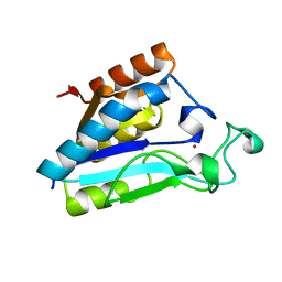
1ILW
DownloadVisualize
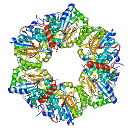
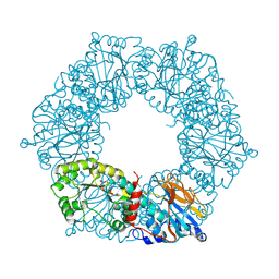
BU of 1ilw by Molmil
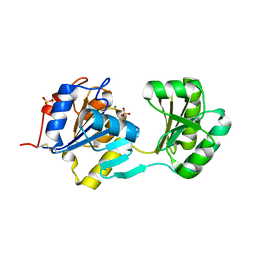
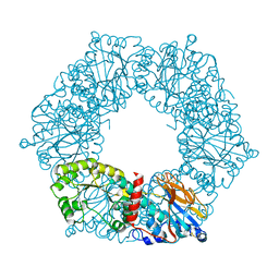
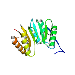
Crystal Structure of Pyrazinamidase/Nicotinamidase of Pyrococcus horikoshii
Descriptor: 180 aa long hypothetical Pyrazinamidase/nicotinamidase
Authors:Du, X, Kim, S.-H, Berkeley Structural Genomics Center (BSGC)
Deposit date:2001-05-08
Release date:2001-12-12
Last modified:2024-04-03
Method:X-RAY DIFFRACTION (2.05 Å)
Cite:Crystal structure and mechanism of catalysis of a pyrazinamidase from Pyrococcus horikoshii.
Biochemistry, 40, 2001
1IM5
DownloadVisualize
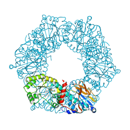
BU of 1im5 by Molmil
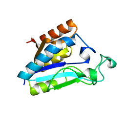
Crystal Structure of Pyrazinamidase of Pyrococcus horikoshii in Complex with Zinc
Descriptor: 180aa long hypothetical Pyrazinamidase/Nicotinamidase, ZINC ION
Authors:Du, X, Kim, S.-H.
Deposit date:2001-05-09
Release date:2001-12-12
Last modified:2024-04-03
Method:X-RAY DIFFRACTION (1.65 Å)
Cite:Crystal structure and mechanism of catalysis of a pyrazinamidase from Pyrococcus horikoshii.
Biochemistry, 40, 2001
1ETL
DownloadVisualize
BU of 1etl by Molmil
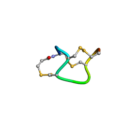
STRUCTURAL CHARACTERISTICS FOR BIOLOGICAL ACTIVITY OF HEAT-STABLE ENTEROTOXIN PRODUCED BY ENTEROTOXIGENIC ESCHERICHIA COLI: X-RAY CRYSTALLOGRAPHY OF WEAKLY TOXIC AND NONTOXIC ANALOGS
Descriptor: 5-BETA-MERCAPTOPROPIONATE HEAT-STABLE ENTEROTOXIN
Authors:Sato, T, Shimonishi, Y.
Deposit date:1994-03-15
Release date:1996-01-29
Last modified:2021-11-03
Method:X-RAY DIFFRACTION (0.89 Å)
Cite:Structural characteristics for biological activity of heat-stable enterotoxin produced by enterotoxigenic Escherichia coli: X-ray crystallography of weakly toxic and nontoxic analogs.
Biochemistry, 33, 1994
1ETM
DownloadVisualize
BU of 1etm by Molmil
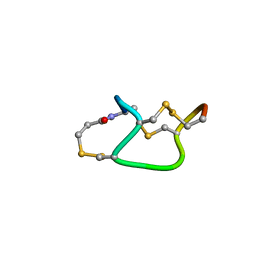
STRUCTURAL CHARACTERISTICS FOR BIOLOGICAL ACTIVITY OF HEAT-STABLE ENTEROTOXIN PRODUCED BY ENTEROTOXIGENIC ESCHERICHIA COLI: X-RAY CRYSTALLOGRAPHY OF WEAKLY TOXIC AND NONTOXIC ANALOGS
Descriptor: 5-BETA-MERCAPTOPROPIONATE HEAT-STABLE ENTEROTOXIN
Authors:Sato, T, Shimonishi, Y.
Deposit date:1994-03-15
Release date:1996-01-29
Last modified:2021-11-03
Method:X-RAY DIFFRACTION (0.89 Å)
Cite:Structural characteristics for biological activity of heat-stable enterotoxin produced by enterotoxigenic Escherichia coli: X-ray crystallography of weakly toxic and nontoxic analogs.
Biochemistry, 33, 1994
3HPE
DownloadVisualize
BU of 3hpe by Molmil
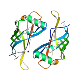
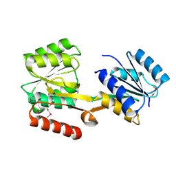
Crystal structure of yceI (HP1286) from Helicobacter pylori
Descriptor: (13Z)-docos-13-enamide, Conserved hypothetical secreted protein
Authors:Sisinni, L, Cendron, L, Zanotti, G.
Deposit date:2009-06-04
Release date:2010-04-14
Last modified:2023-09-06
Method:X-RAY DIFFRACTION (2.1 Å)
Cite:Helicobacter pylori acidic stress response factor HP1286 is a YceI homolog with new binding specificity.
Febs J., 277, 2010
1UKX
DownloadVisualize
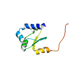
BU of 1ukx by Molmil
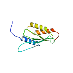
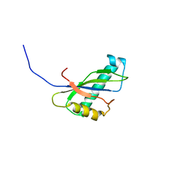
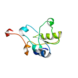
Solution structure of the RWD domain of mouse GCN2
Descriptor: GCN2 eIF2alpha kinase
Authors:Nameki, N, Yoneyama, M, Koshiba, S, Inoue, M, Kigawa, T, Yokoyama, S, RIKEN Structural Genomics/Proteomics Initiative (RSGI)
Deposit date:2003-09-03
Release date:2004-08-03
Last modified:2023-12-27
Method:SOLUTION NMR
Cite:Solution structure of the RWD domain of the mouse GCN2 protein.
Protein Sci., 13, 2004
1T6S
DownloadVisualize
BU of 1t6s by Molmil
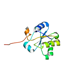
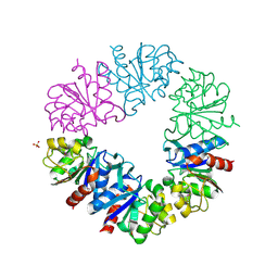
Crystal structure of a conserved hypothetical protein from Chlorobium tepidum
Descriptor: NITRATE ION, conserved hypothetical protein
Authors:Kim, J.S, Shin, D.H, Kim, R, Kim, S.H, Berkeley Structural Genomics Center (BSGC)
Deposit date:2004-05-07
Release date:2004-12-07
Last modified:2024-02-14
Method:X-RAY DIFFRACTION (1.95 Å)
Cite:Crystal structure of ScpB from Chlorobium tepidum, a protein involved in chromosome partitioning.
Proteins, 62, 2006
3UTO
DownloadVisualize
BU of 3uto by Molmil
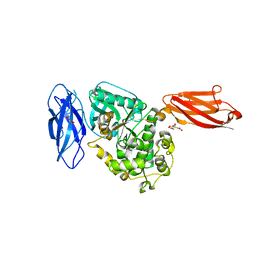
Twitchin kinase region from C.elegans (Fn31-NL-kin-CRD-Ig26)
Descriptor: 3,6,9,12,15,18-HEXAOXAICOSANE-1,20-DIOL, CITRATE ANION, DI(HYDROXYETHYL)ETHER, ...
Authors:Castelmur, E, Barbieri, S, Mayans, O.
Deposit date:2011-11-26
Release date:2012-08-08
Last modified:2024-02-28
Method:X-RAY DIFFRACTION (2.4 Å)
Cite:Identification of an N-terminal inhibitory extension as the primary mechanosensory regulator of twitchin kinase.
Proc.Natl.Acad.Sci.USA, 109, 2012
1N27
DownloadVisualize
BU of 1n27 by Molmil
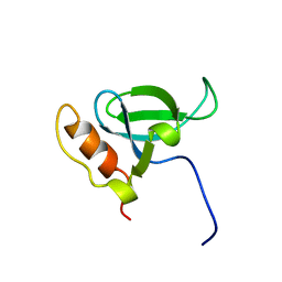
Solution structure of the PWWP domain of mouse Hepatoma-derived growth factor, related protein 3
Descriptor: Hepatoma-derived growth factor, related protein 3
Authors:Nameki, N, Kigawa, T, Koshiba, S, Kobayashi, N, Tochio, N, Inoue, M, Yokoyama, S, RIKEN Structural Genomics/Proteomics Initiative (RSGI)
Deposit date:2002-10-22
Release date:2003-12-23
Last modified:2024-05-29
Method:SOLUTION NMR
Cite:Solution structure of the PWWP domain of the hepatoma-derived growth factor family.
Protein Sci., 14, 2005
2Z7Q
DownloadVisualize
BU of 2z7q by Molmil
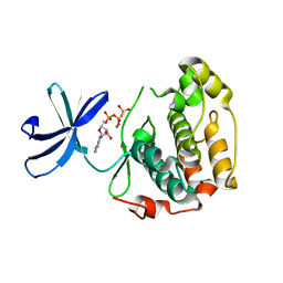
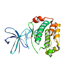
Crystal structure of the N-terminal kinase domain of human RSK-1 bound to AMP-PCP
Descriptor: MAGNESIUM ION, PHOSPHOMETHYLPHOSPHONIC ACID ADENYLATE ESTER, Ribosomal protein S6 kinase alpha-1
Authors:Ikuta, M, Munshi, S.K.
Deposit date:2007-08-28
Release date:2008-05-13
Last modified:2023-11-01
Method:X-RAY DIFFRACTION (2 Å)
Cite:Crystal structures of the N-terminal kinase domain of human RSK1 bound to three different ligands: Implications for the design of RSK1 specific inhibitors.
Protein Sci., 16, 2007
1NF2
DownloadVisualize
BU of 1nf2 by Molmil
X-ray crystal structure of TM0651 from Thermotoga maritima
Descriptor: MAGNESIUM ION, SULFATE ION, phosphatase
Authors:Shin, D.H, Berkeley Structural Genomics Center (BSGC)
Deposit date:2002-12-12
Release date:2003-09-16
Last modified:2011-07-13
Method:X-RAY DIFFRACTION (2.2 Å)
Cite:Crystal structure of a phosphatase with a unique substrate binding domain from Thermotoga maritima
Protein Sci., 12, 2003
2Z7S
DownloadVisualize
BU of 2z7s by Molmil
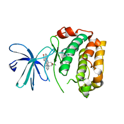
Crystal Structure of the N-terminal Kinase Domain of Human RSK1 bound to Purvalnol A
Descriptor: 2-({6-[(3-CHLOROPHENYL)AMINO]-9-ISOPROPYL-9H-PURIN-2-YL}AMINO)-3-METHYLBUTAN-1-OL, Ribosomal protein S6 kinase alpha-1
Authors:Ikuta, M, Munshi, S.K.
Deposit date:2007-08-28
Release date:2008-05-13
Last modified:2023-11-01
Method:X-RAY DIFFRACTION (2.1 Å)
Cite:Crystal structures of the N-terminal kinase domain of human RSK1 bound to three different ligands: Implications for the design of RSK1 specific inhibitors.
Protein Sci., 16, 2007
2Z7R
DownloadVisualize
BU of 2z7r by Molmil
Crystal Structure of the N-terminal Kinase Domain of Human RSK1 bound to Staurosporine
Descriptor: Ribosomal protein S6 kinase alpha-1, STAUROSPORINE
Authors:Ikuta, M, Munshi, S.K.
Deposit date:2007-08-28
Release date:2008-05-13
Last modified:2023-11-01
Method:X-RAY DIFFRACTION (2 Å)
Cite:Crystal structures of the N-terminal kinase domain of human RSK1 bound to three different ligands: Implications for the design of RSK1 specific inhibitors.
Protein Sci., 16, 2007
2HQB
DownloadVisualize
BU of 2hqb by Molmil
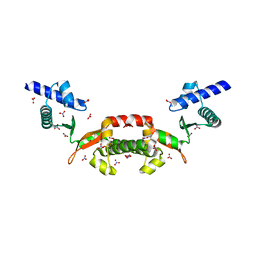
Crystal Structure of a Transcriptional Activator of comK gene from Bacillus halodurans
Descriptor: Transcriptional activator of comK gene
Authors:Xu, Q.S, Berkeley Structural Genomics Center (BSGC)
Deposit date:2006-07-18
Release date:2007-05-29
Last modified:2011-07-13
Method:X-RAY DIFFRACTION (2.7 Å)
Cite:Crystal structure of a transcriptional activator of comK gene from Bacillus halodurans.
Proteins, 69, 2007
2I14
DownloadVisualize
BU of 2i14 by Molmil
Crystal structure of nicotinate-nucleotide pyrophosphorylase from Pyrococcus furiosus
Descriptor: 1-ALPHA-PYROPHOSPHORYL-2-ALPHA,3-ALPHA-DIHYDROXY-4-BETA-CYCLOPENTANE-METHANOL-5-PHOSPHATE, Nicotinate-nucleotide pyrophosphorylase, ZINC ION
Authors:Shin, D.H, Kim, R, Kim, S.-H, Berkeley Structural Genomics Center (BSGC)
Deposit date:2006-08-12
Release date:2006-10-10
Last modified:2023-08-30
Method:X-RAY DIFFRACTION (2.9 Å)
Cite:Crystal structure of nicotinate-nucleotide pyrophosphorylase from Pyrococcus furiosus
To be Published
2JMP
DownloadVisualize
BU of 2jmp by Molmil
Structure for the N-terminus of chromosomal replication initiation protein dnaA from M. genitalium
Descriptor: Chromosomal replication initiator protein dnaA
Authors:Lowery, T.J, Pelton, J.G, Wemmer, D.E, Berkeley Structural Genomics Center (BSGC)
Deposit date:2006-11-27
Release date:2007-07-17
Last modified:2024-05-08
Method:SOLUTION NMR
Cite:NMR structure of the N-terminal domain of the replication initiator protein DnaA.
J.Struct.Funct.Genom., 8, 2007
1YTK
DownloadVisualize
BU of 1ytk by Molmil
Crystal structure of a nicotinate phosphoribosyltransferase from Thermoplasma acidophilum with nicotinate mononucleotide
Descriptor: 2-AMINO-2-HYDROXYMETHYL-PROPANE-1,3-DIOL, NICOTINATE MONONUCLEOTIDE, nicotinate phosphoribosyltransferase from Thermoplasma acidophilum
Authors:Shin, D.H, Berkeley Structural Genomics Center (BSGC)
Deposit date:2005-02-10
Release date:2005-03-08
Last modified:2011-07-13
Method:X-RAY DIFFRACTION (2.65 Å)
Cite:Crystal structure of a nicotinate phosphoribosyltransferase from Thermoplasma acidophilum.
J.Biol.Chem., 280, 2005
1YTE
DownloadVisualize
BU of 1yte by Molmil
Crystal structure of a nicotinate phosphoribosyltransferase from Thermoplasma acidophilum, phosphoribosylpyrophosphate bound structure
Descriptor: 1-ALPHA-PYROPHOSPHORYL-2-ALPHA,3-ALPHA-DIHYDROXY-4-BETA-CYCLOPENTANE-METHANOL-5-PHOSPHATE, nicotinate phosphoribosyltransferase from Thermoplasma acidophilum
Authors:Shin, D.H, Berkeley Structural Genomics Center (BSGC)
Deposit date:2005-02-10
Release date:2005-03-08
Last modified:2011-07-13
Method:X-RAY DIFFRACTION (2.75 Å)
Cite:Crystal structure of a nicotinate phosphoribosyltransferase from Thermoplasma acidophilum.
J.Biol.Chem., 280, 2005
1YTD
DownloadVisualize
BU of 1ytd by Molmil
Crystal structure of a nicotinate phosphoribosyltransferase from Thermoplasma acidophilum, Native Structure
Descriptor: nicotinate phosphoribosyltransferase from Thermoplasma acidophilum
Authors:Shin, D.H, Berkeley Structural Genomics Center (BSGC)
Deposit date:2005-02-10
Release date:2005-03-08
Last modified:2011-07-13
Method:X-RAY DIFFRACTION (2.8 Å)
Cite:Crystal structure of a nicotinate phosphoribosyltransferase from Thermoplasma acidophilum.
J.Biol.Chem., 280, 2005
1IVZ
DownloadVisualize
BU of 1ivz by Molmil
Solution structure of the SEA domain from murine hypothetical protein homologous to human mucin 16
Descriptor: hypothetical protein 1110008I14RIK
Authors:Maeda, T, Inoue, M, Kigawa, T, Yokoyama, S, RIKEN Structural Genomics/Proteomics Initiative (RSGI)
Deposit date:2002-04-02
Release date:2002-10-02
Last modified:2023-12-27
Method:SOLUTION NMR
Cite:Solution structure of the SEA domain from the murine homologue of ovarian cancer antigen CA125 (MUC16)
J.Biol.Chem., 279, 2004
1VEE
DownloadVisualize
BU of 1vee by Molmil
NMR structure of the hypothetical rhodanese domain At4g01050 from Arabidopsis thaliana
Descriptor: proline-rich protein family
Authors:Pantoja-Uceda, D, Lopez-Mendez, B, Koshiba, S, Inoue, M, Kigawa, T, Terada, T, Shirouzu, M, Tanaka, A, Seki, M, Shinozaki, K, Yokoyama, S, Guntert, P, RIKEN Structural Genomics/Proteomics Initiative (RSGI)
Deposit date:2004-03-30
Release date:2005-01-25
Last modified:2023-12-27
Method:SOLUTION NMR
Cite:Solution structure of the rhodanese homology domain At4g01050(175-295) from Arabidopsis thaliana
Protein Sci., 14, 2005
2DCP
DownloadVisualize
BU of 2dcp by Molmil
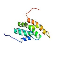
Fully automated NMR structure determination of the ENTH-VHS domain AT3G16270 from Arabidopsis thaliana
Descriptor: hypothetical protein (RAFL09-17-B18)
Authors:Lopez-Mendez, B, Guntert, P.
Deposit date:2006-01-12
Release date:2006-10-17
Last modified:2024-05-29
Method:SOLUTION NMR
Cite:Automated Protein Structure Determination from NMR Spectra
J.Am.Chem.Soc., 128, 2006
2DCQ
DownloadVisualize
BU of 2dcq by Molmil
Fully automated NMR structure determination of the rhodanese homology domain At4g01050(175-295) from Arabidopsis thaliana
Descriptor: Putative protein At4g01050
Authors:Lopez-Mendez, B, Guntert, P.
Deposit date:2006-01-12
Release date:2006-10-17
Last modified:2024-05-29
Method:SOLUTION NMR
Cite:Automated protein structure determination from NMR spectra
J.AM.CHEM.SOC., 128, 2006
1DUS
DownloadVisualize
BU of 1dus by Molmil
MJ0882-A hypothetical protein from M. jannaschii
Descriptor: MJ0882
Authors:Hung, L, Huang, L, Kim, R, Kim, S.H, Berkeley Structural Genomics Center (BSGC)
Deposit date:2000-01-18
Release date:2000-07-19
Last modified:2011-07-13
Method:X-RAY DIFFRACTION (1.8 Å)
Cite:Structure-based experimental confirmation of biochemical function to a methyltransferase, MJ0882, from hyperthermophile Methanococcus jannaschii
J.STRUCT.FUNCT.GENOM., 2, 2002
1G2I
DownloadVisualize
BU of 1g2i by Molmil
CRYSTAL STRUCTURE OF A NOVEL INTRACELLULAR PROTEASE FROM PYROCOCCUS HORIKOSHII AT 2 A RESOLUTION
Descriptor: PROTEASE I, SULFATE ION
Authors:Du, X, Choi, I.-G, Kim, R, Jancarik, J, Kim, S.-H, Berkeley Structural Genomics Center (BSGC)
Deposit date:2000-10-19
Release date:2000-11-08
Last modified:2018-01-24
Method:X-RAY DIFFRACTION (2 Å)
Cite:Crystal structure of an intracellular protease from Pyrococcus horikoshii at 2-A resolution.
Proc.Natl.Acad.Sci.USA, 97, 2000

225946

数据于2024-10-09公开中

PDB statisticsPDBj update infoContact PDBjnumon