1SUO
 
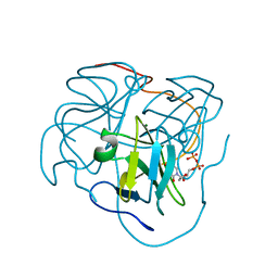
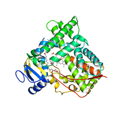
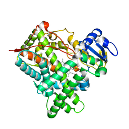 | | Structure of mammalian cytochrome P450 2B4 with bound 4-(4-chlorophenyl)imidazole | | Descriptor: | 4-(4-CHLOROPHENYL)IMIDAZOLE, Cytochrome P450 2B4, PROTOPORPHYRIN IX CONTAINING FE | | Authors: | Scott, E.E, White, M.A, He, Y.A, Johnson, E.F, Stout, C.D, Halpert, J.R. | | Deposit date: | 2004-03-26 | | Release date: | 2004-07-20 | | Last modified: | 2023-08-23 | | Method: | X-RAY DIFFRACTION (1.9 Å) | | Cite: | Structure of mammalian cytochrome P450 2B4 complexed with 4-(4-chlorophenyl)imidazole at 1.9 {angstrom} resolution: Insight into the range of P450 conformations and coordination of redox partner binding.
J.Biol.Chem., 279, 2004
|
|
1PC4
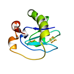
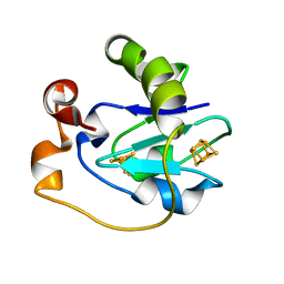 | | Crystal Structure of the P50A mutant of ferredoxin I at 1.65 A Resolution | | Descriptor: | FE3-S4 CLUSTER, Ferredoxin I, IRON/SULFUR CLUSTER | | Authors: | Camba, R, Jung, Y.S, Chen, K, Hunsicker-Wang, L.M, Burgess, B.K, Stout, C.D, Hirst, J, Armstrong, F.A. | | Deposit date: | 2003-05-15 | | Release date: | 2003-09-30 | | Last modified: | 2024-02-14 | | Method: | X-RAY DIFFRACTION (1.65 Å) | | Cite: | Mechanisms of redox-coupled proton transfer in proteins: role of the proximal proline in reactions of the [3Fe-4S] cluster in Azotobacter vinelandii ferredoxin I
Biochemistry, 42, 2003
|
|
1PC5
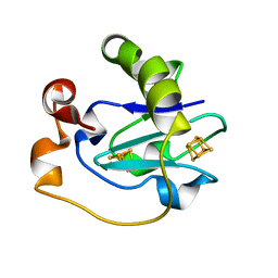 | | Crystal Structure of the P50G Mutant of Ferredoxin I at 1.8 A Resolution | | Descriptor: | FE3-S4 CLUSTER, Ferredoxin I, IRON/SULFUR CLUSTER | | Authors: | Camba, R, Jung, Y.S, Chen, K, Hunsicker-Wang, L.M, Burgess, B.K, Stout, C.D, Hirst, J, Armstrong, F.A. | | Deposit date: | 2003-05-15 | | Release date: | 2003-09-30 | | Last modified: | 2024-02-14 | | Method: | X-RAY DIFFRACTION (1.8 Å) | | Cite: | Mechanisms of redox-coupled proton transfer in proteins: role of the proximal proline in reactions of the [3Fe-4S] cluster in Azotobacter vinelandii ferredoxin I
Biochemistry, 42, 2003
|
|
1F7D
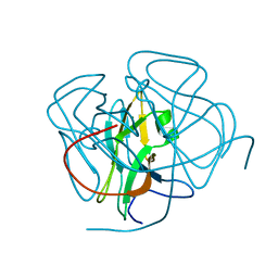 | | CRYSTAL STRUCTURES OF FELINE IMMUNODEFICIENCY VIRUS DUTP PYROPHOSPHATASE AND ITS NUCLEOTIDE COMPLEXES IN THREE CRYSTAL FORMS | | Descriptor: | MAGNESIUM ION, POL POLYPROTEIN | | Authors: | Prasad, G.S, Stura, E.A, Elder, J.H, Stout, C.D. | | Deposit date: | 2000-06-26 | | Release date: | 2000-09-06 | | Last modified: | 2024-02-07 | | Method: | X-RAY DIFFRACTION (1.4 Å) | | Cite: | Structures of feline immunodeficiency virus dUTP pyrophosphatase and its nucleotide complexes in three crystal forms.
Acta Crystallogr.,Sect.D, 56, 2000
|
|
1F7K
 | | CRYSTAL STRUCTURES OF FELINE IMMUNODEFICIENCY VIRUS DUTP PYROPHOSPHATASE AND ITS NUCLEOTIDE COMPLEXES IN THREE CRYSTAL FORMS. | | Descriptor: | 2'-DEOXYURIDINE 5'-MONOPHOSPHATE, MAGNESIUM ION, POL POLYPROTEIN | | Authors: | Prasad, G.S, Stura, E.A, Elder, J.H, Stout, C.D. | | Deposit date: | 2000-06-27 | | Release date: | 2000-09-06 | | Last modified: | 2024-02-07 | | Method: | X-RAY DIFFRACTION (2.2 Å) | | Cite: | Structures of feline immunodeficiency virus dUTP pyrophosphatase and its nucleotide complexes in three crystal forms.
Acta Crystallogr.,Sect.D, 56, 2000
|
|
1F7N
 | | CRYSTAL STRUCTURES OF FELINE IMMUNODEFICIENCY VIRUS DUTP PYROPHOSPHATASE AND ITS NUCLEOTIDE COMPLEXES IN THREE CRYSTAL FORMS. | | Descriptor: | 2'-DEOXYURIDINE 5'-MONOPHOSPHATE, MAGNESIUM ION, POL POLYPROTEIN | | Authors: | Prasad, G.S, Stura, E.A, Elder, J.H, Stout, C.D. | | Deposit date: | 2000-06-27 | | Release date: | 2000-09-06 | | Last modified: | 2024-02-07 | | Method: | X-RAY DIFFRACTION (2.2 Å) | | Cite: | Structures of feline immunodeficiency virus dUTP pyrophosphatase and its nucleotide complexes in three crystal forms.
Acta Crystallogr.,Sect.D, 56, 2000
|
|
1F7O
 | | CRYSTAL STRUCTURES OF FELINE IMMUNODEFICIENCY VIRUS DUTP PYROPHOSPHATASE AND ITS NUCLEOTIDE COMPLEXES IN THREE CRYSTAL FORMS. | | Descriptor: | POL POLYPROTEIN | | Authors: | Prasad, G.S, Stura, E.A, Elder, J.H, Stout, C.D. | | Deposit date: | 2000-06-27 | | Release date: | 2000-09-06 | | Last modified: | 2024-02-07 | | Method: | X-RAY DIFFRACTION (2.2 Å) | | Cite: | Structures of feline immunodeficiency virus dUTP pyrophosphatase and its nucleotide complexes in three crystal forms.
Acta Crystallogr.,Sect.D, 56, 2000
|
|
1F7Q
 | | CRYSTAL STRUCTURES OF FELINE IMMUNODEFICIENCY VIRUS DUTP PYROPHOSPHATASE AND ITS NUCLEOTIDE COMPLEXES IN THREE CRYSTAL FORMS. | | Descriptor: | DEOXYURIDINE-5'-TRIPHOSPHATE, POL POLYPROTEIN | | Authors: | Prasad, G.S, Stura, E.A, Elder, J.H, Stout, C.D. | | Deposit date: | 2000-06-27 | | Release date: | 2000-09-06 | | Last modified: | 2024-02-07 | | Method: | X-RAY DIFFRACTION (2.26 Å) | | Cite: | Structures of feline immunodeficiency virus dUTP pyrophosphatase and its nucleotide complexes in three crystal forms.
Acta Crystallogr.,Sect.D, 56, 2000
|
|
1F7R
 | | CRYSTAL STRUCTURES OF FELINE IMMUNODEFICIENCY VIRUS DUTP PYROPHOSPHATASE AND ITS NUCLEOTIDE COMPLEXES IN THREE CRYSTAL FORMS. | | Descriptor: | MAGNESIUM ION, POL POLYPROTEIN, URIDINE-5'-DIPHOSPHATE | | Authors: | Prasad, G.S, Stura, E.A, Elder, J.H, Stout, C.D. | | Deposit date: | 2000-06-27 | | Release date: | 2000-09-06 | | Last modified: | 2024-02-07 | | Method: | X-RAY DIFFRACTION (2.5 Å) | | Cite: | Structures of feline immunodeficiency virus dUTP pyrophosphatase and its nucleotide complexes in three crystal forms.
Acta Crystallogr.,Sect.D, 56, 2000
|
|
1F7P
 | | CRYSTAL STRUCTURES OF FELINE IMMUNODEFICIENCY VIRUS DUTP PYROPHOSPHATASE AND ITS NUCLEOTIDE COMPLEXES IN THREE CRYSTAL FORMS. | | Descriptor: | POL POLYPROTEIN, URIDINE-5'-DIPHOSPHATE | | Authors: | Prasad, G.S, Stura, E.A, Elder, J.H, Stout, C.D. | | Deposit date: | 2000-06-27 | | Release date: | 2000-09-06 | | Last modified: | 2024-02-07 | | Method: | X-RAY DIFFRACTION (2.3 Å) | | Cite: | Structures of feline immunodeficiency virus dUTP pyrophosphatase and its nucleotide complexes in three crystal forms.
Acta Crystallogr.,Sect.D, 56, 2000
|
|
1D3W
 | | Crystal structure of ferredoxin 1 d15e mutant from azotobacter vinelandii at 1.7 angstrom resolution. | | Descriptor: | FE3-S4 CLUSTER, FERREDOXIN 1, IRON/SULFUR CLUSTER | | Authors: | Chen, K, Hirst, J, Camba, R, Bonagura, C.A, Stout, C.D, Burges, B.K, Armstrong, F.A. | | Deposit date: | 1999-10-01 | | Release date: | 1999-10-14 | | Last modified: | 2024-02-07 | | Method: | X-RAY DIFFRACTION (1.7 Å) | | Cite: | Atomically defined mechanism for proton transfer to a buried redox centre in a protein.
Nature, 405, 2000
|
|
1R0Q
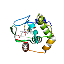 | | Characterization of the conversion of the malformed, recombinant cytochrome rc552 to a 2-formyl-4-vinyl (Spirographis) heme | | Descriptor: | 2-FORMYL-PROTOPORPHRYN IX, Cytochrome c-552 | | Authors: | Fee, J.A, Todaro, T.R, Luna, E, Sanders, D, Hunsicker-Wang, L.M, Patel, K.M, Bren, K.L, Gomez-Moran, E, Hill, M.G, Ai, J, Loehr, T.M, Oertling, W.A, Williams, P.A, Stout, C.D, McRee, D, Pastuszyn, A. | | Deposit date: | 2003-09-22 | | Release date: | 2004-09-28 | | Last modified: | 2024-11-13 | | Method: | X-RAY DIFFRACTION (1.61 Å) | | Cite: | Cytochrome rC552, formed during expression of the truncated, Thermus thermophilus cytochrome c552 gene in the cytoplasm of Escherichia coli, reacts spontaneously to form protein-bound 2-formyl-4-vinyl (Spirographis) heme.
Biochemistry, 43, 2004
|
|
1QYZ
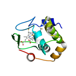 | | Characterization of the malformed, recombinant cytochrome rC552 | | Descriptor: | 2-ACETYL-PROTOPORPHYRIN IX, Cytochrome c-552 | | Authors: | Fee, J.A, Todaro, T.R, Luna, E, Sanders, D, Hunsicker-Wang, L.M, Patel, K.M, Bren, K.L, Gomez-Moran, E, Hill, M.G, Ai, J, Loehr, T.M, Oertling, W.A, Williams, P.A, Stout, C.D, McRee, D, Pastuszyn, A. | | Deposit date: | 2003-09-12 | | Release date: | 2004-09-28 | | Last modified: | 2024-10-30 | | Method: | X-RAY DIFFRACTION (1.4 Å) | | Cite: | Cytochrome rC552, formed during expression of the truncated, Thermus thermophilus cytochrome c552 gene in the cytoplasm of Escherichia coli, reacts spontaneously to form protein-bound 2-formyl-4-vinyl (Spirographis) heme.
Biochemistry, 43, 2004
|
|
2LIS
 | |
1R9O
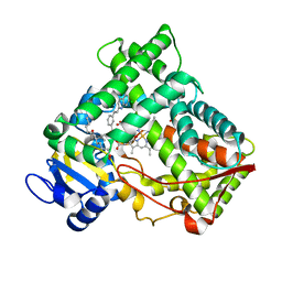
 | | Crystal Structure of P4502C9 with Flurbiprofen bound | | Descriptor: | Cytochrome P450 2C9, FLURBIPROFEN, GLYCEROL, ... | | Authors: | Wester, M.R, Yano, J.K, Schoch, G.A, Griffin, K.J, Stout, C.D, Johnson, E.F. | | Deposit date: | 2003-10-30 | | Release date: | 2004-06-15 | | Last modified: | 2023-08-23 | | Method: | X-RAY DIFFRACTION (2 Å) | | Cite: | The Structure of Human Cytochrome P450 2C9 Complexed with Flurbiprofen at 2.0 A Resolution
J.Biol.Chem., 279, 2004
|
|
1RF9
 | | Crystal structure of cytochrome P450-cam with a fluorescent probe D-4-AD (Adamantane-1-carboxylic acid-5-dimethylamino-naphthalene-1-sulfonylamino-butyl-amide) | | Descriptor: | 1,2-ETHANEDIOL, ADAMANTANE-1-CARBOXYLIC ACID-5-DIMETHYLAMINO-NAPHTHALENE-1-SULFONYLAMINO-BUTYL-AMIDE, Cytochrome P450-cam, ... | | Authors: | Hays, A.-M.A, Dunn, A.R, Gray, H.B, Stout, C.D, Goodin, D.B. | | Deposit date: | 2003-11-07 | | Release date: | 2004-11-16 | | Last modified: | 2023-08-23 | | Method: | X-RAY DIFFRACTION (1.8 Å) | | Cite: | Conformational States of Cytochrome P450cam Revealed by Trapping of synthetic Molecular Wires
J.Mol.Biol., 344, 2004
|
|
2LYN
 | |
1RE9
 | | CRYSTAL STRUCTURE OF CYTOCHROME P450-CAM WITH A FLUORESCENT PROBE D-8-AD (ADAMANTANE-1-CARBOXYLIC ACID-5-DIMETHYLAMINO-NAPHTHALENE-1-SULFONYLAMINO-OCTYL-AMIDE) | | Descriptor: | 1,2-ETHANEDIOL, ADAMANTANE-1-CARBOXYLIC ACID-5-DIMETHYLAMINO-NAPHTHALENE-1-SULFONYLAMINO-OCTYL-AMIDE, Cytochrome P450-cam, ... | | Authors: | Hays, A.-M.A, Dunn, A.R, Gray, H.B, Stout, C.D, Goodin, D.B. | | Deposit date: | 2003-11-06 | | Release date: | 2004-11-16 | | Last modified: | 2023-08-23 | | Method: | X-RAY DIFFRACTION (1.45 Å) | | Cite: | Conformational states of cytochrome P450cam revealed by trapping of synthetic molecular wires.
J.Mol.Biol., 344, 2004
|
|
1PNO
 | | Crystal structure of R. rubrum transhydrogenase domain III bound to NADP | | Descriptor: | NAD(P) transhydrogenase subunit beta, NADP NICOTINAMIDE-ADENINE-DINUCLEOTIDE PHOSPHATE | | Authors: | Sundaresan, V, Yamaguchi, M, Chartron, J, Stout, C.D. | | Deposit date: | 2003-06-12 | | Release date: | 2003-11-11 | | Last modified: | 2024-02-14 | | Method: | X-RAY DIFFRACTION (2.1 Å) | | Cite: | Conformational Change in the NADP(H) Binding Domain of Transhydrogenase Defines Four States
Biochemistry, 42, 2003
|
|
1PNQ
 | | Crystal structure of R. rubrum transhydrogenase domain III bound to NADPH | | Descriptor: | NAD(P) transhydrogenase subunit beta, NADPH DIHYDRO-NICOTINAMIDE-ADENINE-DINUCLEOTIDE PHOSPHATE | | Authors: | Sundaresan, V, Yamaguchi, M, Chartron, J, Stout, C.D. | | Deposit date: | 2003-06-12 | | Release date: | 2003-11-11 | | Last modified: | 2024-02-14 | | Method: | X-RAY DIFFRACTION (2.4 Å) | | Cite: | Conformational Change in the NADP(H) Binding Domain of Transhydrogenase Defines Four States
Biochemistry, 42, 2003
|
|
1PO5
 | | Structure of mammalian cytochrome P450 2B4 | | Descriptor: | Cytochrome P450 2B4, PROTOPORPHYRIN IX CONTAINING FE | | Authors: | Scott, E.E, He, Y.A, Wester, M.R, White, M.A, Chin, C.C, Halpert, J.R, Johnson, E.F, Stout, C.D. | | Deposit date: | 2003-06-13 | | Release date: | 2003-10-07 | | Last modified: | 2023-08-16 | | Method: | X-RAY DIFFRACTION (1.6 Å) | | Cite: | An open conformation of mammalian cytochrome P450 2B4 at 1.6 A resolution
Proc.Natl.Acad.Sci.USA, 100, 2003
|
|
1PQ2
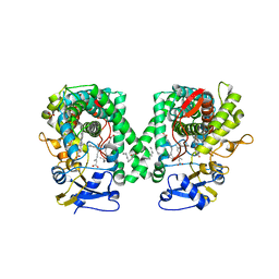
 | | Crystal Structure of Human Drug Metabolizing Cytochrome P450 2C8 | | Descriptor: | Cytochrome P450 2C8, PALMITIC ACID, PHOSPHATE ION, ... | | Authors: | Schoch, G.A, Yano, J.K, Wester, M.R, Griffin, K.J, Stout, C.D, Johnson, E.F. | | Deposit date: | 2003-06-17 | | Release date: | 2004-01-13 | | Last modified: | 2023-08-16 | | Method: | X-RAY DIFFRACTION (2.7 Å) | | Cite: | Structure of human microsomal cytochrome P450 2C8. Evidence for a peripheral fatty acid binding site
J.Biol.Chem., 279, 2004
|
|
2NNI
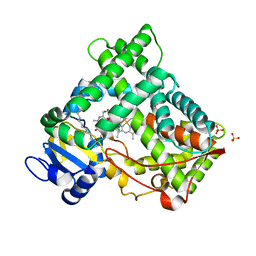
 | | CYP2C8dH complexed with montelukast | | Descriptor: | Cytochrome P450 2C8, MONTELUKAST, PALMITIC ACID, ... | | Authors: | Schoch, G.A, Yano, J.K, Stout, C.D, Johnson, E.F. | | Deposit date: | 2006-10-24 | | Release date: | 2007-10-23 | | Last modified: | 2023-08-30 | | Method: | X-RAY DIFFRACTION (2.8 Å) | | Cite: | Determinants of cytochrome P450 2C8 substrate binding: structures of complexes with montelukast, troglitazone, felodipine, and 9-cis-retinoic acid.
J.Biol.Chem., 283, 2008
|
|
2NNH
 | | CYP2C8dH complexed with 2 molecules of 9-cis retinoic acid | | Descriptor: | (9cis)-retinoic acid, Cytochrome P450 2C8, PALMITIC ACID, ... | | Authors: | Schoch, G.A, Yano, J.K, Stout, C.D, Johnson, E.F. | | Deposit date: | 2006-10-24 | | Release date: | 2007-10-23 | | Last modified: | 2023-08-30 | | Method: | X-RAY DIFFRACTION (2.6 Å) | | Cite: | Determinants of cytochrome P450 2C8 substrate binding: structures of complexes with montelukast, troglitazone, felodipine, and 9-cis-retinoic acid.
J.Biol.Chem., 283, 2008
|
|
2NNJ
 | | CYP2C8dH complexed with felodipine | | Descriptor: | Cytochrome P450 2C8, FELODIPINE, PALMITIC ACID, ... | | Authors: | Schoch, G.A, Yano, J.K, Stout, C.D, Johnson, E.F. | | Deposit date: | 2006-10-24 | | Release date: | 2007-10-23 | | Last modified: | 2023-08-30 | | Method: | X-RAY DIFFRACTION (2.28 Å) | | Cite: | Determinants of cytochrome P450 2C8 substrate binding: structures of complexes with montelukast, troglitazone, felodipine, and 9-cis-retinoic acid.
J.Biol.Chem., 283, 2008
|
|


