Loading
PDBj
MenuPDBj@FacebookPDBj@TwitterPDBj@YouTubewwPDB FoundationwwPDB
RCSB PDBPDBeBMRBAdv. SearchSearch help
Search by PDB author
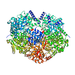
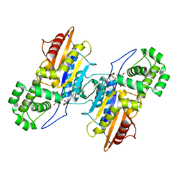
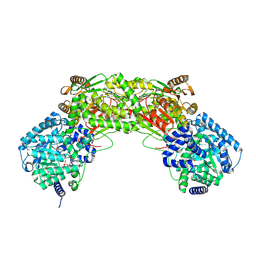
6X0H
DownloadVisualize
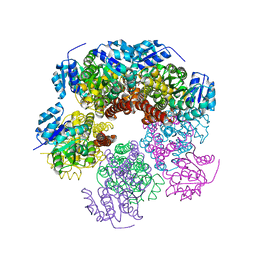
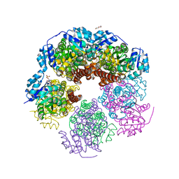
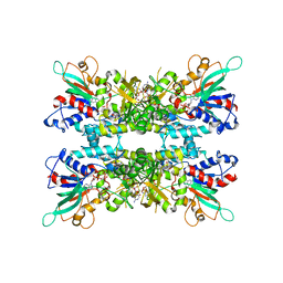
BU of 6x0h by Molmil
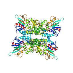
Structure of oxidized SidA ornithine hydroxylase with the FAD in the "out" conformation
Descriptor: ACETATE ION, CALCIUM ION, FLAVIN-ADENINE DINUCLEOTIDE, ...
Authors:Tanner, J.J, Campbell, A.C.
Deposit date:2020-05-15
Release date:2020-08-05
Last modified:2023-10-18
Method:X-RAY DIFFRACTION (2.087 Å)
Cite:Trapping conformational states of a flavin-dependent N -monooxygenase in crystallo reveals protein and flavin dynamics.
J.Biol.Chem., 295, 2020
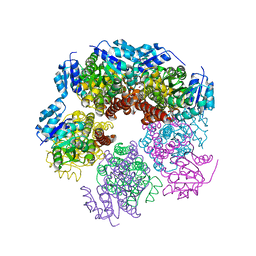
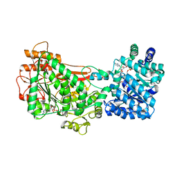
6X0J
DownloadVisualize
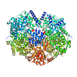
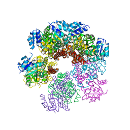
BU of 6x0j by Molmil
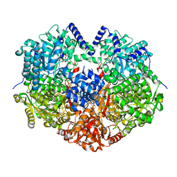
Structure of reduced SidA ornithine hydroxylase with the FAD "in" and complexed with NADP and L-ornithine
Descriptor: DIHYDROFLAVINE-ADENINE DINUCLEOTIDE, L-ornithine, L-ornithine N(5)-monooxygenase, ...
Authors:Tanner, J.J, Campbell, A.C.
Deposit date:2020-05-15
Release date:2020-08-05
Last modified:2023-10-18
Method:X-RAY DIFFRACTION (2.335 Å)
Cite:Trapping conformational states of a flavin-dependent N -monooxygenase in crystallo reveals protein and flavin dynamics.
J.Biol.Chem., 295, 2020
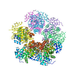
6X0I
DownloadVisualize
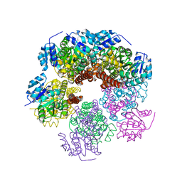
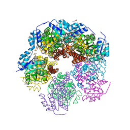
BU of 6x0i by Molmil
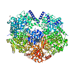
Structure of oxidized SidA ornithine hydroxylase with the FAD "in" and complexed with NADP
Descriptor: ACETATE ION, CALCIUM ION, FLAVIN-ADENINE DINUCLEOTIDE, ...
Authors:Tanner, J.J, Campbell, A.C.
Deposit date:2020-05-15
Release date:2020-08-05
Last modified:2023-10-18
Method:X-RAY DIFFRACTION (1.95 Å)
Cite:Trapping conformational states of a flavin-dependent N -monooxygenase in crystallo reveals protein and flavin dynamics.
J.Biol.Chem., 295, 2020
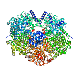
6X9B
DownloadVisualize
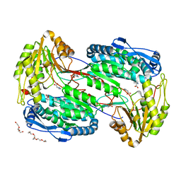
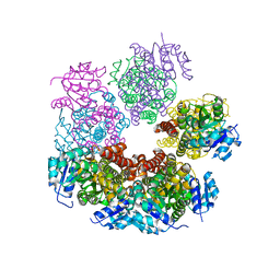
BU of 6x9b by Molmil
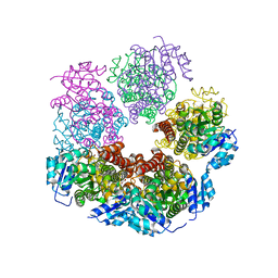
Structure of proline utilization A with cis-4-hydroxy-D-proline bound in the L-glutamate-gamma-semialdehyde dehydrogenase active site
Descriptor: (4R)-4-hydroxy-D-proline, Bifunctional protein PutA, DI(HYDROXYETHYL)ETHER, ...
Authors:Tanner, J.J, Campbell, A.C.
Deposit date:2020-06-02
Release date:2020-12-30
Last modified:2023-10-18
Method:X-RAY DIFFRACTION (1.46 Å)
Cite:Structural analysis of prolines and hydroxyprolines binding to the l-glutamate-gamma-semialdehyde dehydrogenase active site of bifunctional proline utilization A.
Arch.Biochem.Biophys., 698, 2020
6X9C
DownloadVisualize
BU of 6x9c by Molmil
Structure of proline utilization A with L-proline bound in the L-glutamate-gamma-semialdehyde dehydrogenase active site
Descriptor: 1,4-DIHYDRONICOTINAMIDE ADENINE DINUCLEOTIDE, Bifunctional protein PutA, DI(HYDROXYETHYL)ETHER, ...
Authors:Tanner, J.J, Campbell, A.C.
Deposit date:2020-06-02
Release date:2020-12-30
Last modified:2023-10-18
Method:X-RAY DIFFRACTION (1.44 Å)
Cite:Structural analysis of prolines and hydroxyprolines binding to the l-glutamate-gamma-semialdehyde dehydrogenase active site of bifunctional proline utilization A.
Arch.Biochem.Biophys., 698, 2020
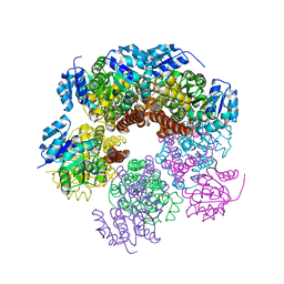
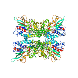
6XOZ
DownloadVisualize
BU of 6xoz by Molmil
Structure of human PYCR1 complexed with L-tetrahydro-2-furoic acid
Descriptor: CHLORIDE ION, Pyrroline-5-carboxylate reductase 1, mitochondrial, ...
Authors:Tanner, J.J, Christensen, E.M.
Deposit date:2020-07-07
Release date:2020-11-04
Last modified:2023-10-18
Method:X-RAY DIFFRACTION (2.35 Å)
Cite:In crystallo screening for proline analog inhibitors of the proline cycle enzyme PYCR1.
J.Biol.Chem., 295, 2020
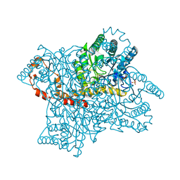
6X99
DownloadVisualize
BU of 6x99 by Molmil
Structure of proline utilization A with D-proline bound in the L-glutamate-gamma-semialdehyde dehydrogenase active site
Descriptor: Bifunctional protein PutA, D-PROLINE, FLAVIN-ADENINE DINUCLEOTIDE, ...
Authors:Tanner, J.J, Campbell, A.C.
Deposit date:2020-06-02
Release date:2020-12-30
Last modified:2023-10-18
Method:X-RAY DIFFRACTION (1.56 Å)
Cite:Structural analysis of prolines and hydroxyprolines binding to the l-glutamate-gamma-semialdehyde dehydrogenase active site of bifunctional proline utilization A.
Arch.Biochem.Biophys., 698, 2020
6XP0
DownloadVisualize
BU of 6xp0 by Molmil
Structure of human PYCR1 complexed with N-formyl L-proline
Descriptor: 1-formyl-L-proline, Pyrroline-5-carboxylate reductase 1, mitochondrial
Authors:Tanner, J.J, Christensen, E.M.
Deposit date:2020-07-07
Release date:2020-11-04
Last modified:2023-11-15
Method:X-RAY DIFFRACTION (1.95 Å)
Cite:In crystallo screening for proline analog inhibitors of the proline cycle enzyme PYCR1.
J.Biol.Chem., 295, 2020
6XP3
DownloadVisualize
BU of 6xp3 by Molmil
Structure of human PYCR1 complexed with cyclopentanecarboxylic acid
Descriptor: Pyrroline-5-carboxylate reductase 1, mitochondrial, SULFATE ION, ...
Authors:Tanner, J.J, Bogner, A.N.
Deposit date:2020-07-07
Release date:2020-11-04
Last modified:2023-10-18
Method:X-RAY DIFFRACTION (1.93 Å)
Cite:In crystallo screening for proline analog inhibitors of the proline cycle enzyme PYCR1.
J.Biol.Chem., 295, 2020
6X9A
DownloadVisualize
BU of 6x9a by Molmil
Structure of proline utilization A with trans-4-hydroxy-D-proline bound in the L-glutamate-gamma-semialdehyde dehydrogenase active site
Descriptor: (4S)-4-hydroxy-D-proline, Bifunctional protein PutA, DI(HYDROXYETHYL)ETHER, ...
Authors:Tanner, J.J, Campbell, A.C.
Deposit date:2020-06-02
Release date:2020-12-30
Last modified:2023-10-18
Method:X-RAY DIFFRACTION (1.41 Å)
Cite:Structural analysis of prolines and hydroxyprolines binding to the l-glutamate-gamma-semialdehyde dehydrogenase active site of bifunctional proline utilization A.
Arch.Biochem.Biophys., 698, 2020
6XP1
DownloadVisualize
BU of 6xp1 by Molmil
Structure of human PYCR1 complexed with L-thiazolidine-2-carboxylate
Descriptor: (2S)-1,3-thiazolidine-2-carboxylic acid, Pyrroline-5-carboxylate reductase 1, mitochondrial, ...
Authors:Tanner, J.J, Christensen, E.M.
Deposit date:2020-07-07
Release date:2020-11-04
Last modified:2023-10-18
Method:X-RAY DIFFRACTION (1.75 Å)
Cite:In crystallo screening for proline analog inhibitors of the proline cycle enzyme PYCR1.
J.Biol.Chem., 295, 2020
6X9D
DownloadVisualize
BU of 6x9d by Molmil
Structure of proline utilization A with trans-4-hydroxy-L-proline bound in the L-glutamate-gamma-semialdehyde dehydrogenase active site
Descriptor: 4-HYDROXYPROLINE, Bifunctional protein PutA, DI(HYDROXYETHYL)ETHER, ...
Authors:Tanner, J.J, Campbell, A.C.
Deposit date:2020-06-02
Release date:2020-12-30
Last modified:2023-10-18
Method:X-RAY DIFFRACTION (1.54 Å)
Cite:Structural analysis of prolines and hydroxyprolines binding to the l-glutamate-gamma-semialdehyde dehydrogenase active site of bifunctional proline utilization A.
Arch.Biochem.Biophys., 698, 2020
6XP2
DownloadVisualize
BU of 6xp2 by Molmil
Structure of human PYCR1 complexed with L-thiazolidine-4-carboxylate
Descriptor: Pyrroline-5-carboxylate reductase 1, mitochondrial, SULFATE ION, ...
Authors:Tanner, J.J, Christensen, E.M.
Deposit date:2020-07-07
Release date:2020-11-04
Last modified:2023-10-18
Method:X-RAY DIFFRACTION (2.3 Å)
Cite:In crystallo screening for proline analog inhibitors of the proline cycle enzyme PYCR1.
J.Biol.Chem., 295, 2020
4E3X
DownloadVisualize
BU of 4e3x by Molmil
Crystal Structure of Mus musculus 1-pyrroline-5-carboxylate dehydrogenase cryoprotected in proline
Descriptor: 3,6,9,12,15,18-HEXAOXAICOSANE, Delta-1-pyrroline-5-carboxylate dehydrogenase, mitochondrial, ...
Authors:Tanner, J.J, Pemberton, T.A.
Deposit date:2012-03-10
Release date:2012-07-25
Last modified:2023-09-13
Method:X-RAY DIFFRACTION (1.24 Å)
Cite:Proline: Mother Nature's cryoprotectant applied to protein crystallography.
Acta Crystallogr.,Sect.D, 68, 2012
4E3W
DownloadVisualize
BU of 4e3w by Molmil
Crystal Structure Francisella tularensis histidine acid phosphatase cryoprotected with proline
Descriptor: Acid phosphatase, PROLINE, SULFATE ION
Authors:Tanner, J.J, Pemberton, T.A.
Deposit date:2012-03-10
Release date:2012-07-25
Last modified:2023-09-13
Method:X-RAY DIFFRACTION (1.75 Å)
Cite:Proline: Mother Nature's cryoprotectant applied to protein crystallography.
Acta Crystallogr.,Sect.D, 68, 2012
4E3V
DownloadVisualize
BU of 4e3v by Molmil
Crystal Structure of XYLOSE ISOMERASE FROM STREPTOMYCES RUBIGINOSUS Cryoprotected in Proline
Descriptor: MAGNESIUM ION, MANGANESE (II) ION, PROLINE, ...
Authors:Tanner, J.J, Pemberton, T.A.
Deposit date:2012-03-10
Release date:2012-07-25
Last modified:2023-09-13
Method:X-RAY DIFFRACTION (1.503 Å)
Cite:Proline: Mother Nature's cryoprotectant applied to protein crystallography.
Acta Crystallogr.,Sect.D, 68, 2012
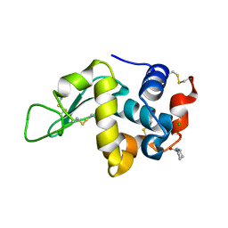
4E3U
DownloadVisualize
BU of 4e3u by Molmil
Crystal Structure of Hen Egg White Lysozyme Cryoprotected in Proline
Descriptor: CHLORIDE ION, Lysozyme C, PROLINE
Authors:Tanner, J.J, Pemberton, T.A.
Deposit date:2012-03-10
Release date:2012-07-25
Last modified:2023-09-13
Method:X-RAY DIFFRACTION (1.5 Å)
Cite:Proline: Mother Nature's cryoprotectant applied to protein crystallography.
Acta Crystallogr.,Sect.D, 68, 2012
5UAW
DownloadVisualize
BU of 5uaw by Molmil
Structure of apo human PYCR-1 crystallized in space group P21212
Descriptor: Pyrroline-5-carboxylate reductase 1, mitochondrial, SULFATE ION
Authors:Tanner, J.J.
Deposit date:2016-12-20
Release date:2017-03-15
Last modified:2023-10-04
Method:X-RAY DIFFRACTION (1.85 Å)
Cite:Resolving the cofactor-binding site in the proline biosynthetic enzyme human pyrroline-5-carboxylate reductase 1.
J. Biol. Chem., 292, 2017
5UAT
DownloadVisualize
BU of 5uat by Molmil
Structure of human PYCR-1 complexed with NADPH
Descriptor: DI(HYDROXYETHYL)ETHER, NADPH DIHYDRO-NICOTINAMIDE-ADENINE-DINUCLEOTIDE PHOSPHATE, Pyrroline-5-carboxylate reductase 1, ...
Authors:Tanner, J.J.
Deposit date:2016-12-20
Release date:2017-03-15
Last modified:2023-10-04
Method:X-RAY DIFFRACTION (1.92 Å)
Cite:Resolving the cofactor-binding site in the proline biosynthetic enzyme human pyrroline-5-carboxylate reductase 1.
J. Biol. Chem., 292, 2017
5UAU
DownloadVisualize
BU of 5uau by Molmil
Structure of human PYCR-1 complexed with proline
Descriptor: PROLINE, Pyrroline-5-carboxylate reductase 1, mitochondrial, ...
Authors:Tanner, J.J.
Deposit date:2016-12-20
Release date:2017-03-15
Last modified:2023-10-04
Method:X-RAY DIFFRACTION (1.9 Å)
Cite:Resolving the cofactor-binding site in the proline biosynthetic enzyme human pyrroline-5-carboxylate reductase 1.
J. Biol. Chem., 292, 2017
5UAV
DownloadVisualize
BU of 5uav by Molmil
Structure of human PYCR-1 complexed with NADPH and L-tetrahydrofuroic acid
Descriptor: DI(HYDROXYETHYL)ETHER, NADPH DIHYDRO-NICOTINAMIDE-ADENINE-DINUCLEOTIDE PHOSPHATE, Pyrroline-5-carboxylate reductase 1, ...
Authors:Tanner, J.J.
Deposit date:2016-12-20
Release date:2017-03-15
Last modified:2023-10-04
Method:X-RAY DIFFRACTION (1.85 Å)
Cite:Resolving the cofactor-binding site in the proline biosynthetic enzyme human pyrroline-5-carboxylate reductase 1.
J. Biol. Chem., 292, 2017
5UAX
DownloadVisualize
BU of 5uax by Molmil
Structure of apo human PYCR-1 crystallized in space group C2
Descriptor: CHLORIDE ION, Pyrroline-5-carboxylate reductase 1, mitochondrial
Authors:Tanner, J.J.
Deposit date:2016-12-20
Release date:2017-03-15
Last modified:2023-10-04
Method:X-RAY DIFFRACTION (1.85 Å)
Cite:Resolving the cofactor-binding site in the proline biosynthetic enzyme human pyrroline-5-carboxylate reductase 1.
J. Biol. Chem., 292, 2017
5UR2
DownloadVisualize
BU of 5ur2 by Molmil
Crystal structure of proline utilization A (PutA) from Bdellovibrio bacteriovorus inactivated by N-propargylglycine
Descriptor: Bifunctional protein PutA, N-propargylglycine-modified flavin adenine dinucleotide
Authors:Tanner, J.J.
Deposit date:2017-02-09
Release date:2017-08-02
Last modified:2023-10-04
Method:X-RAY DIFFRACTION (2.23 Å)
Cite:Biophysical investigation of type A PutAs reveals a conserved core oligomeric structure.
FEBS J., 284, 2017
5UX5
DownloadVisualize
BU of 5ux5 by Molmil
Structure of Proline Utilization A (PutA) from Corynebacterium freiburgense
Descriptor: BIFUNCTIONAL PROTEIN Proline utilization A (PutA), FLAVIN-ADENINE DINUCLEOTIDE, NICOTINAMIDE-ADENINE-DINUCLEOTIDE, ...
Authors:Tanner, J.J.
Deposit date:2017-02-22
Release date:2017-04-26
Last modified:2023-10-04
Method:X-RAY DIFFRACTION (2.7 Å)
Cite:Structure and characterization of a class 3B proline utilization A: Ligand-induced dimerization and importance of the C-terminal domain for catalysis.
J. Biol. Chem., 292, 2017
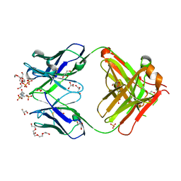
1P7K
DownloadVisualize
BU of 1p7k by Molmil
Crystal structure of an anti-ssDNA antigen-binding fragment (Fab) bound to 4-(2-Hydroxyethyl)piperazine-1-ethanesulfonic acid (HEPES)
Descriptor: 4-(2-HYDROXYETHYL)-1-PIPERAZINE ETHANESULFONIC ACID, DI(HYDROXYETHYL)ETHER, GLYCEROL, ...
Authors:Schuermann, J.P, Henzl, M.T, Deutscher, S.L, Tanner, J.J.
Deposit date:2003-05-02
Release date:2004-05-11
Last modified:2023-08-16
Method:X-RAY DIFFRACTION (1.75 Å)
Cite:Structure of an anti-DNA fab complexed with a non-DNA ligand provides insights into cross-reactivity and molecular mimicry.
Proteins, 57, 2004

220472

数据于2024-05-29公开中

PDB statisticsPDBj update infoContact PDBjnumon