Loading
PDBj
MenuPDBj@FacebookPDBj@TwitterPDBj@YouTubewwPDB FoundationwwPDB
RCSB PDBPDBeBMRBAdv. SearchSearch help
Search by PDB author
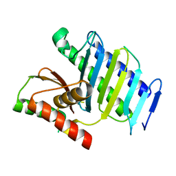
2HBW
DownloadVisualize
BU of 2hbw by Molmil
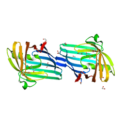
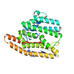
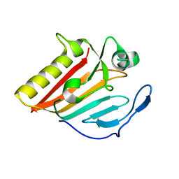
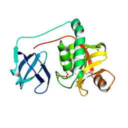
Crystal structure of a putative endopeptidase (ava_3396) from anabaena variabilis atcc 29413 at 1.05 A resolution
Descriptor: ACETATE ION, NLP/P60 protein, UNKNOWN LIGAND
Authors:Joint Center for Structural Genomics (JCSG)
Deposit date:2006-06-14
Release date:2006-08-08
Last modified:2023-01-25
Method:X-RAY DIFFRACTION (1.05 Å)
Cite:Structural Basis of Murein Peptide Specificity of a gamma-D-Glutamyl-L-Diamino Acid Endopeptidase.
Structure, 17, 2009
2GVK
DownloadVisualize
BU of 2gvk by Molmil
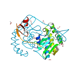
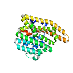
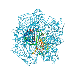
Crystal structure of a dye-decolorizing peroxidase (DyP) from Bacteroides thetaiotaomicron VPI-5482 at 1.6 A resolution
Descriptor: 1,2-ETHANEDIOL, ACETIC ACID, CHLORIDE ION, ...
Authors:Joint Center for Structural Genomics (JCSG)
Deposit date:2006-05-02
Release date:2006-05-16
Last modified:2023-01-25
Method:X-RAY DIFFRACTION (1.6 Å)
Cite:Crystal structures of two novel dye-decolorizing peroxidases reveal a beta-barrel fold with a conserved heme-binding motif.
Proteins, 69, 2007
2H1T
DownloadVisualize
BU of 2h1t by Molmil
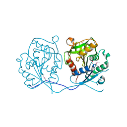
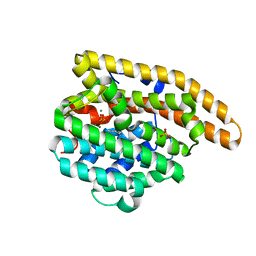
Crystal structure of a duf1089 family protein (pa1994) from pseudomonas aeruginosa at 1.80 A resolution
Descriptor: (4S)-2-METHYL-2,4-PENTANEDIOL, 1,2-ETHANEDIOL, Hypothetical protein
Authors:Joint Center for Structural Genomics (JCSG)
Deposit date:2006-05-16
Release date:2006-05-30
Last modified:2023-01-25
Method:X-RAY DIFFRACTION (1.8 Å)
Cite:The structure of the first representative of Pfam family PF06475 reveals a new fold with possible involvement in glycolipid metabolism.
Acta Crystallogr.,Sect.F, 66, 2010
2GVI
DownloadVisualize
BU of 2gvi by Molmil
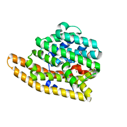
Crystal structure of a putative formylmethanofuran dehydrogenase subunit e (ta1109) from thermoplasma acidophilum at 1.87 A resolution
Descriptor: 1,2-ETHANEDIOL, ACETIC ACID, UNKNOWN LIGAND, ...
Authors:Joint Center for Structural Genomics (JCSG)
Deposit date:2006-05-02
Release date:2006-06-13
Last modified:2023-01-25
Method:X-RAY DIFFRACTION (1.87 Å)
Cite:Structures of three members of Pfam PF02663 (FmdE) implicated in microbial methanogenesis reveal a conserved alpha+beta core domain and an auxiliary C-terminal treble-clef zinc finger.
Acta Crystallogr.,Sect.F, 66, 2010
7SIU
DownloadVisualize
BU of 7siu by Molmil
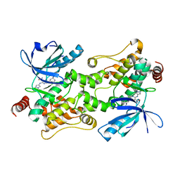
Crystal structure of HPK1 (MAP4K1) complex with inhibitor A-745
Descriptor: 6-[(5R)-5-benzamidocyclohex-1-en-1-yl]-3-[(1-methyl-1H-pyrazol-4-yl)amino]pyrazine-2-carboxamide, Mitogen-activated protein kinase kinase kinase kinase 1, SULFATE ION
Authors:Longenecker, K.L, Korepanova, A, Qiu, W.
Deposit date:2021-10-14
Release date:2022-03-02
Last modified:2023-10-18
Method:X-RAY DIFFRACTION (1.786 Å)
Cite:The HPK1 Inhibitor A-745 Verifies the Potential of Modulating T Cell Kinase Signaling for Immunotherapy.
Acs Chem.Biol., 17, 2022
2GHR
DownloadVisualize
BU of 2ghr by Molmil
Crystal structure of homoserine o-succinyltransferase (NP_981826.1) from Bacillus cereus ATCC 10987 at 2.40 A resolution
Descriptor: Homoserine O-succinyltransferase, SULFATE ION
Authors:Joint Center for Structural Genomics (JCSG)
Deposit date:2006-03-27
Release date:2006-04-11
Last modified:2023-01-25
Method:X-RAY DIFFRACTION (2.4 Å)
Cite:Crystal structure of homoserine O-succinyltransferase from Bacillus cereus at 2.4 A resolution
Proteins, 68, 2007
6AXO
DownloadVisualize
BU of 6axo by Molmil
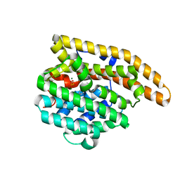
F96S Epi-isozizaene synthase
Descriptor: Epi-isozizaene synthase
Authors:Blank, P.N, Barrow, G.H, Christianson, D.W.
Deposit date:2017-09-07
Release date:2017-10-18
Last modified:2023-10-04
Method:X-RAY DIFFRACTION (2.1 Å)
Cite:Substitution of Aromatic Residues with Polar Residues in the Active Site Pocket of epi-Isozizaene Synthase Leads to the Generation of New Cyclic Sesquiterpenes.
Biochemistry, 56, 2017
6AXM
DownloadVisualize
BU of 6axm by Molmil
F95Y Epi-isozizaene synthase
Descriptor: Epi-isozizaene synthase, MAGNESIUM ION, N-benzyl-N,N-diethylethanaminium, ...
Authors:Blank, P.N, Barrow, G.H, Christianson, D.W.
Deposit date:2017-09-07
Release date:2017-10-18
Last modified:2023-10-04
Method:X-RAY DIFFRACTION (1.8 Å)
Cite:Substitution of Aromatic Residues with Polar Residues in the Active Site Pocket of epi-Isozizaene Synthase Leads to the Generation of New Cyclic Sesquiterpenes.
Biochemistry, 56, 2017
6AXN
DownloadVisualize
BU of 6axn by Molmil
F95C Epi-isozizaene synthase
Descriptor: Epi-isozizaene synthase, MAGNESIUM ION, N-benzyl-N,N-diethylethanaminium, ...
Authors:Blank, P.N, Barrow, G.H, Christianson, D.W.
Deposit date:2017-09-07
Release date:2017-10-18
Last modified:2023-10-04
Method:X-RAY DIFFRACTION (1.9 Å)
Cite:Substitution of Aromatic Residues with Polar Residues in the Active Site Pocket of epi-Isozizaene Synthase Leads to the Generation of New Cyclic Sesquiterpenes.
Biochemistry, 56, 2017
6AXU
DownloadVisualize
BU of 6axu by Molmil
W203Y Epi-isozizaene synthase
Descriptor: Epi-isozizaene synthase, MAGNESIUM ION, N-benzyl-N,N-diethylethanaminium, ...
Authors:Blank, P.N, Barrow, G.H, Christianson, D.W.
Deposit date:2017-09-07
Release date:2017-10-18
Last modified:2023-10-04
Method:X-RAY DIFFRACTION (1.822 Å)
Cite:Substitution of Aromatic Residues with Polar Residues in the Active Site Pocket of epi-Isozizaene Synthase Leads to the Generation of New Cyclic Sesquiterpenes.
Biochemistry, 56, 2017
6AX9
DownloadVisualize
BU of 6ax9 by Molmil
F95N Epi-isozizaene synthase
Descriptor: Epi-isozizaene synthase, MAGNESIUM ION, N-benzyl-N,N-diethylethanaminium, ...
Authors:Blank, P.N, Barrow, G.H, Christianson, D.W.
Deposit date:2017-09-06
Release date:2017-10-18
Last modified:2023-10-04
Method:X-RAY DIFFRACTION (2.403 Å)
Cite:Substitution of Aromatic Residues with Polar Residues in the Active Site Pocket of epi-Isozizaene Synthase Leads to the Generation of New Cyclic Sesquiterpenes.
Biochemistry, 56, 2017
8KAD
DownloadVisualize
BU of 8kad by Molmil
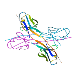
Crystal structure of an antibody light chain tetramer with 3D domain swapping
Descriptor: Antibody light chain
Authors:Sakai, T, Mashima, T, Kobayashi, N, Ogata, H, Uda, T, Hifumi, E, Hirota, S.
Deposit date:2023-08-02
Release date:2023-12-27
Method:X-RAY DIFFRACTION (2 Å)
Cite:Structural and thermodynamic insights into antibody light chain tetramer formation through 3D domain swapping.
Nat Commun, 14, 2023
7E0B
DownloadVisualize
BU of 7e0b by Molmil
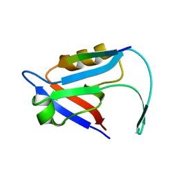
The crystal structure of sorting nexin 27 and PBM complex
Descriptor: PBM, Sorting nexin-27
Authors:Shang, G.J, Qi, J.X.
Deposit date:2021-01-27
Release date:2022-02-02
Last modified:2023-11-29
Method:X-RAY DIFFRACTION (1.29 Å)
Cite:SNX27 suppresses SARS-CoV-2 infection by inhibiting viral lysosome/late endosome entry.
Proc.Natl.Acad.Sci.USA, 119, 2022
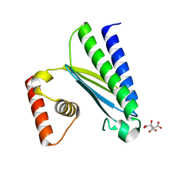
8GUN
DownloadVisualize
BU of 8gun by Molmil
Crystal structure of mutant H528A of EsaD from Staphylococcus aureus
Descriptor: MAGNESIUM ION, Type VII secretion system protein EssD
Authors:Zhang, Z.M, Wang, Y.J.
Deposit date:2022-09-13
Release date:2022-11-09
Last modified:2024-04-03
Method:X-RAY DIFFRACTION (2.30001378 Å)
Cite:A toxin-deformation dependent inhibition mechanism in the T7SS toxin-antitoxin system of Gram-positive bacteria.
Nat Commun, 13, 2022
8GUO
DownloadVisualize
BU of 8guo by Molmil
Crystal structure of the nuclease domain of EsaD in complex with EsaG from Staphylococcus aureus
Descriptor: Type VII secretion system protein EsaG, Type VII secretion system protein EssD
Authors:Zhang, Z.M, Wang, Y.J.
Deposit date:2022-09-13
Release date:2022-11-09
Last modified:2024-04-03
Method:X-RAY DIFFRACTION (2.5939517 Å)
Cite:A toxin-deformation dependent inhibition mechanism in the T7SS toxin-antitoxin system of Gram-positive bacteria.
Nat Commun, 13, 2022
8GUP
DownloadVisualize
BU of 8gup by Molmil
Crystal structure of EsaG from Staphylococcus aureus
Descriptor: CITRIC ACID, Type VII secretion system protein EsaG
Authors:Zhang, Z.M, Wang, Y.J.
Deposit date:2022-09-13
Release date:2022-11-09
Last modified:2024-05-29
Method:X-RAY DIFFRACTION (2.298725 Å)
Cite:A toxin-deformation dependent inhibition mechanism in the T7SS toxin-antitoxin system of Gram-positive bacteria.
Nat Commun, 13, 2022
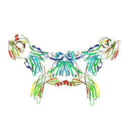
8GO9
DownloadVisualize
BU of 8go9 by Molmil
Structure of beta-arrestin2 in complex with a phosphopeptide corresponding to the human Atypical chemokine receptor 2, ACKR2 (D6R)
Descriptor: Atypical chemokine receptor 2, Beta-arrestin-2, Fab30 Heavy Chain, ...
Authors:Maharana, J, Sarma, P, Yadav, M.K, Banerjee, R, Shukla, A.K.
Deposit date:2022-08-24
Release date:2023-12-27
Last modified:2024-01-17
Method:ELECTRON MICROSCOPY (3.35 Å)
Cite:Molecular insights into atypical modes of beta-arrestin interaction with seven transmembrane receptors.
Science, 383, 2024
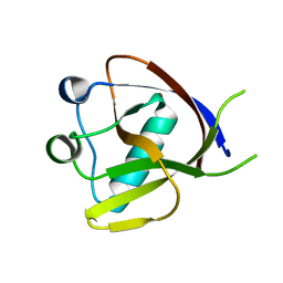
7EQ4
DownloadVisualize
BU of 7eq4 by Molmil
Crystal Structure of the N-terminus of Nonstructural protein 1 from SARS-CoV-2
Descriptor: Host translation inhibitor nsp1
Authors:Liu, Y, Ke, Z, Hu, H, Zhao, K, Xiao, J, Xia, Y, Li, Y.
Deposit date:2021-04-29
Release date:2021-06-09
Last modified:2023-11-29
Method:X-RAY DIFFRACTION (1.25 Å)
Cite:Structural Basis and Function of the N Terminus of SARS-CoV-2 Nonstructural Protein 1.
Microbiol Spectr, 9, 2021
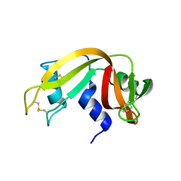
4MXF
DownloadVisualize
BU of 4mxf by Molmil
X-ray structure of the adduct between bovine pancreatic ribonuclease and Auoxo6, a dinuclear gold(III) complex with -dioxo bridges linking the two metal centers
Descriptor: GOLD ION, Ribonuclease pancreatic
Authors:Russo Krauss, I, Vergara, A, Merlino, A.
Deposit date:2013-09-26
Release date:2013-12-25
Last modified:2023-09-20
Method:X-RAY DIFFRACTION (2.25 Å)
Cite:Interactions of gold-based drugs with proteins: crystal structure of the adduct formed between ribonuclease A and a cytotoxic gold(iii) compound.
Metallomics, 6, 2014
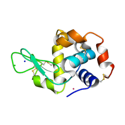
4J1B
DownloadVisualize
BU of 4j1b by Molmil
X-ray structure of the adduct between hen egg white lysozyme and AziRu (black crystal)
Descriptor: CHLORIDE ION, Lysozyme C, RUTHENIUM ION, ...
Authors:Vergara, A, Merlino, A.
Deposit date:2013-02-01
Release date:2013-04-03
Last modified:2013-05-08
Method:X-RAY DIFFRACTION (1.66 Å)
Cite:Interaction of Anticancer Ruthenium Compounds with Proteins: High-Resolution X-ray Structures and Raman Microscopy Studies of the Adduct between Hen Egg White Lysozyme and AziRu.
Inorg.Chem., 52, 2013
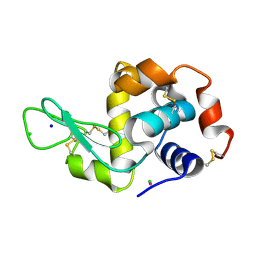
4J1A
DownloadVisualize
BU of 4j1a by Molmil
X-ray structure of the adduct between hen egg white lysozyme and AziRu (green crystal)
Descriptor: CHLORIDE ION, Lysozyme C, RUTHENIUM ION, ...
Authors:Vergara, A, Merlino, A.
Deposit date:2013-02-01
Release date:2013-04-03
Last modified:2013-05-08
Method:X-RAY DIFFRACTION (1.79 Å)
Cite:Interaction of Anticancer Ruthenium Compounds with Proteins: High-Resolution X-ray Structures and Raman Microscopy Studies of the Adduct between Hen Egg White Lysozyme and AziRu.
Inorg.Chem., 52, 2013
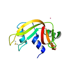
4L55
DownloadVisualize
BU of 4l55 by Molmil
X-ray structure of the adduct between bovine pancreatic ribonuclease and AziRu
Descriptor: RUTHENIUM ION, Ribonuclease pancreatic
Authors:Vergara, A, Russo Krauss, I, Merlino, A.
Deposit date:2013-06-10
Release date:2014-01-29
Last modified:2024-10-09
Method:X-RAY DIFFRACTION (1.65 Å)
Cite:Investigating the ruthenium metalation of proteins: X-ray structure and Raman microspectroscopy of the complex between RNase A and AziRu.
Inorg.Chem., 52, 2013
<1234


225946

数据于2024-10-09公开中

PDB statisticsPDBj update infoContact PDBjnumon