Loading
PDBj
MenuPDBj@FacebookPDBj@TwitterPDBj@YouTubewwPDB FoundationwwPDB
RCSB PDBPDBeBMRBAdv. SearchSearch help
Search by PDB author
7TWZ
DownloadVisualize
BU of 7twz by Molmil
Crystal Structure of NADP-linked putative oxidoreductase from Klebsiella pneumoniae
Descriptor: ACETATE ION, DI(HYDROXYETHYL)ETHER, NADP NICOTINAMIDE-ADENINE-DINUCLEOTIDE PHOSPHATE, ...
Authors:Seattle Structural Genomics Center for Infectious Disease (SSGCID)
Deposit date:2022-02-07
Release date:2022-02-23
Last modified:2023-10-18
Method:X-RAY DIFFRACTION (2.1 Å)
Cite:Crystal Structure of NADP-linked putative oxidoreductase from Klebsiella pneumoniae
to be published
6N41
DownloadVisualize
BU of 6n41 by Molmil
Crystal structure of InvbM.18715.a.KN11: Influenza hemagglutinin from strain A/Netherlands/002P1/1951
Descriptor: 1,2-ETHANEDIOL, 2-acetamido-2-deoxy-beta-D-glucopyranose, 2-acetamido-2-deoxy-beta-D-glucopyranose-(1-4)-2-acetamido-2-deoxy-beta-D-glucopyranose, ...
Authors:Seattle Structural Genomics Center for Infectious Disease (SSGCID)
Deposit date:2018-11-16
Release date:2018-12-05
Last modified:2023-05-24
Method:X-RAY DIFFRACTION (2.5 Å)
Cite:Structural characterisation of hemagglutinin from seven Influenza A H1N1 strains reveal diversity in the C05 antibody recognition site.
Sci Rep, 13, 2023
3K5P
DownloadVisualize
BU of 3k5p by Molmil
Crystal structure of amino acid-binding ACT: D-isomer specific 2-hydroxyacid dehydrogenase catalytic domain from Brucella melitensis
Descriptor: 1,2-ETHANEDIOL, D-3-phosphoglycerate dehydrogenase, GLYCEROL
Authors:Seattle Structural Genomics Center for Infectious Disease (SSGCID)
Deposit date:2009-10-07
Release date:2009-11-03
Last modified:2023-09-06
Method:X-RAY DIFFRACTION (2.15 Å)
Cite:Crystal structure of amino acid-binding ACT: D-isomer specific 2-hydroxyacid dehydrogenase catalytic domain from Brucella melitensis
To be Published
3KNU
DownloadVisualize
BU of 3knu by Molmil
Crystal structure of tRNA (guanine-N1)-methyltransferase from Anaplasma phagocytophilum
Descriptor: tRNA (guanine-N(1)-)-methyltransferase
Authors:Seattle Structural Genomics Center for Infectious Disease (SSGCID)
Deposit date:2009-11-12
Release date:2009-11-24
Last modified:2023-09-06
Method:X-RAY DIFFRACTION (2.25 Å)
Cite:Crystal structure of tRNA (guanine-N1)-methyltransferase from Anaplasma phagocytophilum
To be Published
3K2H
DownloadVisualize
BU of 3k2h by Molmil
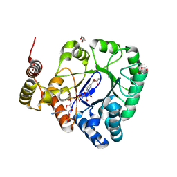
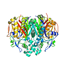
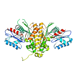
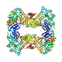
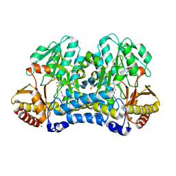
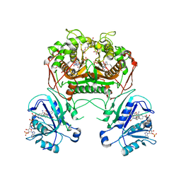
Co-crystal structure of dihydrofolate reductase/thymidylate synthase from Babesia bovis with dUMP, Pemetrexed and NADP
Descriptor: 1,2-ETHANEDIOL, 2'-DEOXYURIDINE 5'-MONOPHOSPHATE, 2-{4-[2-(2-AMINO-4-OXO-4,7-DIHYDRO-3H-PYRROLO[2,3-D]PYRIMIDIN-5-YL)-ETHYL]-BENZOYLAMINO}-PENTANEDIOIC ACID, ...
Authors:Seattle Structural Genomics Center for Infectious Disease (SSGCID)
Deposit date:2009-09-30
Release date:2009-10-13
Last modified:2023-09-06
Method:X-RAY DIFFRACTION (2.2 Å)
Cite:Inhibitor-bound complexes of dihydrofolate reductase-thymidylate synthase from Babesia bovis.
Acta Crystallogr.,Sect.F, 67, 2011
6O3F
DownloadVisualize
BU of 6o3f by Molmil
Crystal Structure of Lysyl-tRNA Synthetase from Chlamydia trachomatis with complexed with L-lysine and a difluoro cyclohexyl chromone ligand
Descriptor: 1,2-ETHANEDIOL, DI(HYDROXYETHYL)ETHER, LYSINE, ...
Authors:Seattle Structural Genomics Center for Infectious Disease (SSGCID)
Deposit date:2019-02-26
Release date:2019-03-27
Last modified:2023-10-11
Method:X-RAY DIFFRACTION (2.4 Å)
Cite:Crystal Structure of Lysyl-tRNA Synthetase from Chlamydia trachomatis with complexed with L-lysine and a difluoro cyclohexyl chromone ligand
to be published
6NS0
DownloadVisualize
BU of 6ns0 by Molmil
Crystal Structure of Lysyl-tRNA Synthetase from Chlamydia trachomatis complexed with L-lysine and Cladosporin
Descriptor: 1,2-ETHANEDIOL, CHLORIDE ION, DI(HYDROXYETHYL)ETHER, ...
Authors:Seattle Structural Genomics Center for Infectious Disease, Seattle Structural Genomics Center for Infectious Disease (SSGCID)
Deposit date:2019-01-24
Release date:2019-02-06
Last modified:2023-10-11
Method:X-RAY DIFFRACTION (2.2 Å)
Cite:Crystal Structure of Lysyl-tRNA Synthetase from Chlamydia trachomatis with complexed with L-lysine and Cladosporin
to be published
7U0M
DownloadVisualize
BU of 7u0m by Molmil
Crystal structure of a enoyl-[acyl-carrier-protein] reductase (InhA) from Mycobacterium abscessus bound to NAD and NITD-916
Descriptor: 6-[(4,4-dimethylcyclohexyl)methyl]-4-hydroxy-3-phenylpyridin-2(1H)-one, Enoyl-[acyl-carrier-protein] reductase [NADH], NICOTINAMIDE-ADENINE-DINUCLEOTIDE, ...
Authors:Seattle Structural Genomics Center for Infectious Disease, Seattle Structural Genomics Center for Infectious Disease (SSGCID)
Deposit date:2022-02-18
Release date:2022-10-05
Last modified:2023-10-18
Method:X-RAY DIFFRACTION (1.45 Å)
Cite:Efficacy and Mode of Action of a Direct Inhibitor of Mycobacterium abscessus InhA.
Acs Infect Dis., 8, 2022
3KZU
DownloadVisualize
BU of 3kzu by Molmil
Crystal structure of 3-oxoacyl-(acyl carrier protein) synthase II from Brucella melitensis
Descriptor: 1,2-ETHANEDIOL, 3-oxoacyl-(Acyl-carrier-protein) synthase ii, SULFATE ION
Authors:Seattle Structural Genomics Center for Infectious Disease (SSGCID)
Deposit date:2009-12-08
Release date:2009-12-15
Last modified:2023-09-06
Method:X-RAY DIFFRACTION (1.75 Å)
Cite:Crystal structure of 3-oxoacyl-(acyl carrier protein) synthase II from Brucella melitensis
TO BE PUBLISHED
3L0D
DownloadVisualize
BU of 3l0d by Molmil
Crystal structure of glyceraldehyde-3-phosphate dehydrogenase from Bartonella henselae with bound NAD
Descriptor: Glyceraldehyde 3-phosphate dehydrogenase, NICOTINAMIDE-ADENINE-DINUCLEOTIDE
Authors:Seattle Structural Genomics Center for Infectious Disease (SSGCID)
Deposit date:2009-12-09
Release date:2009-12-29
Last modified:2023-09-06
Method:X-RAY DIFFRACTION (2.5 Å)
Cite:Crystal structure of glyceraldehyde-3-phosphate dehydrogenase from Bartonella henselae with bound NAD
To be Published
6U3L
DownloadVisualize
BU of 6u3l by Molmil
Crystal structure of Hemerythrin HHE cation binding domain-containing protein: Rv2633c homolog from Mycobacterium kansasii
Descriptor: 1,2-ETHANEDIOL, Hemerythrin HHE cation binding domain protein
Authors:Seattle Structural Genomics Center for Infectious Disease (SSGCID)
Deposit date:2019-08-22
Release date:2020-01-22
Last modified:2024-03-13
Method:X-RAY DIFFRACTION (1.75 Å)
Cite:Crystal structure of a hemerythrin-like protein from Mycobacterium kansasii and homology model of the orthologous Rv2633c protein of M. tuberculosis.
Biochem.J., 477, 2020
6UK3
DownloadVisualize
BU of 6uk3 by Molmil
Crystal Structure of S-adenosyl-L-homocysteine hydrolase from Acanthamoeba castellanii with bound NAD and Adenosine
Descriptor: ACETATE ION, ADENOSINE, Adenosylhomocysteinase, ...
Authors:Seattle Structural Genomics Center for Infectious Disease (SSGCID)
Deposit date:2019-10-04
Release date:2019-10-16
Last modified:2023-10-11
Method:X-RAY DIFFRACTION (1.4 Å)
Cite:Crystal Structure of S-adenosyl-L-homocysteine hydrolase from Acanthamoeba castellanii with bound NAD and Adenosine
to be published
6UJ5
DownloadVisualize
BU of 6uj5 by Molmil
Crystal structure of CAB1 Pantothenate Kinase from Saccharomyces cerevisiae
Descriptor: MAGNESIUM ION, Pantothenate kinase CAB1
Authors:Seattle Structural Genomics Center for Infectious Disease (SSGCID)
Deposit date:2019-10-02
Release date:2020-10-07
Last modified:2024-03-13
Method:X-RAY DIFFRACTION (1.8 Å)
Cite:Crystal structure of CAB1 Pantothenate Kinase from Saccharomyces cerevisiae
to be published
6UJK
DownloadVisualize
BU of 6ujk by Molmil
Crystal Structure of a Probable short-chain type dehydrogenase/reductase (Rv1144) from Mycobacterium tuberculosis with bound NAD
Descriptor: 1,2-ETHANEDIOL, 2-AMINO-2-HYDROXYMETHYL-PROPANE-1,3-DIOL, NICOTINAMIDE-ADENINE-DINUCLEOTIDE, ...
Authors:Seattle Structural Genomics Center for Infectious Disease (SSGCID)
Deposit date:2019-10-03
Release date:2019-10-16
Last modified:2023-10-11
Method:X-RAY DIFFRACTION (1.2 Å)
Cite:Crystal Structure of a Probable short-chain type dehydrogenase/reductase (Rv1144) from Mycobacterium tuberculosis with bound NAD
to be published
6ULD
DownloadVisualize
BU of 6uld by Molmil
Crystal structure of serine hydroxymethyltransferase from Mycobacterium tuberculosis with bound PLP forming a Schiff base with substrate Serine in one monomer and PLP forming a Schiff base with product Glycine in the other monomer
Descriptor: (4S)-2-METHYL-2,4-PENTANEDIOL, GLYCINE, PYRIDOXAL-5'-PHOSPHATE, ...
Authors:Seattle Structural Genomics Center for Infectious Disease (SSGCID)
Deposit date:2019-10-07
Release date:2019-10-16
Last modified:2023-10-11
Method:X-RAY DIFFRACTION (1.5 Å)
Cite:Crystal structure of serine hydroxymethyltransferase from Mycobacterium tuberculosis with bound PLP forming a Schiff base with substrate Serine in one monomer and PLP forming a Schiff base with product Glycine in the other monomer
to be published
6UCZ
DownloadVisualize
BU of 6ucz by Molmil
Crystal structure of dihydropteroate synthase from Anaplasma phagocytophilum with bound 6-hydroxymethylpterin-monophosphate
Descriptor: 1,2-ETHANEDIOL, ACETATE ION, CITRIC ACID, ...
Authors:Seattle Structural Genomics Center for Infectious Disease (SSGCID)
Deposit date:2019-09-18
Release date:2019-10-16
Last modified:2023-10-11
Method:X-RAY DIFFRACTION (2 Å)
Cite:Crystal structure of dihydropteroate synthase from Anaplasma phagocytophilum with bound 6-hydroxymethylpterin-monophosphate
To be Published
6UDE
DownloadVisualize
BU of 6ude by Molmil
Crystal structure of Glycerol kinase from Elizabethkingia anophelis NUHP1 in complex with ADP and glycerol
Descriptor: ADENOSINE-5'-DIPHOSPHATE, GLYCEROL, Glycerol kinase, ...
Authors:Seattle Structural Genomics Center for Infectious Disease (SSGCID)
Deposit date:2019-09-19
Release date:2019-10-16
Last modified:2023-10-11
Method:X-RAY DIFFRACTION (1.95 Å)
Cite:Crystal structure of Glycerol kinase from Elizabethkingia anophelis NUHP1 in complex with ADP and glycerol
TO BE PUBLISHED
6UYN
DownloadVisualize
BU of 6uyn by Molmil
Crystal structure of influenza A virus hemagglutinin from A/Ohio/09/2015 bound to the stalk-binding CR6261 antibody Fab
Descriptor: 2-acetamido-2-deoxy-beta-D-glucopyranose, 2-acetamido-2-deoxy-beta-D-glucopyranose-(1-4)-2-acetamido-2-deoxy-beta-D-glucopyranose, CHLORIDE ION, ...
Authors:Seattle Structural Genomics Center for Infectious Disease, Seattle Structural Genomics Center for Infectious Disease (SSGCID)
Deposit date:2019-11-13
Release date:2020-09-02
Last modified:2023-10-11
Method:X-RAY DIFFRACTION (2.85 Å)
Cite:Crystal structure of influenza A virus hemagglutinin from A/Ohio/09/2015 bound to the stalk-binding CR6261 antibody Fab
To Be Published
6VEL
DownloadVisualize
BU of 6vel by Molmil
Crystal Structure of Human E-cadherin bound by mouse monoclonal antibody 66E8Fab
Descriptor: 66E8 Fab Heavy Chain, 66E8 Fab Light Chain, CALCIUM ION, ...
Authors:Seattle Structural Genomics Center for Infectious Disease, Seattle Structural Genomics Center for Infectious Disease (SSGCID)
Deposit date:2020-01-02
Release date:2020-01-29
Last modified:2023-10-25
Method:X-RAY DIFFRACTION (2.65 Å)
Cite:Regulation of multiple dimeric states of E-cadherin by adhesion activating antibodies revealed through Cryo-EM and X-ray crystallography.
Pnas Nexus, 1, 2022
6VU9
DownloadVisualize
BU of 6vu9 by Molmil
Crystal structure of threonyl-tRNA synthetase (ThrRS) from Stenotrophomonas maltophilia K279a
Descriptor: SULFATE ION, Threonine--tRNA ligase, ZINC ION
Authors:Seattle Structural Genomics Center for Infectious Disease (SSGCID)
Deposit date:2020-02-14
Release date:2020-03-04
Last modified:2023-10-11
Method:X-RAY DIFFRACTION (2.2 Å)
Cite:Crystal structure of threonyl-tRNA synthetase (ThrRS) from Stenotrophomonas maltophilia K279a
To Be Published
6VUD
DownloadVisualize
BU of 6vud by Molmil
Crystal structure of a ribosome recycling factor from Ehrlichia chaffeensis
Descriptor: CHLORIDE ION, Ribosome-recycling factor
Authors:Seattle Structural Genomics Center for Infectious Disease (SSGCID)
Deposit date:2020-02-15
Release date:2020-03-18
Last modified:2024-04-03
Method:X-RAY DIFFRACTION (2.35 Å)
Cite:Crystal structure of a ribosome recycling factor from Ehrlichia chaffeensis
to be published
4JXJ
DownloadVisualize
BU of 4jxj by Molmil
Crystal Structure of Ribosomal RNA small subunit methyltransferase A from Rickettsia bellii Determined by Iodide SAD Phasing
Descriptor: 1,2-ETHANEDIOL, IODIDE ION, Ribosomal RNA small subunit methyltransferase A
Authors:Seattle Structural Genomics Center for Infectious Disease (SSGCID)
Deposit date:2013-03-28
Release date:2013-05-01
Last modified:2024-02-28
Method:X-RAY DIFFRACTION (2 Å)
Cite:Crystal Structure of Ribosomal RNA small subunit methyltransferase A from Rickettsia bellii Determined by Iodide SAD Phasing
TO BE PUBLISHED
4LIH
DownloadVisualize
BU of 4lih by Molmil
The crystal structure of Gamma-glutamyl-gamma-aminobutyraldehyde dehydrogenase from Burkholderia cenocepacia J2315
Descriptor: 1,2-ETHANEDIOL, 2-(N-MORPHOLINO)-ETHANESULFONIC ACID, Gamma-glutamyl-gamma-aminobutyraldehyde dehydrogenase
Authors:Seattle Structural Genomics Center for Infectious Disease, Seattle Structural Genomics Center for Infectious Disease (SSGCID)
Deposit date:2013-07-02
Release date:2013-07-31
Last modified:2023-09-20
Method:X-RAY DIFFRACTION (1.85 Å)
Cite:The crystal structure of Gamma-glutamyl-gamma-aminobutyraldehyde dehydrogenase from Burkholderia cenocepacia J2315
TO BE PUBLISHED
6VJU
DownloadVisualize
BU of 6vju by Molmil
Crystal Structure of Cystathionine beta synthase from Legionella pneumophila with LLP, PLP, and homocysteine
Descriptor: 1,2-ETHANEDIOL, 2-AMINO-4-MERCAPTO-BUTYRIC ACID, ACETATE ION, ...
Authors:Seattle Structural Genomics Center for Infectious Disease (SSGCID)
Deposit date:2020-01-17
Release date:2020-02-05
Last modified:2024-04-03
Method:X-RAY DIFFRACTION (1.3 Å)
Cite:Crystal Structure of Cystathionine beta synthase from Legionella pneumophila with LLP, PLP, and homocysteine
to be published
6WCI
DownloadVisualize
BU of 6wci by Molmil
Crystal structure of a cysteine desulfurase SufS from Stenotrophomonas maltophilia K279a
Descriptor: 1,2-ETHANEDIOL, Cysteine desulfurase, SULFATE ION
Authors:Seattle Structural Genomics Center for Infectious Disease (SSGCID)
Deposit date:2020-03-30
Release date:2020-04-08
Last modified:2023-11-15
Method:X-RAY DIFFRACTION (2.05 Å)
Cite:Crystal structure of a cysteine desulfurase SufS from Stenotrophomonas maltophilia K279a
To Be Published

222415

数据于2024-07-10公开中

PDB statisticsPDBj update infoContact PDBjnumon